Get in touch
555-555-5555

Limited time offer: 20% off all templates ➞


Guide to Using Science Images for Research Papers
One easy way to include science images in your manuscripts is to download and customize them for your figures..
When downloading images from the internet to use in your scientific papers and presentations, you need to be careful that they match the copyright, resolution, and sizing rules that allow them to be used in academic journals. This science image guide provides tips to help you choose the right kinds of files that you can use to create your own impressive designs.

Which image format is best for research papers?
There are two categories of images that can be used for scientific publications: editable and uneditable. Editable images that can be fully customized and scaled without losing resolution are called vector files. Uneditable images don't allow you to adjust the design or color and come in wide range of formats from low to high resolution. Both of these image types can be used in scientific papers as long as you follow the proper copyright and resolution rules. Learn more about these image types and the different uses below.
1. Editable Images
The best kind of science images are editable vector files that allow you to customize the designs to best match the main points of your research. These include image file types such as Scalable Vector Graphics (.svg), Adobe Illustrator (.ai), Affinity Designer (.afdesign), Encapsulated PostScript (.eps), and some files in PowerPoint (.pptx) if they were drawn using PowerPoint shape tools.
Editable Image Tips:
- Editable images are important because some scientific journals, such as Science, require that you provide them with figures that are formatted using editable vector files.
- Be cautious of using images and database tools that only offer limited design customization options such as BioRender. Partially editable images can be difficult to make an illustration that looks professional and seamless with your data and other designs.
- Vector images have customizable sizes, resolution and transparent backgrounds, so you can always scale the image and insert it into any background.
- Make sure you follow the copyright rules associated with your image download. Some vector image databases require attribution and others allow you to use them for any purpose.

How to Find Editable Images
The easiest ways to find editable images is to explore science image databases or use Google search. I recommend using the search terms "drawings", "vector art", or "vector images" paired with the image type keyword.
The example below shows the Google Image search results for "cancer cell drawings" with a variety of different options for downloading different types of science images. You will still need to make sure that the image is available as a vector file type to be fully editable (e.g. SVG, AI, or EPS file types). Most vector images will require some sort of payment or subscription to download the high resolution files and use without copyright issues.

1. Uneditable Images
The second best format is uneditable images. Common uneditable image types are PNG, TIFF, or JPEG formats and these can be incorporated into your scientific figures and presentations as long as they have high enough resolution and have copyright rules that allow you to use them in academic publications.
Uneditable image tips:
- Check the resolution of downloaded images to make sure they are high enough to use in scientific publications without looking grainy or unclear (see the "How to check image resolution" instructions in the section below).
- Try to find PNG images with transparent backgrounds to make it easier to incorporate into your scientific figures and posters.
- Be very careful in checking the source of uneditable images and follow all copyright rules associated with the image. Uneditable image are more likely to have copyright rules associated with them that do not allow their use in scientific journals.

How to Find High Resolution and Transparent Images
The easiest ways to find high resolution and transparent images is to explore science image databases or use Google search. I recommend using the search terms "transparent background" and using the Google "Tools" feature to limit the search for "Large" images.
The examples below shows the Google Large Image search results for "plant cell diagram transparent background" that show a variety of different options for downloading high resolution and transparent science images.

What image sizes are best for scientific publication?
Size and resolution are important because images need to be high resolution enough to show sharp shapes and lines when it is used in a printed or digital figure. Below are tips on how to choose the right image sizes and resolutions.
Image Resolution
Resolution is the most important aspect of a downloaded image or scientific figure and affects the sharpness of the details. A low-resolution image will have around 72 PPI and high resolution images are at least 300 PPI.
- Most scientific journals require images and figures to be at least 300 PPI/DPI.
- The "PPI" stands for Pixels Per Inch and is used when referring to digital file resolution and "DPI" stands for Dots Per Inch and is used for printing resolution.

How to Check Image Resolution:
- Windows computer: Right-click on the file, select Properties, then Details, and you will see the DPI in the Image section, labeled Horizontal Resolution and Vertical Resolution.
- Mac computer: Open the image in Preview and select Tools, then Adjust Size, and find the label Resolution.
The size requirements will depend on how you plan to use the image. Most scientific journals use a maximum figure width of 180mm, so if you only plan to use images in scientific publications, then you only need them large enough to look sharp within a 180 mm wide figure (~600 pixels wide).
If you plan to use the image in presentation or posters slides, you will need to have much larger images to not have resolution issues when shown on a big screen that is 1280 x 720 pixels or printed on a poster that is approximately 48 x 36 inches.
How Can I Find Copyright-Free Images?
Copyright laws ensure that an image is only used in a way that is approved by the image creator. The best way to ensure that you download images with copyright rules that allow you to use them for academic journal submissions is to read the fine print on the image source. The summary below describes how to find images that are allowed for use in scientific papers.
Copyright License Review
Anyone who creates their own original artwork has the right to be acknowledged as the creator of that image. They automatically own the copyright for the image, which means that legally, they have the right to decide where and how that image can be used. In order to be able to use images in scientific papers, you will need to know what kind of copyright license is being used, which you can usually find by looking for the original source of the image or by reading the fine print of the image database.
Types of copyright licenses for scientific use:
- Public Domain - Images generally become public domain after 70 years after the creator's death. If the copyright is not renewed on the creator's behalf, the image can become part of the ‘public domain’, and the copyright no longer applies.
- Creative Commons 4 - You can adapt and share the image in anyway you like, but this license requires attribution, so you will need to include the original creator in the acknowledgements of the research paper, posters, and acknowledged on your presentation slides.
- Stock Images - Image databases that allow you to license the designs. Make sure to read the fine print on how you are allowed to use the image (e.g. personal and commercial uses).

How to Find Copyright-Free Images:
Look for image databases that have copyright licenses that allow you to use the images "For personal, academic, and commercial projects and to modify it" such as:
- FreePik
- Simplified Science
- Wikimedia Commons
NOTE: Some copyright-free image databases may still require that you attribute the illustration to the original author in your scientific publication. Read the fine print to make sure you are using the image correctly!
Here is an example of Simplified Science Usage Rules for comparison to other image databases.
How to Use Downloaded Images in Publications?
After downloading images, the next step is to format them into your scientific designs. Two of the most common software tools that scientists use for figure formatting are Adobe Illustrator and PowerPoint. Below is a link to free online courses that show you how to use the downloaded images in your scientific publications and graphical abstracts.
Create professional science figures with illustration services or use the online courses and templates to quickly learn how to make your own designs.
Interested in free design templates and training.
Explore scientific illustration templates and courses by creating a Simplified Science Publishing Log In. Whether you are new to data visualization design or have some experience, these resources will improve your ability to use both basic and advanced design tools.
Interested in reading more articles on scientific design? Learn more below:

Scientific Presentation Guide: How to Create an Engaging Research Talk

Data Storytelling Techniques: How to Tell a Great Data Story in 4 Steps

Best Science PowerPoint Templates and Slide Design Examples
Content is protected by Copyright license. Website visitors are welcome to share images and articles, however they must include the Simplified Science Publishing URL source link when shared. Thank you!
Online Courses
Stay up-to-date for new simplified science courses, subscribe to our newsletter.
Thank you for signing up!
You have been added to the emailing list and will only recieve updates when there are new courses or templates added to the website.
We use cookies on this site to enhance your user experience and we do not sell data. By using this website, you are giving your consent for us to set cookies: View Privacy Policy
Simplified Science Publishing, LLC
- USC Libraries
- Research Guides
Using Images and Non-Textual Materials in Presentations, Papers, Theses, and Dissertations
- Documenting and Citing Images
- Finding Images - Select Sources
Documenting and Citing Images/Photographs and Their Sources
Please note that this is advice on best practices and considerations in documenting and citing images and non-print materials. It does not represent legal advice on obtaining permissions.
Generally, images copied from other sources should not be used without permissions in publications or for commercial purposes. Many American academic institutions require graduate students to archive their finished and approved theses/dissertations in institutional electronic repositories and/or institutional libraries and repositories, and/or to post them on Proquest's theses database. Unpublished theses and dissertations are a form of scholarly dissemination. Someone else's images, like someone else's ideas, words or music, should be used with critical commentary, and need to be identified and cited. If a thesis/dissertation is revised for publication, waivers or permissions from the copyright holder(s) of the images and non-textual materials must be obtained. Best practices also apply to materials found on the internet and on social media, and, properly speaking, require identification, citation, and clearance of permissions, as relevant.
Use the following elements when identifying and citing an image, depending on the information you have available . It is your responsibility to do due diligence and document as much as possible about the image you are using:
- Artist's/creator's name, if relevant;
- Title of the work/image, if known, or description;
- Ownership information (such as a person, estate, museum, library collection) and source of image;
- Material, if known, particularly for art works;
- Dimensions of the work, if known.
The Chicago Manual of Style online can be searched for norms on appropriate ways to caption illustrations, capitalize titles of visual works, or cite print materials that contain images.
Including images/photographs in a bibliography:
Best practice is to not include images within a bibliography of works cited. It is common, instead, to create a separate list of images (or figures) and their source, such as photographer (even if it's you) or collection. It may be useful to also include location, e.g., museum, geographic reference, address, etc.
Examples of Documenting Images
The image below is scanned from a published book. It can be used in a critical context within a presentation, classroom session, or paper/thesis, as follows:

[ Figure 1. This photograph from 1990 shows the Monument against Fascism designed by Jochen Gerz and Esther Shalev-Gerz, Hamburg, 1986-1993. Image from James Young, ed., Art of Memory: Holocaust Memorials in History (New York: Prestel, 1994), 70]
If you need to use this image in a published work, you will have to seek permission. For example, the book from which this image was scanned should have a section on photo credits which would help you identify the person/archive holding this image.
The image below was found through Google Images and downloaded from the internet. It can be used in a critical context within a presentation, classroom session, or paper/thesis, as follows:

[Figure 2. This image shows the interior of Bibliotheca Alexandrina designed by the Norwegian architecture firm Snøhetta in 2001. Image downloaded from https://mgkhs.com/gallery/alexandria in March 2016.]
If you want to use this image in a published work, you will have to do your best to track down its source to request permission to use. The web site or social media site where you found the image may not be an appropriate source, since it is common for people to repost images without attribution. Just because "everyone does it" does not mean that you should be using such materials without attribution or documentation. In this specific example, you may need to write to the photographer or to the architecture firm. If you have done due diligence and were unable to find the source, or have not received a response, you may be able to use an image found on the internet with appropriate documentation in a publication.
The image below was downloaded from a digitized historic collection of photographs held by an institutional archive. It can be used in a critical context within a presentation, classroom session, or paper/thesis, as follows:

[Figure 3. In the 1920s the urban landscape of Los Angeles started to change, as various developers began building multi-family apartment houses in sections previously zoned for single family dwellings. Seen in this photograph by Dick Whittington is the Warrington apartment building, which was completed in 1928, surrounded by older single family structures. Downloaded from the USC Digital Library in February 2016]
I f you plan to use this photograph in a publication, seek permission from the library/institution from whose digital archive you downloaded the image. Contact information is usually found in the record for the image.
The image below was taken by the author. It can be used in a critical context within a presentation, classroom session , paper/thesis, or a publication* as follows:

[Figure 4. Genex Tower, also known as West City Gate, is a residential tower located in New Belgrade. This example of late 20th century brutalist-style architecture was designed in 1977 by Mihajlo Mitrović. Photographed by the author in 2013.]
*Please note, if you re-photographed someone else's photograph or a work of art, or if you re-photographed a published image, you may not be able to publish your photograph without first seeking permission or credit for its content. If you have done due diligence and were unable to find the source or have not received a response, you may be able to use your image with appropriate documentation.
- << Previous: Fair Use
- Next: Finding Images - Select Sources >>
- Last Updated: Jan 19, 2023 3:12 PM
- URL: https://libguides.usc.edu/fair_use
Research Voyage
Research Tips and Infromation
The Power of Images in Research Papers: How They Enhance the Quality of Your Paper?

Introduction
Why are images important in research papers, the benefits of using images in research papers, using high-resolution images in a research paper, citing the source of the images used in a research paper, using relevant images in a research paper, optimizing the size and placement of images in a research paper, checking the copyright status of images before using them, saving images in a lossless format, compressing images before adding them to a research paper, challenges and limitations of using images in research paper, cameras suitable for taking research images, how can i label my images in research paper inside image itself, popular image labelling tools with their key features:, whether image caption should contain keywords listed in the research paper, whether you need permission to include maps in your research paper .
Research papers must include images and figures because they significantly increase the work’s impact and readability. Images are a useful tool for researchers and authors since, in today’s world, visual information is frequently simpler to absorb and retain than text-only information. Images in research papers can do more than just serve as illustrations; they can also help to clarify difficult concepts, offer further details, and even enhance the text in a way that makes the article more interesting and memorable.
In this post, we’ll examine the use of photographs in research papers and the reasons they’re crucial to academic and scientific writing. We will also go over the many kinds of photos that can be utilised in research papers, their advantages, and the best ways to use them. If you want to write engaging and effective research papers, whether you’re a researcher, student, or scientific enthusiast, you must grasp the significance of images in research papers.
Images are a useful tool for researchers and authors in the scientific community because of their capacity to enthral and instruct. Research papers can benefit from the addition of figures like photographs, drawings, and block diagrams, whether they are taken with a camera or made using software like Canva. To make sure that images have the desired effect, it is crucial to use them efficiently. The advantages of include photos in research papers, the optimal usage methods, as well as the difficulties and restrictions that must be taken into account, will all be covered in this article.
From relevance and clarity to captioning and accessibility, we will examine the key factors that can impact the use and impact of images in research papers. Whether you are a seasoned researcher or just starting out, this discussion will provide valuable insights and guidance for using images effectively in your scientific work.
This article focuses only about the images which are captured from the cameras and block diagrams drawn by the researchers to show the methodology or any other aspect related to research. I have written separate articles on charts/graphs and Tables which you can refer below for further details.
- Maximizing the Impact of Your Research Paper with Graphs and Charts
- Best Practices for Designing and Formatting Tables in Research Papers
Images in research papers serve a variety of functions, from promoting reading and engagement to strengthening comprehension and memory. Many times, using graphics can make it easier for readers to understand complicated ideas and information. For instance, pictures can give a clear visual representation of the research topic, while sketches and block diagrams can help explain intricate systems and processes.
Images can enhance the paper’s readability and comprehension in addition to acting as a textual supplement. A picture can give the research a context in the real world, while a diagram can aid to demonstrate a topic or process that is mentioned in the text. When used well, photographs can create a seamless transition between written and visual data, strengthening the study paper’s impact and retention.
Cross-disciplinary communication can also be facilitated by the use of images in research articles. A block diagram, for instance, can be used to explain a complicated concept to a non-expert audience, while a photograph can draw in readers from many cultural backgrounds. Images can contribute to the accessibility and impact of research articles by bridging the gap between text and visual information.
In conclusion, graphics play a variety of roles in research papers and can significantly improve the work by bringing complicated concepts into focus, enhancing the language, and adding to its readability and retention. Images are a useful tool for researchers and authors who want to produce work that is impactful and accessible, whether they are used to depict study subjects, clarify procedures, or provide context.
The use of images in research papers can bring many benefits, making them valuable tools for researchers and authors. Some of the key benefits include:
- Enhancing readability and engagement: Images can make research papers more visually appealing and engaging, encouraging readers to stay focused and interested in the work. They can also help to break up text-heavy sections and make the paper more visually appealing, which can improve the overall reading experience.
- Improving understanding and retention of information: Research has shown that people tend to remember information better when it is presented in a visual format. By incorporating images into research papers, authors can help readers to better understand and retain information, which can increase the impact of the work.
- Facilitating cross-disciplinary communication: Images can help to bridge the gap between text and visual information, making research papers more accessible to a wider audience. This can be especially useful when communicating complex ideas to non-experts or individuals from different cultural backgrounds.
- Making the paper stand out and be more memorable: Research papers with high-quality, relevant, and clear images are more likely to be remembered and have a greater impact. By using images effectively, authors can make their work stand out from the crowd and increase its impact.
Best Practices to Follow when Adding Pictures to a Research Paper
There are a few best practices to follow when adding pictures to a research paper:
- Use high-resolution images: Make sure the images you use are of high quality and resolution. This will ensure that they look clear and crisp when printed or viewed on a screen.
- Cite the source of the image: Always include a caption for the image and cite the source. This is important for academic integrity and to give credit to the original creator of the image.
- Use relevant images: Choose images that are directly related to the content of the paper and will help to enhance the reader’s understanding.
- Optimize the size and placement of the images: Make sure the images are appropriately sized and placed in the document to ensure they do not detract from the text.
- Check copyright: Make sure the image you are using is not copyrighted and that you have permission to use it.
- Save images in a lossless format: To ensure that images maintain their quality, save them in a lossless format, such as TIFF or PNG.
- Compress images: Reduce the file size of the images before adding them to the paper, this will make the paper more manageable.
Sure, when it comes to using high-resolution images in a research paper, there are a few key things to keep in mind:
- Resolution refers to the number of pixels in an image and is typically measured in dots per inch (dpi) or pixels per inch (ppi). The higher the resolution, the more pixels an image contains, and the sharper and more detailed it will appear.
- For printed materials, a resolution of at least 300 dpi is generally recommended. This will ensure that the images look clear and crisp when printed, even at a larger size.
- For images that will be viewed primarily on a screen, a resolution of 72 dpi is typically sufficient. Keep in mind that higher-resolution images will have larger file sizes, which may slow down loading times.
- It’s also important to keep in mind the size of the image when using it in a research paper. Larger images will take up more space and may cause the paper to be larger in size.
- Always check the resolution and size of the image before using it in your paper. If the resolution is too low, the image may appear pixelated or blurry.
In summary, by using high-resolution images, you ensure that they look clear and crisp when printed or viewed on a screen, also the size of the image should be considered to not make the paper too large.

Citing the source of the images used in a research paper is an important aspect of academic integrity. It gives credit to the original creator of the image and allows readers to locate the image themselves if they wish to see it in more detail.
When including an image in a research paper, it is important to include a caption for the image. The caption should include the following information:
- Image number: This is a number or letter that corresponds to the image, typically in the format “Figure 1” or “Image A.”
- Title: A brief title that describes the image.
- Source: The source of the image, including the name of the creator, the title of the work, and the date of creation.
- Copyright information: If the image is copyrighted, it is important to include the copyright information along with the source. This includes the name of the copyright holder and the year the image was copyrighted.
- Permission: If you obtained permission to use the image, include the name of the individual or organization that granted permission.
It is also important to include a list of figures or a bibliography at the end of the paper that includes all the images used in the paper with the same information provided in the caption.
Citing the source of the image is not only important for academic integrity, but also it gives credit to the original creator and allows the readers to locate the image if they want to see it in more detail. Additionally, it also demonstrates that you have done the necessary research to support the claims made in the paper.
Including pertinent graphics in a research paper can both improve the reader’s comprehension of the material and make the document more interesting.
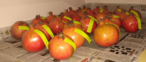
When choosing photographs, it’s crucial to pick ones that directly relate to the paper’s subject matter and that will enrich the text. An photograph of that species, for instance, would be pertinent if the paper is on that type of animal, as opposed to an image of an entirely different animal.
Example : Pomegranate Fruit Quality Assessment using Image Processing techniques
It’s crucial to take the context of the photographs into account when inserting them in the document. For the reader to grasp the connection between the image and the text, the images should be positioned close to the words to which they are related.
It’s also a good idea to take the image and text’s formats into account; the text should be readable and the image should be presented in an understandable manner.
In summary, using relevant images in a research paper can greatly enhance the reader’s understanding of the content and make the paper more engaging. It is important to choose images that are directly related to the content of the paper and to place them close to the text that they are related to. The format of the image and the text should also be considered to ensure the reader can easily understand the relationship between the image and the text.
Making sure that photographs in a research paper are the right size and location can ensure that they do not distract from the content and that readers can easily understand them.
It’s crucial to make sure that the photos are proportionately sized to the text when it comes to size. Small images could be challenging to see and interpret, while large images might take up a lot of space and make the paper appear cluttered.
It’s crucial to take the paper’s flow into account when deciding where to position things. The sequence of the images should make sense and correspond to the flow of the text. In order for the reader to grasp the connection between the image and the text, they should also be placed close to the text to which they are related.
In order to avoid obstructing the text’s flow and causing the document to appear cluttered, it’s crucial to take the page layout into account while adding photos.
In conclusion, making sure that photographs in a research paper are the right size and placed properly will assist to guarantee that they do not take away from the text and that the reader can easily understand them. To ensure that the reader can easily understand the relationship between the image and the text, images should be appropriately sized in relation to the text, placed in a logical order that follows the progression of the text, and placed close to the text that they are related to. The format of the image and the text should also be taken into consideration. The page layout should also be taken into account to prevent the graphics from obstructing the text’s flow and from giving the document a cluttered appearance.

Before utilising any photos in your research report, it’s crucial to check their copyright status to make sure you have permission to use them.
While each country has its own copyright regulations, generally speaking, an image is protected by copyright if it was made by someone who also owns the rights to it.
You must request permission from the owner of the copyright to use an image in your research work. Usually, you can do this by getting in touch with the copyright owners personally or using a copyright clearance centre.
Additionally, it’s crucial to keep in mind that some photographs can be subject to Creative Commons licences, which permit the image’s restricted use in exchange for correct acknowledgement. It’s crucial to read and comprehend the terms of the licence before utilising the image because these licences can be found on the website where the image is posted.
In conclusion, it’s crucial to verify the copyright status of photographs before utilising them in a research paper to make sure you have the legal right to do so. If you want to use an image in your research work, you must first get the owner’s permission. You should also read and comprehend any Creative Commons licences that may be applicable before utilising the image.
To guarantee that the photographs retain their quality when utilised in a research article, it is crucial to save them in a lossless format.
When an image is saved and opened, there is no loss of image quality thanks to a lossless format because it does not compress the image’s data. TIFF, PNG, and GIF are popular lossless image formats. Compared to “lossy” formats like JPEG, these formats often have greater file sizes, but they maintain the image’s integrity and guarantee that it will seem just as crisp and detailed when opened as when it was saved.
Contrarily, lossy formats, like JPEG, are intended to minimise the file size of an image but do so at the expense of part of the image’s data, which might degrade the image’s quality. This is inappropriate for research articles because they call for high-quality photographs.
Additionally, it’s crucial to keep in mind that when you save an image in a lossless format, you can modify it more than once without it losing quality. This is crucial since you might need to crop or resize the image for the publication.
In summary, Saving images in a lossless format is important to ensure that the images maintain their quality when used in a research paper. Common lossless image formats include TIFF, PNG and GIF, and it’s also important to note that when you save an image in a lossless format, you can open and edit the image multiple times without losing quality.
Compressing images before adding them to a research paper is important to reduce the file size of the images and make the paper more manageable.
File size can be an issue when working with images in a research paper, as large image files can slow down the loading times of the paper and make it more difficult to share or upload. Compressing images can help to reduce the file size of the images and make the paper more manageable.
There are several ways to compress images:
- Lossless compression : This type of compression reduces the file size of the image without losing any image quality. Common lossless compression formats include PNG and GIF.
- Lossy compression : This type of compression reduces the file size of the image by discarding some of the image data. Common lossy compression formats include JPEG.
- Photoshop : you can use photoshop to save for web, this option will give you more control on how much you want to compress the image and the quality of the image.
It’s important to note that lossy compression can result in a loss of image quality, so it’s best to use lossless compression if possible. Additionally, you should always check the image quality after compressing it to make sure that it’s still suitable for the paper.
In summary, compressing images before adding them to a research paper is important to reduce the file size of the images and make the paper more manageable. There are several ways to compress images, such as lossless compression, lossy compression and using photoshop to save for web, but it’s important to keep in mind that lossy compression can result in a loss of image quality, so it’s best to use lossless compression if possible. Additionally, you should always check the image quality after compressing it to make sure that it’s still suitable for the paper.
Challenges and Limitations of Using Images in Research Papers Despite the many benefits of using images in research papers, there are also challenges and limitations to consider. Some of the key challenges and limitations include:
- Cost : Creating high-quality images can be expensive, especially if specialized software or equipment is required. This can be a challenge for researchers and authors working with limited budgets.
- Technical proficiency : Creating clear and effective images requires technical proficiency, which may not be available to all researchers and authors. This can limit the use of images in research papers and the impact they have.
- Copyright and intellectual property issues : Using images from other sources can raise questions of copyright and intellectual property. It is important to be aware of these issues and ensure that all images used in research papers are properly cited and attributed.
- Space limitations : Research papers often have limited space, which can impact the use and impact of images. This may require authors to carefully consider the number and size of images used in their work.
- Accessibility : Some images may not be accessible to all readers, especially those with visual impairments. This can limit the reach and impact of research papers and should be considered when using images.
In conclusion, while the use of images in research papers can bring many benefits, it is important to be aware of the challenges and limitations associated with their use. Whether related to cost, technical proficiency, intellectual property, space limitations, or accessibility, these factors can impact the use and impact of images in research papers. Careful consideration and planning can help to mitigate these challenges and ensure that images are used effectively to enhance the impact of research papers.
When it comes to taking images for a research paper, the most important factor to consider is the quality of the images. While there are many cameras on the market, not all cameras are equally suitable for taking research-related images. Here are some key components to consider when choosing a camera for research purposes:
- Image Resolution : High image resolution is essential for capturing images that are clear, detailed, and suitable for publication. Look for a camera with at least 12 megapixels, but higher is better.
- Image Sensor : The image sensor is the part of the camera that captures light and converts it into a digital image. The larger the image sensor, the more light it can capture, which can result in better image quality. Look for a camera with a full-frame image sensor.
- Lens Quality : The quality of the lens will greatly impact the sharpness and detail of your images. Look for a camera with high-quality lenses, or consider purchasing additional lenses to meet your specific needs.
- Shooting Modes : Research-related images often require specialized shooting modes, such as macro, time-lapse, or slow-motion. Make sure the camera you choose has the shooting modes you need for your research.
- Image Stabilization : Image stabilization helps to reduce camera shake, which can result in blurry images. If you plan on hand-holding the camera, consider a camera with built-in image stabilization.
- Cost : Research cameras can be expensive, so consider your budget when choosing a camera. Some lower-cost cameras may still meet your needs, so it’s important to research your options.
Labelling images within the image itself is a common practice in research papers and is used to provide additional information about the image or to highlight specific parts of the image. There are several methods to label images in a research paper:

- Use annotations or callouts : These are text boxes or shapes that can be added to the image to provide additional information or to highlight specific parts of the image. Annotation tools are available in most photo editing software, including Adobe Photoshop or GIMP.
- Use arrows, lines or shapes : You can use arrows, lines or shapes to draw attention to specific parts of the image or to show relationships between different parts of the image. This is especially useful in images that show complex structures or relationships.
- Use overlay text : You can add overlay text to the image to provide additional information. This is useful in cases where you want to provide information about the image that is not immediately obvious from the image itself.
- Label the axes : In images that represent data, it is important to label the axes to help the reader understand the data being represented. This can be done using annotation tools or by using overlay text.
- Use colour coding : You can use colour coding to highlight specific parts of the image or to show relationships between different parts of the image. This is especially useful in images that show complex structures or relationships.

It is important to use labelling and annotations in a clear and concise manner, as they help to provide additional information about the image and to make the image easier to understand. Labels and annotations should also be placed in a consistent manner throughout the research paper to help maintain visual consistency.
Sometimes you have huge amount of image data with data labelling requirements for your research tasks. Then my advice to you is to outsource the image data for data labelling expert while taking proper care regarding your data protection. I have written a blog post on outsourcing images for data labelling. You can visit the post below.
Outsourcing Research Data Labelling: Risks and Rewards for Researchers
Sometimes research scholars are badly in need of financial assistance. They can not take regular job due to the research work and stress they may come across because of the new job. They can take up data labelling jobs which is a pure mechanical work rather a mental stress. I have written an article on data labelling jobs for researchers. Please visit the blog post below for further details.
Data Annotation (Data Labelling): A Part-Time Job for Research Scholars
Following is the list of image labelling tools that are commonly used in research papers. The choice of tool will depend on the specific needs of the research paper and the level of detail required in the labelling and annotations.
- Adobe Photoshop : Adobe Photoshop is a professional-grade image editing software that has robust features for annotating and labelling images. It allows you to add text boxes, shapes, arrows, and lines to an image and also has a variety of brush tools for more detailed labelling.
- GIMP : GIMP is a free and open-source image editing software that has similar features to Adobe Photoshop. It allows you to add text boxes, shapes, and arrows to an image and also has a variety of brush tools for detailed labelling.
- Inkscape : Inkscape is a free and open-source vector graphics editor that is often used for annotating and labelling images. It has robust features for adding text boxes, shapes, and lines to an image, and also allows you to import and export images in a variety of file formats.
- Microsoft PowerPoint : Microsoft PowerPoint is a presentation software that has basic image labelling tools. It allows you to add text boxes, shapes, and arrows to an image but is limited in its capabilities compared to Adobe Photoshop or GIMP.
- Canva : Canva is a user-friendly design tool that has a variety of features for annotating and labelling images. It allows you to add text boxes, shapes, and arrows to an image and also has a variety of design elements that can be added to an image.
These are just a few examples of image-labelling tools that are commonly used in research papers. The choice of tool will depend on the specific needs of the research paper and the level of detail required in the labelling and annotations.
It is recommended to include relevant keywords in the caption of images in a research paper. Keywords are an important aspect of search engine optimization (SEO) and can help increase the visibility of the paper online. By including keywords in the caption, you make it easier for readers to understand the context and content of the image, and you also help search engines better understand the context of the paper. Additionally, including keywords in the caption can also help establish a clear connection between the image and the rest of the paper, making it easier for readers to understand the overall narrative of the research.
For maps created by government agencies (e.g. USGS, NASA), copyright restrictions may apply, but generally these maps can be used for educational and research purposes without obtaining permission.
For maps created by commercial map providers (e.g. Google Maps, Mapbox), the use of the map may be subject to licensing agreements and usage restrictions. In these cases, it is important to review the terms and conditions of use and to obtain the necessary permission before including the map in your research paper.
For custom maps created by individuals or organizations, it is important to obtain permission from the creator before using the map in your research paper. This includes both maps created by yourself as well as maps created by others that you would like to include in your paper.
It is always best to check the copyright and usage restrictions for any maps you plan to include in your research paper, and to obtain the necessary permission if required, in order to ensure that you are using the maps in a legal and ethical manner.
In conclusion, the inclusion of graphics in research articles can significantly affect their clarity, interest level, and overall impact. Images can enhance research papers’ clarity, depth, and visual appeal, allowing authors to convey their findings and concepts. But it’s crucial to use images wisely, keeping in mind things like relevancy, clarity, captioning, and accessibility. Researchers and authors should be aware of both the advantages and disadvantages of utilising images in their work, whether they choose to do so with photographs, drawings, or block diagrams. Researchers can improve the impact of their research papers and more effectively explain their findings by using photographs strategically.
Researchers can improve the impact of their research papers and more effectively explain their findings by using photographs strategically. It is ultimately up to each researcher and author to harness the power of images in research papers in order to make their work stand out.
Upcoming Events
- Visit the Upcoming International Conferences at Exotic Travel Destinations with Travel Plan
- Visit for Research Internships Worldwide

Recent Posts
- 04 Reasons for Outsourcing Academic Conference Management
- How to Put Research Grants on Your CV ?
- How to Request for Journal Publishing Charge (APC) Discount or Waiver?
- Do Review Papers Count for the Award of a PhD Degree?
- Vinay Kabadi, University of Melbourne, Interview on Award-Winning Research
- All Blog Posts
- Research Career
- Research Conference
- Research Internship
- Research Journal
- Research Tools
- Uncategorized
- Research Conferences
- Research Journals
- Research Grants
- Internships
- Research Internships
- Email Templates
- Conferences
- Blog Partners
- Privacy Policy
Copyright © 2024 Research Voyage
Design by ThemesDNA.com

Have a language expert improve your writing
Run a free plagiarism check in 10 minutes, generate accurate citations for free.
- Knowledge Base
- Citing sources
How to Cite an Image | Photographs, Figures, Diagrams
Published on March 25, 2021 by Jack Caulfield . Revised on June 28, 2022.
To cite an image, you need an in-text citation and a corresponding reference entry. The reference entry should list:
- The creator of the image
- The year it was published
- The title of the image
- The format of the image (e.g., “photograph”)
- Its location or container (e.g. a website , book , or museum)
The format varies depending on where you accessed the image and which citation style you’re using: APA , MLA , or Chicago .
Instantly correct all language mistakes in your text
Upload your document to correct all your mistakes in minutes

Table of contents
Citing an image in apa style, citing an image in mla style, citing an image in chicago style, frequently asked questions about citations.
In an APA Style reference entry for an image found on a website , write the image title in italics, followed by a description of its format in square brackets. Include the name of the site and the URL. The APA in-text citation just includes the photographer’s name and the year.
The information included after the title and format varies for images from other containers (e.g. books , articles ).
When you include the image itself in your text, you’ll also have to format it as a figure and include appropriate copyright/permissions information .
Images viewed in person
For an artwork viewed at a museum, gallery, or other physical archive, include information about the institution and location. If there’s a page on the institution’s website for the specific work, its URL can also be included.
Scribbr Citation Checker New
The AI-powered Citation Checker helps you avoid common mistakes such as:
- Missing commas and periods
- Incorrect usage of “et al.”
- Ampersands (&) in narrative citations
- Missing reference entries

In an MLA Works Cited entry for an image found online , the title of the image appears in quotation marks, the name of the site in italics. Include the full publication date if available, not just the year.
The MLA in-text citation normally just consists of the author’s last name.
The information included after the title and format differs for images contained within other source types, such as books and articles .
If you include the image itself as a figure, make sure to format it correctly .
A citation for an image viewed in a museum (or other physical archive, e.g. a gallery) includes the name and location of the institution instead of website information.
In Chicago style , images may just be referred to in the text without need for a citation or bibliography entry.
If you have to include a full Chicago style image citation , however, list the title in italics, add relevant information about the image format, and add a URL at the end of the bibliography entry for images consulted online.
Chicago also offers an alternative author-date citation style . Examples of image citations in this style can be found here .
For an image viewed in a museum, gallery, or other physical archive, you can again just refer to it in the text without a formal citation. If a citation is required, list the institution and the city it is located in at the end of the bibliography entry.
The main elements included in image citations across APA , MLA , and Chicago style are the name of the image’s creator, the image title, the year (or more precise date) of publication, and details of the container in which the image was found (e.g. a museum, book , website ).
In APA and Chicago style, it’s standard to also include a description of the image’s format (e.g. “Photograph” or “Oil on canvas”). This sort of information may be included in MLA too, but is not mandatory.
Untitled sources (e.g. some images ) are usually cited using a short descriptive text in place of the title. In APA Style , this description appears in brackets: [Chair of stained oak]. In MLA and Chicago styles, no brackets are used: Chair of stained oak.
For social media posts, which are usually untitled, quote the initial words of the post in place of the title: the first 160 characters in Chicago , or the first 20 words in APA . E.g. Biden, J. [@JoeBiden]. “The American Rescue Plan means a $7,000 check for a single mom of four. It means more support to safely.”
MLA recommends quoting the full post for something short like a tweet, and just describing the post if it’s longer.
In APA , MLA , and Chicago style citations for sources that don’t list a specific author (e.g. many websites ), you can usually list the organization responsible for the source as the author.
If the organization is the same as the website or publisher, you shouldn’t repeat it twice in your reference:
- In APA and Chicago, omit the website or publisher name later in the reference.
- In MLA, omit the author element at the start of the reference, and cite the source title instead.
If there’s no appropriate organization to list as author, you will usually have to begin the citation and reference entry with the title of the source instead.
Check if your university or course guidelines specify which citation style to use. If the choice is left up to you, consider which style is most commonly used in your field.
- APA Style is the most popular citation style, widely used in the social and behavioral sciences.
- MLA style is the second most popular, used mainly in the humanities.
- Chicago notes and bibliography style is also popular in the humanities, especially history.
- Chicago author-date style tends to be used in the sciences.
Other more specialized styles exist for certain fields, such as Bluebook and OSCOLA for law.
The most important thing is to choose one style and use it consistently throughout your text.
Cite this Scribbr article
If you want to cite this source, you can copy and paste the citation or click the “Cite this Scribbr article” button to automatically add the citation to our free Citation Generator.
Caulfield, J. (2022, June 28). How to Cite an Image | Photographs, Figures, Diagrams. Scribbr. Retrieved April 9, 2024, from https://www.scribbr.com/citing-sources/cite-an-image/
Is this article helpful?

Jack Caulfield
Other students also liked, how to cite a youtube video | mla, apa & chicago, how to cite a website | mla, apa & chicago examples, how to cite a book | apa, mla, & chicago examples, what is your plagiarism score.
Images in Research Paper
Have you ever wondered how to insert a picture in a research paper? Well, you are not alone. Our article will explore the top strategies for including pictures in a research paper.

Can you put pictures in a research paper? The answer is Yes.
It's true that pictures in research papers are not really necessary.
However, one photo is the equivalent of a thousand words. In this way, illustrations improve an article’s appearance and add valuable insights. Furthermore, images in research paper complement verbal discussions and are even more valuable for presenting data or analysis.
But, since images in research papers are not an integral part of writing, many struggle with finding & integrating pictures. The reason is that, beyond including photos, the writer must cite them to avoid copyright infringement. So, check out how to cite an image in a research paper and where to find the right images in the sections below.
What is a Research Paper?
A research paper is an academic writing that presents the results of an investigation or study. Unlike most writing types, it is a formal piece. In other words, it is not based on a researcher’s subjective opinion. As a result, the article provides a detailed analysis, interpretation, and evaluation of a topic. There are different research paper types that include:
- Term papers.
- Seminar presentations.
- Undergraduate projects.
- Pot-graduate theses or dissertations.
- Journal entries.
- Conference or workshop papers.
Using Homework Help for Research Papers
Researchers structure articles in different ways, but, despite this, they all revolve around similar processes:
- The scientific paper starts with identifying a problem or interest area.
- The scientist formulates some research questions or hypotheses and reviews existing field literature.
- Additionally, the paper writer measures and analyzes relevant data, discusses findings, and concludes.
- Optionally, the author adds pictures in research paper.
- It ends with recommendations and suggestions on possible directions for future research.
Due to the sheer workload, it can be hard to balance it with all types of academic work and personal life. A good alternative is to hire a writer from PapersOwl research papers writing service, to help you with your homework. Those professionals have experience in different fields, and you can choose the perfect writer for your assignment. With this, you have enough time to manage deadlines and keep your grades up.
Research papers are written by scientists, scholars and researchers in different fields. They include social sciences, humanities, natural sciences, and engineering. So, hire a professional writer or ask for help with homework from your professor when you encounter issues.
How to Include Images in a Research Paper Correctly
Including pictures in scientific papers enhances data presentation, provides concept illustration or demonstrates examples. Use this guide to learn how to include pictures in a research paper accurately:
1. Where to Find Pictures To Include in Research Papers
Start by defining the purpose of the picture in your article. Do you want it to illustrate a concept, support data, or provide an example?
After understanding the specific purpose, investigate different sources. Reputable image sources include online image repositories, academic databases, websites, and scholarly journals .
Public Domain Images
Public domain images are photos and graphics that are available for anyone to use, free of charge. These images have no copyright, either because the copyright expired, or it never had one to begin with.
In the United States, copyright protection usually expires 70 years after the author’s death.
So, if you are looking for free images for educational use, free images for presentations or free images for academic use, check out the sources below.
Websites or platforms such as JSTOR , Wikimedia Commons or Library of Congress have several high-quality free-to-use photo collections.
Free Stock Images
Alternatively, you can use stock images from sites like Pixabay or Unsplash that are free for all types of projects including commercial projects.
IMPORTANT: Some free image databases may still require that you give attribution to the original author in your scientific publication. Track license & author details so you can reference the owner later.
2. What Types of Images To Include in Research Papers
Select a pic that is relevant to the research paper. These pictures must be high-quality, clear, and legible. Below are the types of illustrations you can use:
- Graphs in research paper.
- Photographs in research paper.
- Charts in research paper.
- Screenshots in research paper.
How to create figures for scientific papers? Choose charts & graphs, tables, infographics to present your data visually. There are lots of software that can help you create infographics.
Any visuals you choose must shed light on verbal analysis and deepen the reader’s understanding.
Optimize the image and resize it to fit within the margins. We recommend you use image editing software to adjust contrast, enhance clarity, annotate the picture, add other elements like highlighted text, arrows, etc.
There are different types of image formats that you can choose for your scientific paper:
- Editable images like SVG (Scalable Vector Graphics), AI (Adobe Illustrator), EPS (Encapsulated PostScript) . These vector images have high resolution and you can resize them without losing quality.
- Uneditable images like PNG, TIFF, or JPEG . Among these, PNG images with transparent backgrounds are probably the best choice. Try to find images at 300 DPI (Dots Per Inch) that have high resolution and are the right images for printing. If you need to add text, check out my post on how to edit text in image .
It's important that the images you use in a research paper to have high-quality and good resolution (300 dpi for print is recommended) .
3. Where & How to Include Pictures in a Research Paper
Most times, researchers determine where they want a picture to be placed inside a scientific publication.
The most important thing is to use it in a section to promote clarity. For example, close to texts or in a separate section like an appendix. A tabular data presentation is often on the fourth chapter’s first page.
Don’t forget to provide a clear description or image caption to explain the pic’s purpose. Also, assign a label to each illustration for easy in-text referencing. For example, figure 1, 2, or any other desired labeling. Some ways to format a research paper with pictures include:
- Directly embed them within the text.
- Create a separate space in the Figure Section.
- Put them in the appendix.
- Online repository or hyperlinks.
- Supplementary file or digital companion.
4. Refer to the Image in the Text
Assigning numbers to visuals is not sufficient to convey its message. If the goal is to provide clarity, there is a high chance that readers will need to reference the information while reading. Hence, carry the images along.
You can use descriptive text like:
- “As shown in Figure 1, the data shows a relationship between A and B.”.
- “Figure 1 is a pictorial representation of the variables in the experiment.”
5. How to Cite Images to Avoid Copyright Infringement
Do not use images in your research paper without a proper license to avoid copyright infringement.
Copyright infringement is the use or production of copyright-protected material without the permission of the copyright holder. Most teaching and paper-writing uses of images fall under the famous “fair use” provision (single use for scholarly purposes). However, it’s better to use free images or to get permission from the authors to use their image(s) in your scientific work.
How to cite an image in research paper? Follow these steps to reference images in scientific papers:
How to Cite an Image MLA Style (Modern Language Association)
- The owner’s name.
- Image name, title, or description.
- Source or website where it was first published.
- Contributor's name, if any.
- Serial number, if any.
- Publisher’s detail.
- Full date (DD:MM: YYYY) of the first published image.
- Link to the original picture
Format: Image Creator’s Last Name, First Name. “Image Title”. Website Name, Day Month Year Published, URL.
Example: Trent, Paul. “McMinnville UFO photographs”. McMinnville Telephone-Register Newspaper, 11 May 1950, https://en.wikipedia.org/wiki/McMinnville_UFO_photographs .
How to Cite an Image APA style (American Psychological Association)
- Image owner’s name.
- The complete date or year alone where the date or month is unknown.
- The place where it was first published.
- Image title or name (Optional).
- Publisher’s name or organization.
Format: Author’s last name. First Initial. (Publication date). Image Title [Type of image]. Publisher’s Name. Museum or university. URL.
Example: Trent, P. 1950. McMinnville UFO photographs.[Photo]. McMinnville Telephone-Register Newspaper. https://en.wikipedia.org/wiki/McMinnville_UFO_photographs
Visualization of Information
Information visualization , often referred to as data visualization or simply visualization, is the practice of representing data in a graphical or pictorial format to help people understand complex information more easily. It involves the use of visual elements such as charts, graphs, maps, and diagrams to convey patterns, trends, and insights that might be difficult to discern from raw data or text alone.
Types of Research Papers with Pictures
Research paper with pictures usually focus on data visualization. Below you have a list of different types of research papers that benefit from using photos, graphics, charts, infographics and other visuals.
Academic and research papers that benefit from the inclusion of images and graphics can vary widely depending on the subject matter and the specific goals of the paper. So, here are some types of student papers and research papers where images and graphics are commonly used:
- Scientific Research Papers: In scientific papers, graphs, charts, diagrams, and images are often included to illustrate data, experimental setups, and results. This helps in conveying complex scientific information more effectively.
- Engineering Reports: Papers related to engineering disciplines frequently use images, CAD drawings, schematics, and charts to explain designs, processes, and technical concepts.
- Geological Studies: Geology papers often incorporate maps, cross-sections, and images of rock formations to support geological findings and observations.
- Medical and Healthcare Research: Research papers in the medical field often include medical images, such as X-rays, MRI scans, and microscopy images to illustrate conditions, procedures, or research findings.
- Environmental Studies: Papers in the field of environmental science may use maps, satellite images, and charts to visualize data related to environmental changes, ecosystems, and geography.
- History and Archaeology Papers: Historical research papers may include images of historical artifacts, maps, photographs, and archival materials to support historical narratives and analysis.
- Art and Art History Papers: Papers in art-related fields may include images of artworks for analysis, as well as illustrations and diagrams to explain artistic techniques or concepts.
- Architecture and Design Papers: Papers related to architecture and design often feature sketches, CAD drawings, and architectural diagrams to demonstrate design concepts and plans.
- Social Sciences and Psychology Research: Research papers in psychology and social sciences may incorporate charts, graphs, and infographics to display research data and statistical analysis.
- Educational Papers: Educational research papers may use graphics, flowcharts, and illustrations to explain teaching methodologies or to visually represent educational theories and concepts.
- Computer Science and IT Research: Papers in these fields can include code snippets, flowcharts, network diagrams, and screenshots to illustrate algorithms, software, and technology concepts.
- Literary Analysis and Humanities Papers: While less common, literary analysis papers may include images of book covers, artwork, or manuscripts as visual aids in discussing literary themes or historical context.
How to Make Images for Research Paper
In the reasearch paper Detecting Photoshopped Faces by Scripting Photoshop" that I found on Arxiv, you can see lots of examples of images that are used to better illustrate the data. Arxiv is a free online archive of preprint and postprint manuscripts and reaserch papers in physics, mathematics, computer science, etc.
You can use Photoshop to make images in research paper. Photoshop is a great tool if you want to merge images, combine multiple photos, add text to image , create custom illustrations and diagrams, edit and enhance photographs, create maps, overlays, image annotations , etc.
Here on PSDDude we have lots of tutorials and resources that will help you create images for research papers. In my opinion more and more research papers use pictures and focus on visual data display. So, it is important that you do the same!
In Conclusion: Use Pictures to Boost Your Research Paper’s Quality
The use of images such as graphs and photos in a presentation enhances its quality and impact. Thus, consider utilizing tables, charts, graphs, or infographics to present findings. Graphs in research paper give value to your work and increase trust. Insert images to illustrate concepts, provide examples, or support comparisons.
Images in research paper enhance storytelling. But, strike a balance. Utilize imagery to boost your article’s appearance and not overwhelm it. Likewise, the ones that reinforce the research. Most importantly, follow the proper citation guidelines to reference pics to avoid punishment. Check with your academic institution or target journal for more information before publishing your work.
Credit: Freepik & Unsplash
If you enjoyed reading this you can get new posts by email:
Cheaper Adobe
Books on How to Write a Dissertation
Be a Cool Dude!
With your donation you can help us maintain this website and keep producing free resources many months to come! Any amount counts
No comment(s) for "Images in Research Paper"
Have something to say leave us a comment.
Share your Psd Dude inspired works
Get new posts by email:
- Pop Art Photoshop Tutorials and Action
- The Best Online Text Editor
- [FREE] Textures for Photoshop
Using Images in Publications
Many scholarly publications are enhanced with images, ranging from reproductions of fine art to graphs showing the results of scientific research. Including images in books and articles can complement the text, visually demonstrate the author's analysis, and engage the reader. Using images in publications, however, raises copyright issues, which can be complex, time-consuming, and expensive. To help authors navigate this process, publishers often provide specific guidance, including what rights must be requested, acceptable file formats, image resolution, etc. See Requesting 3rd party Permissions from Oxford Journals or Image Guidelines from Johns Hopkins University Press as examples.
The primary issues that you need to aware of when incorporating images in your publication are:
The right to publish a copyrighted image is controlled by the copyright owner, so each copyrighted image that you use must have permission or fall within an exception to the general copyright statue, such as public domain, fair use, or open access. Copyright permission fees are sometimes waived or reduced for scholarly publications; if not, however, they can be quite expensive as well as time-consuming to obtain. We recommend that you begin the permissions process early to avoid any last-minute complications that may delay publication of your work. In addition to copyright permission, some museums and other providers of images charge a fee for the production or use of a digital image from their collections, even if the underlying work is in the public domain. Like permissions fees, use fees are sometimes waived or reduced for scholarly publications.
High resolution images
Publishers will require a high resolution image for publication (usually at least 300 ppi). These may come from museums, archives, other collections, your own work, or suppliers of stock photos. There may be a fee assessed for use, the amount of which can vary significantly depending on who is supplying the image and how you are using it.
Printing costs
The cost of printing images can be substantial for the publisher, so be sure to discuss with your editor how many images they will publish, whether they will be in color, and whether a subvention will be required if the manuscript contains a large number of images.
Privacy and publicity rights
If you have a photograph with people in it, there may be privacy or publicity rights that need to be addressed.
- Susan Bielstein, Copyright Clearance: A Publisher's Perspective (2005) (article begins on page 19)
- Susan Bielstein, Permissions, A Survival Guide: Blunt Talk about Art as Intellectual Property (2006) (ebook - Georgetown NetID required for off-campus access)
- Lois Farfel Stark, Obtaining Image Permissions for Your Book: An Author’s Perspective (2018)
Copyright Principles
Public domain.
If you can find a usable image in a book or journal article published before 1927, it will be in the public domain , and therefore free of any copyright restrictions. Certain images published between 1927 and 1989 may also be in the public domain, depending on if they were published with a copyright notice and if the copyright was renewed. For more information, use this public domain chart or contact [email protected] .
Works of the United States government are also in the public domain and may be used freely.
Some museums, libraries, and archives make public domain images freely available with few or no restrictions. Read more in the Finding Images section.
Open Access / Creative Commons
Wikimedia Commons has a large collection of images that are licensed using the Creative Commons licensing system . Restrictions, if any, are listed with the image. It is important to recognize that if you use Wikimedia, you are relying on copyright information provided by the person uploading the image. You should review the copyright information carefully to be sure it appears to be accurate.
Many of the licenses in Wikimedia permit noncommercial uses only. The definition of noncommercial for purposes of the CC BY-NC license is, “NonCommercial means not primarily intended for or directed towards commercial advantage or monetary compensation.” Creative Commons provides some further guidance on how to interpret the NC license.
Under certain circumstances, publishers may be comfortable with relying on fair use when publishing images accompanying scholarly works.
The guidelines in the College Art Association’s Code of Best Practices in Fair Use for the Visual Arts set out the fair use arguments for using art for educational purposes:
PRINCIPLE In their analytic writing about art, scholars and other writers (and, by extension, their publishers) may invoke fair use to quote, excerpt, or reproduce copyrighted works, subject to certain limitations:
Limitations
- The writer’s use of the work, whether in part or in whole, should be justified by the analytic objective, and the user should be prepared to articulate that justification.
- The writer’s analytic objective should predominate over that of merely representing the work or works used.
- The amount and kind of material used and (where images are concerned) the size and resolution of the published reproduction should not exceed that appropriate to the analytic objective.
- Justifications for use and the amount used should be considered especially carefully in connection with digital-format reproductions of born-digital works, where there is a heightened risk that reproductions may function as substitutes for the originals.
- Reproductions of works should represent the original works as accurately as can be achieved under the circumstances.
- The writing should provide attribution of the original work as is customary in the field, to the extent possible.
Your own work
If you have your own high resolution photograph, you may use it freely since you own the copyright in your photograph. If, however, your photograph is of a copyrighted work of art, permission of the artist will be required unless it is a fair use . Note that many museums do not allow photography of works in their collections, so obtaining your own image of a work of art may not be an option. While architectural works are subject to copyright protection, photographs of publicly viewable buildings may be used. 17 U.S.C. § 120(a) .
If your image does not fall into any of the above categories, you will need to request permission from the copyright holder for use of the image. You may be able to obtain permission from one of the sites listed in the next section, or you may need to request permission from the artists or their representatives. The Artists Rights Society represents the intellectual property rights interests of visual artists and their estates worldwide and covers works in private collections as well as museums and galleries. ARS has a request form for permissions requests. Note that ARS handles permission requests only and does not supply images of the works.
For more general information on requesting permission, visit our Requesting Permission page.
Finding Images
Museums, libraries, and archives.
Some museums, libraries, and archives have collections of public domain images available for use in scholarly publications. The content of the collections and the permitted uses vary among institutions. Many do not allow images to be used as cover art since that is usually considered to be a commercial use, and some limit use to print publications. Below is a list of libraries and museums that make works available with few or no restrictions.
- British Library - The British Library’s collection on flickr allows access to millions of public domain images from the Library's collections. Higher quality images, if required, are available for purchase through the British Library. For more information, visit the Library's Images Online page.
- J. Paul Getty Museum - The Getty makes available, without charge, all available digital images to which the Getty holds the rights or that are in the public domain to be used for any purpose. More information about the content of the collections is available on their Open Content Program page.
- Library of Congress - Prints and Photographs - This collection has over 1,200,000 digitized images from the Library's collections. Rights information is available for each image - look for the field marked "Rights Advisory." Many collections have no known restrictions on use. For further information about using the collection, read the Copyright and Other Restrictions That Apply to Publication/Distribution of Images . Information on restrictions on use by collection is also available.
- National Gallery of Art - NGA Images is a repository of images presumed to be in the public domain from the collections of the National Gallery of Art. Users may download— free of charge and without seeking authorization from the Gallery— any image of a work in the Gallery’s collection that the Gallery believes is in the public domain and is free of other known restrictions.
- New York Public Library - This collection contains more than 180,000 photographs, postcards, maps and other public-domain items from the library’s special collections in downloadable high-resolution files. High resolutions downloads are available with no permission required and no restrictions on use.
- Victoria & Albert Museum - These images of art from the collections of the V&A are available for academic publishing with some limitations (print runs up to 4,000 copies or 5 years online use). Read the full terms and conditions to see if your use qualifies.
Stock image sites
There are many companies that provide both a high quality image for publication and a license for publication. These sites usually have good selection of images, the images are high quality, and the search features are sophisticated. Licensing fees vary considerably and can be high, though you may be able to negotiate a discount for use in a scholarly publication.
For some of the sites listed below, the price will vary depending on which rights you need for publication: print/electronic, region of the world, number of languages, number of books, where the image will be placed (inside/cover), and size of the image. After entering that information, a license fee will display based on your use. The license fee is not automatically available for some images; for those, you will usually receive an email message after submitting your request. You should consult with your editor when selecting options to be sure you have selected the appropriate options for your book or article.
- Art Resource (license fee based on rights needed)
- Bridgeman Images (license fee based on rights needed)
- Getty Images (license fee based on rights needed)
- iStock (flat fee)
- Shutterstock (flat fee)
Artstor (Georgetown NetID required for off-campus access) is a subscription database that includes some images specifically licensed for academic publishing. These images are identified with “IAP” (Images for Academic Publishing) under the thumbnail image in your search results. Details of the use, including size of print run and credit line, vary among IAP images. You can view these by clicking on the IAP icon under the thumbnail image. The Terms and Conditions agreement displays when you download the image. Most Artstor images, however, are not in the IAP program and are not licensed for use in scholarly publishing. To use a non-IAP image in a book or article, you will usually need to request permission or go through a fee-based stock photo archive, often Art Resource, for a license. Artstor provides contact information for permissions in the "Rights" section of image information page.
You may also find usable images for publication on the sites listed on.
Additional options
- College Art Association's list of image sources
- Georgetown Library's Copyright and Multimedia: Images page
- Georgetown Library's Images LibGuide
Specific Uses
Cover images.
Images that appear on the cover of a book often require specific permission for that use and a higher fee.
Film frames
The Association of University Presses has this statement on fair use and film frames in their Permissions FAQ :
You may use frame enlargements and publicity stills (both from films and from television shows) when you can justify their inclusion in the work under fair use guidelines—for example, when it can be argued that the illustration serves as a quote from the filmic “text” to illustrate a point. Be conservative in selecting material—if the still or frame illuminates a point you are making or is specifically discussed, then the use may qualify as fair use. Where possible, limit the number of frames reprinted from any one film and from different films that represent the subject of your work. If your use is decorative, you must seek permission from the rightsholder to include it. When purchasing material from a photo agency, read the conditions stated on the agreement and on the back of the photo very carefully (particularly the fine print). In all cases, acknowledge the original copyright holder. For a more in-depth analysis of fair use as related to stills and frame enlargements, the fair use section of the Society for Cinema and Media Studies website offers a number of policy statements and disciplinary guidelines that may be useful.
If your use goes beyond fair use, or if your publisher has a more restrictive policy, you will need to get permission from the copyright owner. Most major film studios have a licensing division where you can submit a request – MGM , Sony , Warner Brothers , Paramount Pictures , Universal , and Walt Disney Studios , for example. For smaller producers, you will need to contact them directly with your request.
Charts, graphs, and figures
There are differences among publishers with respect to what permissions they require for graphs, so a good first step is to consult with your editor on their policies. A few sample policies are:
- Princeton University Press - "Where a chart, graph, or table is being reproduced in a critical study of the work or to buttress an argument of the writer, no permission is needed. Data is not copyrightable. Unless there is a creative element to data depiction that is being reproduced without alteration, fair use can be asserted, with attribution."
- Harvard University Press - "Data is not protected by copyright. However, graphics like tables and charts are copyright protected if the data is organized or presented in a unique way or if the graphic provides interpretation of the data. If you plan to reprint a graphic from another source that is protected by copyright, please clear permission. If you plan to reprint existing tables and charts, adapt existing tables and charts, or create your own tables and charts that will not be subject to copyright protection, please refer to the following guidelines for credit: The standard way to credit tables and charts you are reprinting is: Source: Credit."
- Oxford University Press - "As a guide, you should always seek permission for: . . . Pictures (paintings, drawings, charts, engravings, photographs, cartoons, and so on); Figures and maps; Tables."
There are permissions guidelines that many STM publishers use in setting policies for the reuse of images from their publications. The guidelines include gratis permission for the use of limited numbers of figures/tables/images from journal articles or books, though note that not all members have adopted policies exactly as written in the guidelines.
Many publishers who follow the STM guidelines, or who have similar policies, provide free permissions through the Copyright Clearance Center's Marketplace so those requests are usually quick, easy, and free. The Marketplace system requires information about your publication and exactly what rights you are seeking. For charts, graphs, or figures that fall outside the guidelines, the license fees are often in the $20-$50 range, although that depends on many factors and could be higher or lower.
If you have questions about using images in a scholarly publication, please email [email protected] .

Designing High-Quality Images for Research Papers and Theses: The Available Tools
Manuscripts express data collected from months or years of careful experimentation. However, raw data or narratives alone don’t make good journal articles. Data visualization tools and free drawing software enable scientists to explain their scientific story. By using tools to perfect scientific illustration, your manuscript can grab reviewers’ attention. More importantly, it will help your readers understand data quickly, increasing the likelihood of citing and sharing your research paper .
Why Image Quality Matters
Journals have strict guidelines regarding figure/image quality (e.g. “dots per inch”/DPI or number of pixels per image). Editors and their staff will turn down manuscripts prior to review if the images are of insufficient quality. Furthermore, poor figure quality can leave a bad impression on readers and reviewers. So, when editing and creating scientific images, be sure to use scientific illustration software or drawing tools to make your data clear and understandable!
Tables can help communicate data quickly to readers, who are often short on time. For this reason, when you have a well-designed table, your paper can have a far greater impact. For this reason, your tables should have clear and descriptive titles, well-defined headings , aligned data entries in each cell, and clearly defined units for all data entries. Meanwhile, when designing figures, there are many tools available to researchers to create publication-ready images.
Related: Creating images for your research paper? Check out these resources and avoid image manipulation now!
Uses and Limitations of Common Tools
There is a myriad of tools available for scientists. Picking which one to work with depends on your computer literacy, budget, and desired outcomes.
R is a free statistics computing program that also facilitates graphics development. It works on a variety of operating systems. Furthermore, the default design choices for image rendering were made to generate publication-quality plots with ease. While it is free, it is not as user-friendly as subscription services, such as Prism, which allows for both data analysis and figure development.
ImageJ is a freely available software developed by the National Institutes of Health . In short, it is an image-processing program that allows users to edit, analyze, process, save, print, modify colors, and quantitate images . One of the more exciting features is its ability to generate stacks (a series of images) from videos or convert photos into videos. This is helpful for live cell imaging.
Inkspace is a quality vector graphics editor that is open sourced and provides flexible drawing tools . It has broad file format compatibility and a powerful text tool.
GIMP is a free image manipulation program that can be combined with plugins to enhance features. It requires greater computer literacy than most other image formatting tools.
Cytospace is an open source network for complex network analysis that helps users integrate, analyze, and visualize data. While it is free to use, it is not as user-friendly as Ingenuity Pathway Analysis, which allows for pathways to be designed and rendered artistically with great ease (for a fee, of course).
ImageMagick is another tool that can be used to read and write images in many commonly used formats (e.g., PNG, JPEG, FIG, TIFF, PDF, etc). For this reason, it can modify images in nearly any manner. It allows users to composite images, animate, manage color, decorate, draw, and delineate image features (e.g., edges of colors). Furthermore, it is compatible with multiple coding languages.
- While creating figures can be a fun process, it’s important to always do it correctly. First, check the required format for images prior to submitting. If you have to convert the image file, check to ensure that your DPI is still at least 300.
- Once ready to submit, carefully review figures for errors prior to publishing. One method of doing this is to print your figures in color and review them manually. This will help you spot oddities that may have otherwise been missed by an electronic review.
- When modifying your images for publication, never manipulate your images in a manner that is fraudulent. Western blots are often the most suspicious images available that will carefully be scrutinized by your reviewers.
- Finally, while a lot of data is helpful to have, be sure to reduce the presence of “chartjunk” – the unnecessary visual elements that distract the reader from what really matters…your data!
Rate this article Cancel Reply
Your email address will not be published.

Enago Academy's Most Popular Articles

- Figures & Tables
- Reporting Research
Scientific Illustrations: A Complete Guide for Researchers
As a researcher, writing reports and articles is a big part of your job. Academics…

InkScape: Create Eye-Catching Scientific Illustrations for Your Manuscript
Scientific researchers know how important it is to communicate their findings via illustrations and diagrams.…

Analyze and Process Your Images in Seconds With ImageJ!
ImageJ is a software package developed by the National Institutes of Health (NIH). Formerly known…

Canva–Present Your PhD Research in Style & Get a Job!
Canva is a design program that began with an idea back in 2007. Founder Melanie…

Softwares for Creating Scientific Images and Figures
In the process of academic research, the data obtained by researchers can only mean something…

Sign-up to read more
Subscribe for free to get unrestricted access to all our resources on research writing and academic publishing including:
- 2000+ blog articles
- 50+ Webinars
- 10+ Expert podcasts
- 50+ Infographics
- 10+ Checklists
- Research Guides
We hate spam too. We promise to protect your privacy and never spam you.
I am looking for Editing/ Proofreading services for my manuscript Tentative date of next journal submission:

What should universities' stance be on AI tools in research and academic writing?
- Resources Home 🏠
- Try SciSpace Copilot
- Search research papers
- Add Copilot Extension
- Try AI Detector
- Try Paraphraser
- Try Citation Generator
- April Papers
- June Papers
- July Papers

How to cite images and graphs in your research paper

Table of Contents

If you are confused about whether you should include pictures, images, charts, and other non-textual elements in your research paper or not, I would suggest you must insert such elements in your research paper. Including non-textual elements like images and charts in the research paper helps extract a higher acceptance of your proposed theories.
An image or chart will make your research paper more attractive, interesting, explanatory, and understandable for the audience. In addition, when you cite an image or chart, it helps you describe your research and its parts with far more precision than simple, long paragraphs.
There are plenty of reasons why you should cite images in your research paper. However, most scholars and academicians avoid it altogether, losing the opportunity to make their research papers more interesting and garner higher readership.
Additionally, it has been observed that there are many misconceptions around the use or citation of images in research papers. For example, it is widely believed and practiced that using pictures or any graphics in the research papers will render it unprofessional or non-academic. However, in reality, no such legit rules or regulations prohibit citing images or any graphic elements in the research papers.
You will find it much easier once you know the appropriate way to cite images or non-textual elements in your research paper. But, it’s important to keep in mind some rules and regulations for using different non-textual elements in your research paper. You can easily upgrade your academic/ research writing skills by leveraging various guides in our repository.
In this guide, you will find clear explanations and guidelines that will teach you how to identify appropriate images and other non-textual elements and cite them in your research paper. So, cut the clutter; let’s start.
Importance of citing images in a research paper
Although it’s not mandatory to cite images in a research paper, however, if you choose to include them, it will help showcase your deep understanding of the research topic. It can even represent the clarity you carry for your research topic and help the audience navigate your paper easily.

There are several reasons why you must cite images in your research paper like:
(i) A better explanation for the various phenomenon
While writing your research paper, certain topics will be comparatively more complex than others. In such a scenario where you find out that words are not providing the necessary explanation, you can always switch to illustrating the process using images. For example, you can write paragraphs describing climate change and its associated factors and/or cite a single illustration to describe the complete process with its embedded factors.
(ii) To simplify examples
To create an impeccable research paper, you need to include evidence and examples supporting your argument for the research topic. Rather than always explaining the supporting evidence and examples through words, it will be better to depict them through images. For example, to demonstrate climate change's effects on a region, you can always showcase and cite the “before and after” images.
(iii) Easy Classification
If your research topic requires segregation into various sub-topics and further, you can easily group and classify them in the form of a classification tree or a chart. Providing such massive information in the format of a classification tree will save you a lot of words and present the information in a more straightforward and understandable form to your audience.
(iv) Acquire greater attention from the audience
Including images in your research paper, theses, and dissertations will help you garner the audience's greater attention. If you add or cite images in the paper, it will provide a better understanding and clarification of the topics covered in your research. Additionally, it will make your research paper visually attractive.
Types of Images that you can use or cite in your research paper
Using and citing images in a research paper as already explained can make your research paper more understanding and structured in appearance. For this, you can use photos, drawings, charts, graphs, infographics, etc. However, there are no mandatory regulations to use or cite images in a research paper, but there are some recommendations as per the journal style.
Before including any images in your research paper, you need to ensure that it fits the research topic and syncs with your writing style. As already mentioned, there are no strict regulations around the usage of images. However, you should make sure that it satisfies certain parameters like:
- Try using HD quality images for better picture clarity in both print and electronic formats
- It should not be copyrighted, and if it is, you must obtain the license to use it. In short cite the image properly by providing necessary credits to its owner
- The image should satisfy the context of the research topic
You can cite images in your research paper either at the end, in between the topics, or in a separate section for all the non-textual elements used in the paper. You can choose to insert images in between texts, but you need to provide the in-text citations for every image that has been used.
Additionally, you need to attach the name, description and image number so that your research paper stays structured. Moreover, you must cite or add the copyright details of the image if you borrow images from other platforms to avoid any copyright infringement.
Graphs and Charts
You can earn an advantage by providing better and simple explanations through graphs and charts rather than wordy descriptions. There are several reasons why you must cite or include graphs and charts in your research paper:
- To draw a comparison between two events, phenomena, or any two random parameters
- Illustration of statistics through charts and graphs are most significant in drawing audience attention towards your research topic
- Classification tree or pie charts goes best to show off the degree of influence of a specific event, or phenomenon in your research paper
With the usage of graphs and charts, you can answer several questions of your readers without them even questioning. With charts and graphs, you can provide an immense amount of information in a brief yet attractive manner to your readers, as these elements keep them interested in your research topic.
Providing these non-textual elements in your research paper increases its readability. Moreover, the graphs and charts will drive the reader’s attention compared to text-heavy paragraphs.
You can easily use the graphs or charts of some previously done research in your chosen domain, provided that you cite them appropriately, or else you can create your graphs through different tools like Canva, Excel, or MS PowerPoint. Additionally, you must provide supporting statements for the graphs and charts so that readers can understand the meaning of these illustrations easily.
Similarly, like pictures or images, you can choose one of the three possible methods of placement in your research paper, i.e., either after the text or on a different page right after the corresponding paragraph or inside the paragraph itself.
How to Cite Images and Graphs in a Research Paper?

Once you have decided the type of images you will be using in your paper, understand the rules of various journals for the fair usage of these elements. Using pictures or graphs as per these rules will help your reader navigate and understand your research paper easily. If you borrow or cite previously used pictures or images, you need to follow the correct procedure for that citation.
Usage or citation of pictures or graphs is not prohibited in any academic writing style, and it just differs from each other due to their respective formats.
Cite an Image/Graphs in APA (American Psychological Association) style
Most of the scientific works, society, and media-based research topics are presented in the APA style. It is usually followed by museums, exhibitions, galleries, libraries, etc. If you create your research paper in APA style and cite already used images or graphics, you need to provide complete information about the source.
In APA style, the list of the information that you must provide while citing an element is as follows:
- Owner of the image (artist, designer, photographer, etc.)
- Complete Date of the Image: Follow the simple DD/MM/YYYY to provide the details about the date of the image. If you have chosen a certain historical image, you can choose to provide the year only, as the exact date or month may be unknown
- Country or City where the Image was first published
- A Name or Title of the Image (Optional: Means If it is not available, you can skip it)
- Publisher Name: Organization, association, or the person to whom the image was first submitted
If you want to cite some images from the internet, try providing its source link rather than the name or webpage.
Format/Example of Image Citation:
Johanson, M. (Photographer). (2017, September, Vienna, Austria. Rescued bird. National gallery.
Cite an Image/Graphs in MLA (Modern Language Association) style
MLA style is again one of the most preferred styles worldwide for research paper publication. You can easily use or cite images in this style provided no rights of the image owner get violated. Additionally, the format or the information required for citation or usage is very brief yet precise.
In the MLA style, the following are the details that a used image or graph must carry:
- Name of the creator of the owner
- Title, Name, or the Description of the Image
- Website Or the Source were first published
- Contributors Name (if any)
- Version or Serial Number (if any)
- Publisher’s Details; at least Name must be provided
- Full Date (DD:MM: YYYY) of the first published Image
- Link to the original image
Auteur, Henry. “Abandoned gardens, Potawatomi, Ontario.” Historical Museum, Reproduction no. QW-YUJ78-1503141, 1989, www.flickr.com/pictures/item/609168336/
Final Words
It is easy to cite images in your research paper, and you should add different forms of non-textual elements in the paper. There are different rules for using or citing images in research papers depending on writing styles to ensure that your paper doesn’t fall for copyright infringement or the owner's rights get violated.
No matter which writing style you choose to write your paper, make sure that you provide all the details in the appropriate format. Once you have all the details and understanding of the format of usage or citation, feel free to use as many images that make your research paper intriguing and interesting enough.
If you still have doubts about how to use or cite images, join our SciSpace (Formerly Typeset) Community and post your questions there. Our experts will address your queries at the earliest. Explore the community to know what's buzzing and be a part of hot discussion topics in the academic domain.
Learn more about SciSpace's dedicated research solutions by heading to our product page. Our suite of products can simplify your research workflows so that you can focus more on what you do best: advance science.
With a best-in-class solution, you can handle everything from literature search and discovery to profile management, research writing, and formatting.
But Before You Go,
You might also like.

Consensus GPT vs. SciSpace GPT: Choose the Best GPT for Research

Literature Review and Theoretical Framework: Understanding the Differences

Types of Essays in Academic Writing - Quick Guide (2024)
Loading metrics
Open Access
Peer-reviewed
Meta-Research Article
Meta-Research Articles feature data-driven examinations of the methods, reporting, verification, and evaluation of scientific research.
See Journal Information »
Creating clear and informative image-based figures for scientific publications
Roles Conceptualization, Investigation, Methodology, Resources, Visualization, Writing – original draft, Writing – review & editing
¶ ‡ These authors share first authorship on this work.
Affiliation Mildred Scheel Early Career Center, Medical Faculty, Technische Universität Dresden, Dresden, Germany
Roles Data curation, Formal analysis, Investigation, Methodology, Visualization, Writing – review & editing
Affiliations Department of Electronics, Information and Bioengineering, Politecnico di Milano, Italy, Department of Brain and Behavioral Sciences, University of Pavia, Pavia, Italy
Roles Investigation, Methodology, Writing – review & editing
Affiliation Orthogonal Research and Education Laboratory, Champaign, IL, United States of America
Affiliation Evolutionary Genomics Unit, Okinawa Institute of Science and Technology, Okinawa, Japan
Roles Investigation, Methodology, Resources, Writing – review & editing
Affiliation Department of Plant Physiology, Faculty of Biology, Technische Universität Dresden, Dresden, Germany
Affiliations Max Plank Institute of Immunology and Epigenetics, Freiburg, Germany, Hubrecht Institute, Utrecht, the Netherlands
Affiliation Carl R Woese Institute for Genomic Biology, University of Illinois at Urbana-Champaign, Urbana, IL, United States of America
Roles Investigation, Methodology, Resources, Visualization, Writing – review & editing
Affiliation Junior Research Group Evolution of Microbial Interactions, Leibniz Institute for Natural Product Research and Infection Biology—Hans Knöll Institute (HKI), Jena, Germany
Affiliations CIBIO/InBIO, Centro de Investigação em Biodiversidade e Recursos Genéticos, Campus Agrário de Vairão, Universidade do Porto, Vairão, Portugal, Departamento de Biologia, Faculdade de Ciências, Universidade do Porto, Porto, Portugal
Affiliations The Hormel Institute, University of Minnesota, Austin, MN, United States of America, The Masonic Cancer Center, University of Minnesota, Minneapolis, MN, United States of America
Affiliation Aarhus University, Aarhus, Denmark
Affiliations Neuroscience Research Center, Charité—Universitätsmedizin Berlin, Corporate member of Freie Universität Berlin, Humboldt—Universität zu Berlin, Berlin Institute of Health, Berlin, Germany, Einstein Center for Neurosciences Berlin, Berlin, Germany
Affiliation Section of Plant Biology, School of Integrative Plant Science, Cornell University, Ithaca, NY, United States of America
Affiliation Gastroenterology and Hepatology Unit, Internal Medicine Department, Faculty of Medicine, University of Zagazig, Zagazig, Egypt
Affiliation Institute for Computational Medicine and the Department of Biomedical Engineering, Johns Hopkins University, Baltimore, MD, United States of America
Affiliation National Centre for Biological Sciences (NCBS), Tata Institute of Fundamental Research (TIFR), Bangalore, Karnataka, India
Affiliation Department of Molecular Biology and Genetics, Cornell University, Ithaca, NY, United States of America
- [ ... ],
Roles Conceptualization, Data curation, Investigation, Methodology, Project administration, Resources, Supervision, Validation, Visualization, Writing – original draft, Writing – review & editing
* E-mail: [email protected]
Affiliation Berlin Institute of Health at Charité–Universitätsmedizin Berlin, QUEST Center, Berlin, Germany
- [ view all ]
- [ view less ]
- Helena Jambor,
- Alberto Antonietti,
- Bradly Alicea,
- Tracy L. Audisio,
- Susann Auer,
- Vivek Bhardwaj,
- Steven J. Burgess,
- Iuliia Ferling,
- Małgorzata Anna Gazda,
- Published: March 31, 2021
- https://doi.org/10.1371/journal.pbio.3001161
- See the preprint
- Peer Review
- Reader Comments
Scientists routinely use images to display data. Readers often examine figures first; therefore, it is important that figures are accessible to a broad audience. Many resources discuss fraudulent image manipulation and technical specifications for image acquisition; however, data on the legibility and interpretability of images are scarce. We systematically examined these factors in non-blot images published in the top 15 journals in 3 fields; plant sciences, cell biology, and physiology ( n = 580 papers). Common problems included missing scale bars, misplaced or poorly marked insets, images or labels that were not accessible to colorblind readers, and insufficient explanations of colors, labels, annotations, or the species and tissue or object depicted in the image. Papers that met all good practice criteria examined for all image-based figures were uncommon (physiology 16%, cell biology 12%, plant sciences 2%). We present detailed descriptions and visual examples to help scientists avoid common pitfalls when publishing images. Our recommendations address image magnification, scale information, insets, annotation, and color and may encourage discussion about quality standards for bioimage publishing.
Citation: Jambor H, Antonietti A, Alicea B, Audisio TL, Auer S, Bhardwaj V, et al. (2021) Creating clear and informative image-based figures for scientific publications. PLoS Biol 19(3): e3001161. https://doi.org/10.1371/journal.pbio.3001161
Academic Editor: Jason R. Swedlow, University of Dundee, UNITED KINGDOM
Received: October 19, 2020; Accepted: February 26, 2021; Published: March 31, 2021
Copyright: © 2021 Jambor et al. This is an open access article distributed under the terms of the Creative Commons Attribution License , which permits unrestricted use, distribution, and reproduction in any medium, provided the original author and source are credited.
Data Availability: The authors confirm that all data underlying the findings are fully available without restriction. The abstraction protocol, data, code and slides for teaching are available on an OSF repository ( https://doi.org/10.17605/OSF.IO/B5296 ).
Funding: TLW was funded by American Heart Association grant 16GRNT30950002 ( https://www.heart.org/en/professional/institute/grants ) and a Robert W. Fulk Career Development Award (Mayo Clinic Division of Nephrology & Hypertension; https://www.mayoclinic.org/departments-centers/nephrology-hypertension/sections/overview/ovc-20464571 ). LHH was supported by The Hormel Foundation and National Institutes of Health grant CA187035 ( https://www.nih.gov ). The funders had no role in study design, data collection and analysis, decision to publish, or preparation of the manuscript.
Competing interests: The authors have declared that no competing interests exist.
Abbreviations: GFP, green fluorescent protein; LUT, lookup table; OSF, Open Science Framework; RRID, research resource identifier
Introduction
Images are often used to share scientific data, providing the visual evidence needed to turn concepts and hypotheses into observable findings. An analysis of 8 million images from more than 650,000 papers deposited in PubMed Central revealed that 22.7% of figures were “photographs,” a category that included microscope images, diagnostic images, radiology images, and fluorescence images [ 1 ]. Cell biology was one of the most visually intensive fields, with publications containing an average of approximately 0.8 photographs per page [ 1 ]. Plant sciences papers included approximately 0.5 photographs per page [ 1 ].
While there are many resources on fraudulent image manipulation and technical requirements for image acquisition and publishing [ 2 – 4 ], data examining the quality of reporting and ease of interpretation for image-based figures are scarce. Recent evidence suggests that important methodological details about image acquisition are often missing [ 5 ]. Researchers generally receive little or no training in designing figures; yet many scientists and editors report that figures and tables are one of the first elements that they examine when reading a paper [ 6 , 7 ]. When scientists and journals share papers on social media, posts often include figures to attract interest. The PubMed search engine caters to scientists’ desire to see the data by presenting thumbnail images of all figures in the paper just below the abstract [ 8 ]. Readers can click on each image to examine the figure, without ever accessing the paper or seeing the introduction or methods. EMBO’s Source Data tool (RRID:SCR_015018) allows scientists and publishers to share or explore figures, as well as the underlying data, in a findable and machine readable fashion [ 9 ].
Image-based figures in publications are generally intended for a wide audience. This may include scientists in the same or related fields, editors, patients, educators, and grants officers. General recommendations emphasize that authors should design figures for their audience rather than themselves and that figures should be self-explanatory [ 7 ]. Despite this, figures in papers outside one’s immediate area of expertise are often difficult to interpret, marking a missed opportunity to make the research accessible to a wide audience. Stringent quality standards would also make image data more reproducible. A recent study of fMRI image data, for example, revealed that incomplete documentation and presentation of brain images led to nonreproducible results [ 10 , 11 ].
Here, we examined the quality of reporting and accessibility of image-based figures among papers published in top journals in plant sciences, cell biology, and physiology. Factors assessed include the use of scale bars, explanations of symbols and labels, clear and accurate inset markings, and transparent reporting of the object or species and tissue shown in the figure. We also examined whether images and labels were accessible to readers with the most common form of color blindness [ 12 ]. Based on our results, we provide targeted recommendations about how scientists can create informative image-based figures that are accessible to a broad audience. These recommendations may also be used to establish quality standards for images deposited in emerging image data repositories.
Using a science of science approach to investigate current practices
This study was conducted as part of a participant-guided learn-by-doing course, in which eLife Community Ambassadors from around the world worked together to design, complete, and publish a meta-research study [ 13 ]. Participants in the 2018 Ambassadors program designed the study, developed screening and abstraction protocols, and screened papers to identify eligible articles (HJ, BA, SJB, VB, LHH, VI, SS, EMW). Participants in the 2019 Ambassadors program refined the data abstraction protocol, completed data abstraction and analysis, and prepared the figures and manuscript (AA, SA, TLA, IF, MAG, HL, SYM, MO, AV, KW, HJ, TLW).
To investigate current practices in image publishing, we selected 3 diverse fields of biology to increase generalizability. For each field, we examined papers published in April 2018 in the top 15 journals, which publish original research ( S1 – S3 Tables). All full-length original research articles that contained at least one photograph, microscope image, electron microscope image, or clinical image (MRI, ultrasound, X-ray, etc.) were included in the analysis ( S1 Fig ). Blots and computer-generated images were excluded, as some of the criteria assessed do not apply to these types of images. Two independent reviewers assessed each paper, according to the detailed data abstraction protocol (see methods and information deposited on the Open Science Framework (OSF) (RRID:SCR_017419) at https://doi.org/10.17605/OSF.IO/B5296 ) [ 14 ]. The repository also includes data, code, and figures.
Image analysis
First, we confirmed that images are common in the 3 biology subfields analyzed. More than half of the original research articles in the sample contained images (plant science: 68%, cell biology: 72%, physiology: 55%). Among the 580 papers that included images, microscope images were very common in all 3 fields (61% to 88%, Fig 1A ). Photographs were very common in plant sciences (86%), but less widespread in cell biology (38%) and physiology (17%). Electron microscope images were less common in all 3 fields (11% to 19%). Clinical images, such as X-rays, MRI or ultrasound, and other types of images were rare (2% to 9%).
- PPT PowerPoint slide
- PNG larger image
- TIFF original image
(A) Microscope images and photographs were common, whereas other types of images were used less frequently. ( B) Complete scale information was missing in more than half of the papers examined. Partial scale information indicates that scale information was presented in some figures, but not others, or that the authors reported magnification rather than including scale bars on the image. ( C) Problems with labeling and describing insets are common. Totals may not be exactly 100% due to rounding.
https://doi.org/10.1371/journal.pbio.3001161.g001
Scale information is essential to interpret biological images. Approximately half of papers in physiology (49%) and cell biology (55%) and 28% of plant science papers provided scale bars with dimensions (in the figure or legend) for all images in the paper ( Fig 1B , S4 Table ). Approximately one-third of papers in each field contained incomplete scale information, such as reporting magnification or presenting scale information for a subset of images. Twenty-four percent of physiology papers, 10% of cell biology papers, and 29% of plant sciences papers contained no scale information on any image.
Some publications use insets to show the same image at 2 different scales (cell biology papers: 40%, physiology: 17%, plant sciences: 12%). In this case, the authors should indicate the position of the high-magnification inset in the low-magnification image. The majority of papers in all 3 fields clearly and accurately marked the location of all insets (53% to 70%; Fig 1C , left panel); however, one-fifth of papers appeared to have marked the location of at least one inset incorrectly (17% to 22%). Clearly visible inset markings were missing for some or all insets in 13% to 28% of papers ( Fig 1C , left panel). Approximately half of papers (43% to 53%; Fig 1C , right panel) provided legend explanations or markings on the figure to clearly show that an inset was used, whereas this information was missing for some or all insets in the remaining papers.
Many images contain information in color. We sought to determine whether color images were accessible to readers with deuteranopia, the most common form of color blindness, by using the color blindness simulator Color Oracle ( https://colororacle.org/ , RRID: SCR_018400). We evaluated only images in which the authors selected the image colors (e.g., fluorescence microscopy). Papers without any colorblind accessible figures were uncommon (3% to 6%); however, 45% of cell biology papers and 21% to 24% of physiology and plant science papers contained some images that were inaccessible to readers with deuteranopia ( Fig 2A ). Seventeen percent to 34% of papers contained color annotations that were not visible to someone with deuteranopia.
(A) While many authors are using colors and labels that are visible to colorblind readers, the data show that improvement is needed. (B) Most papers explain colors in image-based figures; however, explanations are less common for the species and tissue or object shown, and labels and annotations. Totals may not be exactly 100% due to rounding.
https://doi.org/10.1371/journal.pbio.3001161.g002
Figure legends and, less often, titles typically provide essential information needed to interpret an image. This text provides information on the specimen and details of the image, while also explaining labels and annotations used to highlight structures or colors. Fifty-seven percent of physiology papers, 48% of cell biology papers, and 20% of plant papers described the species and tissue or object shown completely. Five percent to 17% of papers did not provide any such information ( Fig 2B ). Approximately half of the papers (47% to 58%; Fig 1C , right panel) also failed or partially failed to adequately explain that insets were used. Annotations of structures were better explained. Two-thirds of papers across all 3 fields clearly stated the meaning of all image labels, while 18% to 24% of papers provided partial explanations. Most papers (73% to 83%) completely explained the image colors by stating what substance each color represented or naming the dyes or staining technique used.
Finally, we examined the number of papers that used optimal image presentation practices for all criteria assessed in the study. Twenty-eight (16%) physiology papers, 19 (12%) cell biology papers, and 6 (2%) plant sciences papers met all criteria for all image-based figures in the paper. In plant sciences and physiology, the most common problems were with scale bars, insets, and specifying in the legend the species and tissue or object shown. In cell biology, the most common problems were with insets, colorblind accessibility, and specifying in the legend the species and tissue or object shown.
Designing image-based figures: How can we improve?
Our results obtained by examining 580 papers from 3 fields provide us with unique insights into the quality of reporting and the accessibility of image-based figures. Our quantitative description of standard practices in image publication highlights opportunities to improve transparency and accessibility to readers from different backgrounds. We have therefore outlined specific actions that scientists can take when creating images, designing multipanel figures, annotating figures, and preparing figure legends.
Throughout the paper, we provide visual examples to illustrate each stage of the figure preparation process. Other elements are often omitted to focus readers’ attention on the step illustrated in the figure. For example, a figure that highlights best practices for displaying scale bars may not include annotations designed to explain key features of the image. When preparing image-based figures in scientific publications, readers should address all relevant steps in each figure. All steps described below (image cropping and insets, adding scale bars and annotation, choosing color channel appearances, figure panel layout) can be implemented with standard image processing software such as FIJI [ 15 ] (RRID:SCR_002285) and ImageJ2 [ 16 ] (RRID:SCR_003070), which are open source, free programs for bioimage analysis. A quick guide on how to do basic image processing for publications with FIJI is available in a recent cheat sheet publication [ 17 ], and a discussion forum and wiki are available for FIJI and ImageJ ( https://imagej.net/ ).
1. Choose a scale or magnification that fits your research question.
Scientists should select an image scale or magnification that allows readers to clearly see features needed to answer the research question. Fig 3A [ 18 ] shows Drosophila melanogaster at 3 different microscopic scales. The first focuses on the ovary tissue and might be used to illustrate the appearance of the tissue or show stages of development. The second focuses on a group of cells. In this example, the “egg chamber” cells show different nucleic acid distributions. The third example focuses on subcellular details in one cell, for example, to show finer detail of RNA granules or organelle shape.
(A) Magnification and display detail of images should permit readers to see features related to the main message that the image is intended to convey. This may be the organism, tissue, cell, or a subcellular level. Microscope images [ 18 ] show D . melanogaster ovary (A1), ovarian egg chamber cells (A2), and a detail in egg chamber cell nuclei (A3). (B ) Insets or zoomed-in areas are useful when 2 different scales are needed to allow readers to see essential features. It is critical to indicate the origin of the inset in the full-scale image. Poor and clear examples are shown. Example images were created based on problems observed by reviewers. Images show B1, B2, B3, B5: Protostelium aurantium amoeba fed on germlings of Aspergillus fumigatus D141-GFP (green) fungal hyphae, dead fungal material stained with propidium iodide (red), and acidic compartments of amoeba marked with LysoTracker Blue DND-22 dye (blue); B4: Lendrum-stained human lung tissue (Haraszti, Public Health Image Library); B6: fossilized Orobates pabsti [ 19 ].
https://doi.org/10.1371/journal.pbio.3001161.g003
When both low and high magnifications are necessary for one image, insets are used to show a small portion of the image at higher magnification ( Fig 3B , [ 19 ]). The inset location must be accurately marked in the low-magnification image. We observed that the inset position in the low-magnification image was missing, unclear, or incorrectly placed in approximately one-third of papers. Inset positions should be clearly marked by lines or regions of interest in a high-contrast color, usually black or white. Insets may also be explained in the figure legend. Care must be taken when preparing figures outside vector graphics suits, as insert positions may move during file saving or export.
2. Include a clearly labeled scale bar.
Scale information allows audiences to quickly understand the size of features shown in images. This is especially important for microscopic images where we have no intuitive understanding of scale. Scale information for photographs should be considered when capturing images as rulers are often placed into the frame. Our analysis revealed that 10% to 29% of papers screened failed to provide any scale information and that another third only provided incomplete scale information ( Fig 1B ). Scientists should consider the following points when displaying scale bars:
- Every image type needs a scale bar: Authors usually add scale bars to microscope images but often leave them out in photos and clinical images, possibly because these depict familiar objects such a human or plant. Missing scale bars, however, adversely affect reproducibility. A size difference of 20% in between a published study and the reader’s lab animals, for example, could impact study results by leading to an important difference in phenotype. Providing scale bars allows scientists to detect such discrepancies and may affect their interpretation of published work. Scale bars may not be a standard feature of image acquisition and processing software for clinical images. Authors may need to contact device manufacturers to determine the image size and add height and width labels.
- Scale bars and labels should be clearly visible: Short scale bars, thin scale bars, and scale bars in colors that are similar to the image color can easily be overlooked ( Fig 4 ). In multicolor images, it can be difficult to find a color that makes the scale bar stand out. Authors can solve this problem by placing the scale bar outside the image or onto a box with a more suitable background color.
- Annotate scale bar dimensions on the image: Stating the dimensions along with the scale bar allows readers to interpret the image more quickly. Despite this, dimensions were typically stated in the legend instead ( Fig 1B ), possibly a legacy of printing processes that discouraged text in images. Dimensions should be in high resolution and large enough to be legible. In our set, we came across small and/or low-resolution annotations that were illegible in electronic versions of the paper, even after zooming in. Scale bars that are visible on larger figures produced by authors may be difficult to read when the size of the figure is reduced to fit onto a journal page. Authors should carefully check page proofs to ensure that scale bars and dimensions are clearly visible.
Scale bars provide essential information about the size of objects, which orients readers and helps them to bridge the gap between the image and reality. Scales may be indicated by a known size indicator such as a human next to a tree, a coin next to a rock, or a tape measure next to a smaller structure. In microscope images, a bar of known length is included. Example images were created based on problems observed by reviewers. Poor scale bar examples (1 to 6), clear scale bar examples (7 to 12). Images 1, 4, 7: Microscope images of D . melanogaster nurse cell nuclei [ 18 ]; 2: Microscope image of Dictyostelium discoideum expressing Vps32-GFP (Vps32-green fluorescent protein shows broad signal in cells) and stained with dextran (spotted signal) after infection with conidia of Aspergillus fumigatus ; 3, 5, 8, 10: Electron microscope image of mouse pancreatic beta-islet cells (Andreas Müller); 6, 11: Microscope image of Lendrum-stained human lung tissue (Haraszti, Public Health Image Library); 9: Photo of Arabidopsis thaliana ; 12: Photograph of fossilized Orobates pabsti [ 19 ].
https://doi.org/10.1371/journal.pbio.3001161.g004
3. Use color wisely in images.
Colors in images are used to display the natural appearance of an object or to visualize features with dyes and stains. In the scientific context, adapting colors is possible and may enhance readers’ understanding, while poor color schemes may distract or mislead. Images showing the natural appearance of a subject, specimen, or staining technique (e.g., images showing plant size and appearance, or histopathology images of fat tissue from mice on different diets) are generally presented in color ( Fig 5 ). Images showing electron microscope images are captured in black and white (“grayscale”) by default and may be kept in grayscale to leverage the good contrast resulting from a full luminescence spectrum.
Shown are examples of the types of images that one might find in manuscripts in the biological or biomedical sciences: photograph, fluorescent microscope images with 1 to 3 color hues/LUT, electron microscope images. The relative visibility is assessed in a colorblind rendering for deuteranopia, and in grayscale. Grayscale images offer the most contrast (1-color microscope image) but cannot show several structures in parallel (multicolor images, color photographs). Color combinations that are not colorblind accessible were used in rows 3 and 4 to illustrate the importance of colorblind simulation tests. Scale bars are not included in this figure, as they could not be added in a nondistracting way that would not detract from the overall message of the figure. Images show: Row 1: Darth Vader being attacked, Row 2: D . melanogaster salivary glands [ 18 ], Row 3: D . melanogaster egg chambers [ 18 ], Row 4: D . melanogaster nurse cell nuclei [ 18 ], and Row 5: mouse pancreatic beta-islet cells. LUT, lookup table.
https://doi.org/10.1371/journal.pbio.3001161.g005
In some instances, scientists can choose whether to show grayscale or color images. Assigning colors may be optional, even though it is the default setting in imaging programs. When showing only one color channel, scientists may consider presenting this channel in grayscale to optimally display fine details. This may include variations in staining intensity or fine structures. When opting for color, authors should use grayscale visibility tests ( Fig 6 ) to determine whether visibility is compromised. This can occur when dark colors, such as magenta, red, or blue, are shown on a black background.
The best contrast is achieved with grayscale images or dark hues on a light background (first row). Dark color hues, such as red and blue, on a dark background (last row), are least visible. Visibility can be tested with mock grayscale. Images show actin filaments in Dictyostelium discoideum (LifeAct-GFP). All images have the same scale. GFP, green fluorescent protein.
https://doi.org/10.1371/journal.pbio.3001161.g006
4. Choose a colorblind accessible color palette.
Fluorescent images with merged color channels visualize the colocalization of different markers. While many readers find these images to be visually appealing and informative, these images are often inaccessible to colorblind coauthors, reviewers, editors, and readers. Deuteranopia, the most common form of colorblindness, affects up to 8% of men and 0.5% of women of northern European ancestry [ 12 ]. A study of articles published in top peripheral vascular disease journals revealed that 85% of papers with color maps and 58% of papers with heat maps used color palettes that were not colorblind safe [ 20 ]. We show that approximately half of cell biology papers, and one-third of physiology papers and plant science papers, contained images that were inaccessible to readers with deuteranopia. Scientists should consider the following points to ensure that images are accessible to colorblind readers.
- Select colorblind safe colors: Researchers should use colorblind safe color palettes for fluorescence and other images where color may be adjusted. Fig 7 illustrates how 4 different color combinations would look to viewers with different types of color blindness. Green and red are indistinguishable to readers with deuteranopia, whereas green and blue are indistinguishable to readers with tritanopia, a rare form of color blindness. Cyan and magenta are the best options, as these 2 colors look different to viewers with normal color vision, deuteranopia, or tritanopia. Green and magenta are also shown, as scientists often prefer to show colors close to the excitation value of the fluorescent dyes, which are often green and red.
- Display separate channels in addition to the merged image: Selecting a colorblind safe color palette becomes increasingly difficult as more colors are added. When the image includes 3 or more colors, authors are encouraged to show separate images for each channel, followed by the merged image ( Fig 8 ). Individual channels may be shown in grayscale to make it easier for readers to perceive variations in staining intensity.
- Use simulation tools to confirm that essential features are visible to colorblind viewers: Free tools, such as Color Oracle (RRID:SCR_018400), quickly simulate different forms of color blindness by adjusting the colors on the computer screen to simulate what a colorblind person would see. Scientists using FIJI (RRID:SCR002285) can select the “Simulate colorblindness” option in the “Color” menu under “Images.”
The figure illustrates how 4 possible color combinations for multichannel microscope images would appear to someone with normal color vision, the most common form of colorblindness (deuteranopia), and a rare form of color blindness (tritanopia). Some combinations that are accessible to someone with deuteranopia are not accessible to readers with tritanopia, for example, green/blue combinations. Microscope images show Dictyostelium discoideum expressing Vps32-GFP (Vps32-green fluorescent protein shows broad signal in cells) and stained with dextran (spotted signal) after infection with conidia of Aspergillus fumigatus . All images have the same scale. GFP, green fluorescent protein.
https://doi.org/10.1371/journal.pbio.3001161.g007
Images in the first row are not colorblind safe. Readers with the most common form of colorblindness would not be able to identify key features. Possible accessible solutions are shown: changing colors/LUTs to colorblind-friendly combinations, showing each channel in a separate image, showing colors in grayscale and inverting grayscale images to maximize contrast. Solutions 3 and 4 (show each channel in grayscale, or in inverted grayscale) are more informative than solutions 1 and 2. Regions of overlap are sometimes difficult to see in merged images without split channels. When splitting channels, scientists often use colors that have low contrast, as explained in Fig 6 (e.g., red or blue on black). Microscope images show D . melanogaster egg chambers (2 colors) and nurse cell nuclei (3 colors) [ 18 ]. All images of egg chambers and nurse cells respectively have the same scale. LUT, lookup table.
https://doi.org/10.1371/journal.pbio.3001161.g008
5. Design the figure.
Figures often contain more than one panel. Careful planning is needed to convey a clear message, while ensuring that all panels fit together and follow a logical order. A planning table ( Fig 9A ) helps scientists to determine what information is needed to answer the research question. The table outlines the objectives, types of visualizations required, and experimental groups that should appear in each panel. A planning table template is available on OSF [ 14 ]. After completing the planning table, scientists should sketch out the position of panels and the position of images, graphs, and titles within each panel ( Fig 9B ). Audiences read a page either from top to bottom and/or from left to right. Selecting one reading direction and arranging panels in rows or columns helps with figure planning. Using enough white space to separate rows or columns will visually guide the reader through the figure. The authors can then assemble the figure based on the draft sketch.
Planning tables and layout sketches are useful tools to efficiently design figures that address the research question. ( A) Planning tables allow scientists to select and organize elements needed to answer the research question addressed by the figure. ( B) Layout sketches allow scientists to design a logical layout for all panels listed in the planning table and ensure that there is adequate space for all images and graphs.
https://doi.org/10.1371/journal.pbio.3001161.g009
6. Annotate the figure.
Annotations with text, symbols, or lines allow readers from many different backgrounds to rapidly see essential features, interpret images, and gain insight. Unfortunately, scientists often design figures for themselves, rather than their audience [ 7 ]. Examples of annotations are shown in Fig 10 . Table 1 describes important factors to consider for each annotation type.
Text descriptions alone are often insufficient to clearly point to a structure or region in an image. Arrows and arrowheads, lines, letters, and dashed enclosures can help if overlaid on the respective part of the image. Microscope images show D . melanogaster egg chambers [ 18 ], with the different labeling techniques in use. The table provides an overview of their applicability and common pitfalls. All images have the same scale.
https://doi.org/10.1371/journal.pbio.3001161.g010
https://doi.org/10.1371/journal.pbio.3001161.t001
When adding annotations to an image, scientists should consider the following steps.
- Choose the right amount of labeling. Fig 11 shows 3 levels of annotation. The barely annotated image ( Fig 11A ) is only accessible to scientists already familiar with the object and technique, whereas the heavily annotated version ( Fig 11C ) contains numerous annotations that obstruct the image and a legend that is time consuming to interpret. Fig 11B is more readable; annotations of a few key features are shown, and the explanations appear right below the image for easy interpretation. Explanations of labels are often placed in the figure legend. Alternating between examining the figure and legend is time consuming, especially when the legend and figure are on different pages. Fig 11D shows one option for situations where extensive annotations are required to explain a complex image. An annotated image is placed as a legend next to the original image. A semitransparent white layer mutes the image to allow annotations to stand out.
- Use abbreviations cautiously: Abbreviations are commonly used for image and figure annotation to save space but inevitably require more effort from the reader. Abbreviations are often ambiguous, especially across fields. Authors should run a web search for the abbreviation [ 21 ]. If the intended meaning is not a top result, authors should refrain from using the abbreviation or clearly define the abbreviation on the figure itself, even if it is already defined elsewhere in the manuscript. Note that in Fig 11 , abbreviations have been written out below the image to reduce the number of legend entries.
- Explain colors and stains: Explanations of colors and stains were missing in around 20% of papers. Fig 12 illustrates several problematic practices observed in our dataset, as well as solutions for clearly explaining what each color represents. This figure uses fluorescence images as an example; however, we also observed many histology images in which authors did not mention which stain was used. Authors should describe how stains affect the tissue shown or use annotations to show staining patterns of specific structures. This allows readers who are unfamiliar with the stain to interpret the image.
- Ensure that annotations are accessible to colorblind readers: Confirming that labels or annotations are visible to colorblind readers is important for both color and grayscale images ( Fig 13 ). Up to one-third of papers in our dataset contained annotations or labels that would not have been visible to someone with deuteranopia. This occurred because the annotations blended in with the background (e.g., red arrows on green plants) or the authors use the same symbol in colors that are indistinguishable to someone with deuteranopia to mark different features. Fig 13 illustrates how to annotate a grayscale image so that it is accessible to color blind readers. Using text to describe colors is also problematic for colorblind readers. This problem can be alleviated by using colored symbols in the legend or by using distinctly shaped annotations such as open versus closed arrows, thin versus wide lines, or dashed versus solid lines. Color blindness simulators help in determining whether annotations are accessible to all readers.
Annotations help to orient the audience but may also obstruct parts of the image. Authors must find the right balance between too few and too many annotations. (1) Example with no annotations. Readers cannot determine what is shown. (2) Example with a few annotations to orient readers to key structures. (3) Example with many annotations, which obstruct parts of the image. The long legend below the figure is confusing. (4) Example shows a solution for situations where many annotations are needed to explain the image. An annotated version is placed next to an unannotated version of the image for comparison. The legend below the image helps readers to interpret the image, without having to refer to the figure legend. Note the different requirements for space. Electron microscope images show mouse pancreatic beta-islet cells.
https://doi.org/10.1371/journal.pbio.3001161.g011
Cells and their structures are almost all transparent. Every dye, stain, and fluorescent label therefore should be clearly explained to the audience. Labels should be colorblind safe. Large labels that stand out against the background are easy to read. Authors can make figures easier to interpret by placing the color label close to the structure; color labels should only be placed in the figure legend when this is not possible. Example images were created based on problems observed by reviewers. Microscope images show D . melanogaster egg chambers stained with the DNA dye DAPI (4′,6-diamidino-2-phenylindole) and probe for a specific mRNA species [ 18 ]. All images have the same scale.
https://doi.org/10.1371/journal.pbio.3001161.g012
(1) The annotations displayed in the first image are inaccessible to colorblind individuals, as shown with the visibility test below. This example was created based on problems observed by reviewers. (2, 3) Two colorblind safe alternative annotations, in color (2) and in grayscale (3). The bottom row shows a test rendering for deuteranopia colorblindness. Note that double-encoding of different hues and different shapes (e.g., different letters, arrow shapes, or dashed/nondashed lines) allows all audiences to interpret the annotations. Electron microscope images show mouse pancreatic beta-cell islet cells. All images have the same scale.
https://doi.org/10.1371/journal.pbio.3001161.g013
7. Prepare figure legends.
Each figure and legend are meant to be self-explanatory and should allow readers to quickly assess a paper or understand complex studies that combine different methodologies or model systems. To date, there are no guidelines for figure legends for images, as the scope and length of legends varies across journals and disciplines. Some journals require legends to include details on object, size, methodology, or sample size, while other journals require a minimalist approach and mandate that information should not be repeated in subsequent figure legends.
Our data suggest that important information needed to interpret images was regularly missing from the figure or figure legend. This includes the species and tissue type, or object shown in the figure, clear explanations of all labels, annotations and colors, and markings or legend entries denoting insets. Presenting this information on the figure itself is more efficient for the reader; however, any details that are not marked in the figure should be explained in the legend.
While not reporting species and tissue information in every figure legend may be less of an issue for papers that examine a single species and tissue, this is a major problem when a study includes many species and tissues, which may be presented in different panels of the same figure. Additionally, the scientific community is increasingly developing automated data mining tools, such as the Source Data tool, to collect and synthesize information from figures and other parts of scientific papers. Unlike humans, these tools cannot piece together information scattered throughout the paper to determine what might be shown in a particular figure panel. Even for human readers, this process wastes time. Therefore, we recommend that authors present information in a clear and accessible manner, even if some information may be repeated for studies with simple designs.
A flood of images is published every day in scientific journals and the number is continuously increasing. Of these, around 4% likely contain intentionally or accidentally duplicated images [ 3 ]. Our data show that, in addition, most papers show images that are not fully interpretable due to issues with scale markings, annotation, and/or color. This affects scientists’ ability to interpret, critique, and build upon the work of others. Images are also increasingly submitted to image archives to make image data widely accessible and permit future reanalyses. A substantial fraction of images that are neither human nor machine-readable lowers the potential impact of such archives. Based on our data examining common problems with published images, we provide a few simple recommendations, with examples illustrating good practices. We hope that these recommendations will help authors to make their published images legible and interpretable.
Limitations: While most results were consistent across the 3 subfields of biology, findings may not be generalizable to other fields. Our sample included the top 15 journals that publish original research for each field. Almost all journals were indexed in PubMed. Results may not be generalizable to journals that are unindexed, have low impact factors, or are not published in English. Data abstraction was performed manually due to the complexity of the assessments. Error rates were 5% for plant sciences, 4% for physiology, and 3% for cell biology. Our assessments focused on factors that affect readability of image-based figures in scientific publications. Future studies may include assessments of raw images and meta-data to examine factors that affect reproducibility, such as contrast settings, background filtering, and processing history.
Actions journals can take to make image-based figures more transparent and easier to interpret
The role of journals in improving the quality of reporting and accessibility of image-based figures should not be overlooked. There are several actions that journals might consider.
- Screen manuscripts for figures that are not colorblind safe: Open source automated screening tools [ 22 ] may help journals to efficiently identify common color maps that are not colorblind safe.
- Update journal policies: We encourage journal editors to update policies regarding colorblind accessibility, scale bars, and other factors outlined in this manuscript. Importantly, policy changes should be accompanied by clear plans for implementation and enforcement. Meta-research suggests that changing journal policy, without enforcement or implementation plans, has limited effects on author behavior. Amending journal policies to require authors to report research resource identifiers (RRIDs), for example, increases the number of papers reporting RRIDs by 1% [ 23 ]. In a study of life sciences articles published in Nature journals, the percentage of animal studies reporting the Landis 4 criteria (blinding, randomization, sample size calculation, exclusions) increased from 0% to 16.4% after new guidelines were released [ 24 ]. In contrast, a randomized controlled trial of animal studies submitted to PLOS ONE demonstrated that randomizing authors to complete the ARRIVE checklist during submission did not improve reporting [ 25 ]. Some improvements in reporting of confidence intervals, sample size justification, and inclusion and exclusion criteria were noted after Psychological Science introduced new policies [ 26 ], although this may have been partially due to widespread changes in the field. A joint editorial series published in the Journal of Physiology and British Journal of Pharmacology did not improve the quality of data presentation or statistical reporting [ 27 ].
- Reevaluate limits on the number of figures: Limitations on the number of figures originally stemmed from printing costs calculations, which are becoming increasingly irrelevant as scientific publishing moves online. Unintended consequences of these policies include the advent of large, multipanel figures. These figures are often especially difficult to interpret because the legend appears on a different page, or the figure combines images addressing different research questions.
- Reduce or eliminate page charges for color figures: As journals move online, policies designed to offset the increased cost of color printing are no longer needed. The added costs may incentivize authors to use grayscale in cases where color would be beneficial.
- Encourage authors to explain labels or annotations in the figure, rather than in the legend: This is more efficient for readers.
- Encourage authors to share image data in public repositories: Open data benefits authors and the scientific community [ 28 – 30 ].
How can the scientific community improve image-based figures?
The role of scientists in the community is multifaceted. As authors, scientists should familiarize themselves with guidelines and recommendations, such as ours provided above. As reviewers, scientists should ask authors to improve erroneous or uninformative image-based figures. As instructors, scientists should ensure that bioimaging and image data handling is taught during undergraduate or graduate courses, and support existing initiatives such as NEUBIAS (Network of EUropean BioImage AnalystS) [ 31 ] that aim to increase training opportunities in bioimage analysis.
Scientists are also innovators. As such, they should support emerging image data archives, which may expand to automatically source images from published figures. Repositories for other types of data are already widespread; however, the idea of image repositories has only recently gained traction [ 32 ]. Existing image databases, which are mainly used for raw image data and meta-data, include the Allen Brain Atlas, the Image Data Resource [ 33 ], and the emerging BioImage Archives [ 32 ]. Springer Nature encourages authors to submit imaging data to the Image Data Resource [ 33 ]. While scientists have called for common quality standards for archived images and meta-data [ 32 ], such standards have not been defined, implemented, or taught. Examining standard practices for reporting images in scientific publications, as outlined here, is one strategy for establishing common quality standards.
In the future, it is possible that each image published electronically in a journal or submitted to an image data repository will follow good practice guidelines and will be accompanied by expanded “meta-data” or “alt-text/attribute” files. Alt-text is already published in html to provide context if an image cannot be accessed (e.g., by blind readers). Similarly, images in online articles and deposited in archives could contain essential information in a standardized format. The information could include the main objective of the figure, specimen information, ideally with RRID [ 34 ], specimen manipulation (dissection, staining, RRID for dyes and antibodies used), as well as the imaging method including essential items from meta-files of the microscope software, information about image processing and adjustments, information about scale, annotations, insets, and colors shown, and confirmation that the images are truly representative.
Conclusions
Our meta-research study of standard practices for presenting images in 3 fields highlights current shortcomings in publications. Pubmed indexes approximately 800,000 new papers per year, or 2,200 papers per day ( https://www.nlm.nih.gov/bsd/index_stats_comp.html ). Twenty-three percent [ 1 ], or approximately 500 papers per day, contain images. Our survey data suggest that most of these papers will have deficiencies in image presentation, which may affect legibility and interpretability. These observations lead to targeted recommendations for improving the quality of published images. Our recommendations are available as a slide set via the OSF and can be used in teaching best practice to avoid misleading or uninformative image-based figures. Our analysis underscores the need for standardized image publishing guidelines. Adherence to such guidelines will allow the scientific community to unlock the full potential of image collections in the life sciences for current and future generations of researchers.
Systematic review
We examined original research articles that were published in April of 2018 in the top 15 journals that publish original research for each of 3 different categories (physiology, plant science, cell biology). Journals for each category were ranked according to 2016 impact factors listed for the specified categories in Journal Citation Reports. Journals that only publish review articles or that did not publish an April issue were excluded. We followed all relevant aspects of the PRISMA guidelines [ 35 ]. Items that only apply to meta-analyses or are not relevant to literature surveys were not followed. Ethical approval was not required.
Search strategy
Articles were identified through a PubMed search, as all journals were PubMed indexed. Electronic search results were verified by comparison with the list of articles published in April issues on the journal website. The electronic search used the following terms:
Physiology: ("Journal of pineal research"[Journal] AND 3[Issue] AND 64[Volume]) OR ("Acta physiologica (Oxford, England)"[Journal] AND 222[Volume] AND 4[Issue]) OR ("The Journal of physiology"[Journal] AND 596[Volume] AND (7[Issue] OR 8[Issue])) OR (("American journal of physiology. Lung cellular and molecular physiology"[Journal] OR "American journal of physiology. Endocrinology and metabolism"[Journal] OR "American journal of physiology. Renal physiology"[Journal] OR "American journal of physiology. Cell physiology"[Journal] OR "American journal of physiology. Gastrointestinal and liver physiology"[Journal]) AND 314[Volume] AND 4[Issue]) OR (“American journal of physiology. Heart and circulatory physiology”[Journal] AND 314[Volume] AND 4[Issue]) OR ("The Journal of general physiology"[Journal] AND 150[Volume] AND 4[Issue]) OR ("Journal of cellular physiology"[Journal] AND 233[Volume] AND 4[Issue]) OR ("Journal of biological rhythms"[Journal] AND 33[Volume] AND 2[Issue]) OR ("Journal of applied physiology (Bethesda, Md.: 1985)"[Journal] AND 124[Volume] AND 4[Issue]) OR ("Frontiers in physiology"[Journal] AND ("2018/04/01"[Date—Publication]: "2018/04/30"[Date—Publication])) OR ("The international journal of behavioral nutrition and physical activity"[Journal] AND ("2018/04/01"[Date—Publication]: "2018/04/30"[Date—Publication])).
Plant science: ("Nature plants"[Journal] AND 4[Issue] AND 4[Volume]) OR ("Molecular plant"[Journal] AND 4[Issue] AND 11[Volume]) OR ("The Plant cell"[Journal] AND 4[Issue] AND 30[Volume]) OR ("Plant biotechnology journal"[Journal] AND 4[Issue] AND 16[Volume]) OR ("The New phytologist"[Journal] AND (1[Issue] OR 2[Issue]) AND 218[Volume]) OR ("Plant physiology"[Journal] AND 4[Issue] AND 176[Volume]) OR ("Plant, cell & environment"[Journal] AND 4[Issue] AND 41[Volume]) OR ("The Plant journal: for cell and molecular biology"[Journal] AND (1[Issue] OR 2[Issue]) AND 94[Volume]) OR ("Journal of experimental botany"[Journal] AND (8[Issue] OR 9[Issue] OR 10[Issue]) AND 69[Volume]) OR ("Plant & cell physiology"[Journal] AND 4[Issue] AND 59[Volume]) OR ("Molecular plant pathology"[Journal] AND 4[Issue] AND 19[Volume]) OR ("Environmental and experimental botany"[Journal] AND 148[Volume]) OR ("Molecular plant-microbe interactions: MPMI"[Journal] AND 4[Issue] AND 31[Volume]) OR (“Frontiers in plant science”[Journal] AND ("2018/04/01"[Date—Publication]: "2018/04/30"[Date—Publication])) OR (“The Journal of ecology” ("2018/04/01"[Date—Publication]: "2018/04/30"[Date—Publication])).
Cell biology: ("Cell"[Journal] AND (2[Issue] OR 3[Issue]) AND 173[Volume]) OR ("Nature medicine"[Journal] AND 24[Volume] AND 4[Issue]) OR ("Cancer cell"[Journal] AND 33[Volume] AND 4[Issue]) OR ("Cell stem cell"[Journal] AND 22[Volume] AND 4[Issue]) OR ("Nature cell biology"[Journal] AND 20[Volume] AND 4[Issue]) OR ("Cell metabolism"[Journal] AND 27[Volume] AND 4[Issue]) OR ("Science translational medicine"[Journal] AND 10[Volume] AND (435[Issue] OR 436[Issue] OR 437[Issue] OR 438[Issue])) OR ("Cell research"[Journal] AND 28[Volume] AND 4[Issue]) OR ("Molecular cell"[Journal] AND 70[Volume] AND (1[Issue] OR 2[Issue])) OR("Nature structural & molecular biology"[Journal] AND 25[Volume] AND 4[Issue]) OR ("The EMBO journal"[Journal] AND 37[Volume] AND (7[Issue] OR 8[Issue])) OR ("Genes & development"[Journal] AND 32[Volume] AND 7–8[Issue]) OR ("Developmental cell"[Journal] AND 45[Volume] AND (1[Issue] OR 2[Issue])) OR ("Current biology: CB"[Journal] AND 28[Volume] AND (7[Issue] OR 8[Issue])) OR ("Plant cell"[Journal] AND 30[Volume] AND 4[Issue]).
Screening for each article was performed by 2 independent reviewers (Physiology: TLW, SS, EMW, VI, KW, MO; Plant science: TLW, SJB; Cell biology: EW, SS) using Rayyan software (RRID:SCR_017584), and disagreements were resolved by consensus. A list of articles was uploaded into Rayyan. Reviewers independently examined each article and marked whether the article was included or excluded, along with the reason for exclusion. Both reviewers screened all articles published in each journal between April 1 and April 30, 2018, to identify full length, original research articles ( S1 – S3 Tables, S1 Fig ) published in the print issue of the journal. Articles for online journals that do not publish print issues were included if the publication date was between April 1 and April 30, 2018. Articles were excluded if they were not original research articles, or if an accepted version of the paper was posted as an “in press” or “early release” publication; however, the final version did not appear in the print version of the April issue. Articles were included if they contained at least one eligible image, such as a photograph, an image created using a microscope or electron microscope, or an image created using a clinical imaging technology such as ultrasound or MRI. Blot images were excluded, as many of the criteria in our abstraction protocol cannot easily be applied to blots. Computer generated images, graphs, and data figures were also excluded. Papers that did not contain any eligible images were excluded.
Abstraction
All abstractors completed a training set of 25 articles before abstracting data. Data abstraction for each article was performed by 2 independent reviewers (Physiology: AA, AV; Plant science: MO, TLA, SA, KW, MAG, IF; Cell biology: IF, AA, AV, KW, MAG). When disagreements could not be resolved by consensus between the 2 reviewers, ratings were assigned after a group review of the paper. Eligible manuscripts were reviewed in detail to evaluate the following questions according to a predefined protocol (available at: https://doi.org/10.17605/OSF.IO/B5296 ) [ 14 ]. Supplemental files were not examined, as supplemental images may not be held to the same peer review standards as those in the manuscript.
The following items were abstracted:
- Types of images included in the paper (photograph, microscope image, electron microscope image, image created using a clinical imaging technique such as ultrasound or MRI, other types of images)
- Did the paper contain appropriately labeled scale bars for all images?
- Were all insets clearly and accurately marked?
- Were all insets clearly explained in the legend?
- Is the species and tissue, object, or cell line name clearly specified in the figure or legend for all images in the paper?
- Are any annotations, arrows, or labels clearly explained for all images in the paper?
- Among images where authors can control the colors shown (e.g., fluorescence microscopy), are key features of the images visible to someone with the most common form of colorblindness (deuteranopia)?
- If the paper contains colored labels, are these labels visible to someone with the most common form of color blindness (deuteranopia)?
- Are colors in images explained either on the image or within the legend?
Questions 7 and 8 were assessed by using Color Oracle [ 36 ] (RRID:SCR_018400) to simulate the effects of deuteranopia.
Verification
Ten percent of articles in each field were randomly selected for verification abstraction, to ensure that abstractors in different fields were following similar procedures. Data were abstracted by a single abstractor (TLW). The question on species and tissue was excluded from verification abstraction for articles in cell biology and plant sciences, as the verification abstractor lacked the field-specific expertise needed to assess this question. Results from the verification abstractor were compared with consensus results from the 2 independent abstractors for each paper, and discrepancies were resolved through discussion. Error rates were calculated as the percentage of responses for which the abstractors’ response was incorrect. Error rates were 5% for plant sciences, 4% for physiology, and 3% for cell biology.
Data processing and creation of figures
Data are presented as n (%). Summary statistics were calculated using Python (RRID:SCR_008394, version 3.6.9, libraries NumPy 1.18.5 and Matplotlib 3.2.2). Charts were prepared with a Python-based Jupyter Notebook (Jupyter-client, RRID:SCR_018413 [ 37 ], Python version 3.6.9, RRID:SCR_008394, libraries NumPy 1.18.5 [ 38 ], and Matplotlib 3.2.2 [ 39 ]) and assembled into figures with vector graphic software. Example images were previously published or generously donated by the manuscript authors as indicated in the figure legends. Image acquisition was described in references ( D . melanogaster images [ 18 ], mouse pancreatic beta islet cells: A. Müller personal communication, and Orobates pabsti [ 19 ]). Images were cropped, labeled, and color-adjusted with FIJI [ 15 ] (RRID:SCR_002285) and assembled with vector-graphic software. Colorblind and grayscale rendering of images was done using Color Oracle [ 36 ] (RRID:SCR_018400). All poor and clear images presented here are “mock examples” prepared based on practices observed during data abstraction.
Supporting information
S1 fig. flow chart of study screening and selection process..
This flow chart illustrates the number of included and excluded journals or articles, along with reasons for exclusion, at each stage of the study.
https://doi.org/10.1371/journal.pbio.3001161.s001
S1 Table. Number of articles examined by journal in physiology.
Values are n, or n (% of all articles). Screening was performed to exclude articles that were not full-length original research articles (e.g., reviews, editorials, perspectives, commentaries, letters to the editor, short communications, etc.), were not published in April 2018, or did not include eligible images. AJP, American Journal of Physiology.
https://doi.org/10.1371/journal.pbio.3001161.s002
S2 Table. Number of articles examined by journal in plant science.
Values are n, or n (% of all articles). Screening was performed to exclude articles that were not full-length original research articles (e.g., reviews, editorials, perspectives, commentaries, letters to the editor, short communications, etc.), were not published in April 2018, or did not include eligible images. *This journal was also included on the cell biology list (Table S3). **No articles from the Journal of Ecology were screened as the journal did not publish an April 2018 issue.
https://doi.org/10.1371/journal.pbio.3001161.s003
S3 Table. Number of articles examined by journal in cell biology.
Values are n, or n (% of all articles). Screening was performed to exclude articles that were not full-length original research articles (e.g., reviews, editorials, perspectives, commentaries, letters to the editor, short communications, etc.), were not published in April 2018, or did not include eligible images. *This journal was also included on the plant science list (Table S2).
https://doi.org/10.1371/journal.pbio.3001161.s004
S4 Table. Scale information in papers.
Values are percent of papers.
https://doi.org/10.1371/journal.pbio.3001161.s005
Acknowledgments
We thank the eLife Community Ambassadors program for facilitating this work, and Andreas Müller and John A. Nyakatura for generously sharing example images. Falk Hillmann and Thierry Soldati provided the amoeba strains used for imaging. Some of the early career researchers who participated in this research would like to thank their principal investigators and mentors for supporting their efforts to improve science.
- View Article
- Google Scholar
- PubMed/NCBI
- 37. Kluyver T, Ragan-Kelley B, Pérez F and Granger B. Jupyter Notebooks—a publishing format for reproducible computational workflows. In: Scmidt F. L. a. B., ed. Positioning and Power in Academic Publishing : Players, Agents and Agendas Netherlands: IOS Press; 2016.

An official website of the United States government
The .gov means it’s official. Federal government websites often end in .gov or .mil. Before sharing sensitive information, make sure you’re on a federal government site.
The site is secure. The https:// ensures that you are connecting to the official website and that any information you provide is encrypted and transmitted securely.
- Publications
- Account settings
Preview improvements coming to the PMC website in October 2024. Learn More or Try it out now .
- Advanced Search
- Journal List
- v.19(3); 2021 Mar
Creating clear and informative image-based figures for scientific publications
Helena jambor.
1 Mildred Scheel Early Career Center, Medical Faculty, Technische Universität Dresden, Dresden, Germany
Alberto Antonietti
2 Department of Electronics, Information and Bioengineering, Politecnico di Milano, Italy
3 Department of Brain and Behavioral Sciences, University of Pavia, Pavia, Italy
Bradly Alicea
4 Orthogonal Research and Education Laboratory, Champaign, IL, United States of America
Tracy L. Audisio
5 Evolutionary Genomics Unit, Okinawa Institute of Science and Technology, Okinawa, Japan
Susann Auer
6 Department of Plant Physiology, Faculty of Biology, Technische Universität Dresden, Dresden, Germany
Vivek Bhardwaj
7 Max Plank Institute of Immunology and Epigenetics, Freiburg, Germany
8 Hubrecht Institute, Utrecht, the Netherlands
Steven J. Burgess
9 Carl R Woese Institute for Genomic Biology, University of Illinois at Urbana-Champaign, Urbana, IL, United States of America
Iuliia Ferling
10 Junior Research Group Evolution of Microbial Interactions, Leibniz Institute for Natural Product Research and Infection Biology—Hans Knöll Institute (HKI), Jena, Germany
Małgorzata Anna Gazda
11 CIBIO/InBIO, Centro de Investigação em Biodiversidade e Recursos Genéticos, Campus Agrário de Vairão, Universidade do Porto, Vairão, Portugal
12 Departamento de Biologia, Faculdade de Ciências, Universidade do Porto, Porto, Portugal
Luke H. Hoeppner
13 The Hormel Institute, University of Minnesota, Austin, MN, United States of America
14 The Masonic Cancer Center, University of Minnesota, Minneapolis, MN, United States of America
Vinodh Ilangovan
15 Aarhus University, Aarhus, Denmark
16 Neuroscience Research Center, Charité—Universitätsmedizin Berlin, Corporate member of Freie Universität Berlin, Humboldt—Universität zu Berlin, Berlin Institute of Health, Berlin, Germany
17 Einstein Center for Neurosciences Berlin, Berlin, Germany
Mischa Olson
18 Section of Plant Biology, School of Integrative Plant Science, Cornell University, Ithaca, NY, United States of America
Salem Yousef Mohamed
19 Gastroenterology and Hepatology Unit, Internal Medicine Department, Faculty of Medicine, University of Zagazig, Zagazig, Egypt
Sarvenaz Sarabipour
20 Institute for Computational Medicine and the Department of Biomedical Engineering, Johns Hopkins University, Baltimore, MD, United States of America
Aalok Varma
21 National Centre for Biological Sciences (NCBS), Tata Institute of Fundamental Research (TIFR), Bangalore, Karnataka, India
Kaivalya Walavalkar
Erin m. wissink.
22 Department of Molecular Biology and Genetics, Cornell University, Ithaca, NY, United States of America
Tracey L. Weissgerber
23 Berlin Institute of Health at Charité–Universitätsmedizin Berlin, QUEST Center, Berlin, Germany
Associated Data
The authors confirm that all data underlying the findings are fully available without restriction. The abstraction protocol, data, code and slides for teaching are available on an OSF repository ( https://doi.org/10.17605/OSF.IO/B5296 ).
Scientists routinely use images to display data. Readers often examine figures first; therefore, it is important that figures are accessible to a broad audience. Many resources discuss fraudulent image manipulation and technical specifications for image acquisition; however, data on the legibility and interpretability of images are scarce. We systematically examined these factors in non-blot images published in the top 15 journals in 3 fields; plant sciences, cell biology, and physiology ( n = 580 papers). Common problems included missing scale bars, misplaced or poorly marked insets, images or labels that were not accessible to colorblind readers, and insufficient explanations of colors, labels, annotations, or the species and tissue or object depicted in the image. Papers that met all good practice criteria examined for all image-based figures were uncommon (physiology 16%, cell biology 12%, plant sciences 2%). We present detailed descriptions and visual examples to help scientists avoid common pitfalls when publishing images. Our recommendations address image magnification, scale information, insets, annotation, and color and may encourage discussion about quality standards for bioimage publishing.
Introduction
Images are often used to share scientific data, providing the visual evidence needed to turn concepts and hypotheses into observable findings. An analysis of 8 million images from more than 650,000 papers deposited in PubMed Central revealed that 22.7% of figures were “photographs,” a category that included microscope images, diagnostic images, radiology images, and fluorescence images [ 1 ]. Cell biology was one of the most visually intensive fields, with publications containing an average of approximately 0.8 photographs per page [ 1 ]. Plant sciences papers included approximately 0.5 photographs per page [ 1 ].
While there are many resources on fraudulent image manipulation and technical requirements for image acquisition and publishing [ 2 – 4 ], data examining the quality of reporting and ease of interpretation for image-based figures are scarce. Recent evidence suggests that important methodological details about image acquisition are often missing [ 5 ]. Researchers generally receive little or no training in designing figures; yet many scientists and editors report that figures and tables are one of the first elements that they examine when reading a paper [ 6 , 7 ]. When scientists and journals share papers on social media, posts often include figures to attract interest. The PubMed search engine caters to scientists’ desire to see the data by presenting thumbnail images of all figures in the paper just below the abstract [ 8 ]. Readers can click on each image to examine the figure, without ever accessing the paper or seeing the introduction or methods. EMBO’s Source Data tool (RRID:SCR_015018) allows scientists and publishers to share or explore figures, as well as the underlying data, in a findable and machine readable fashion [ 9 ].
Image-based figures in publications are generally intended for a wide audience. This may include scientists in the same or related fields, editors, patients, educators, and grants officers. General recommendations emphasize that authors should design figures for their audience rather than themselves and that figures should be self-explanatory [ 7 ]. Despite this, figures in papers outside one’s immediate area of expertise are often difficult to interpret, marking a missed opportunity to make the research accessible to a wide audience. Stringent quality standards would also make image data more reproducible. A recent study of fMRI image data, for example, revealed that incomplete documentation and presentation of brain images led to nonreproducible results [ 10 , 11 ].
Here, we examined the quality of reporting and accessibility of image-based figures among papers published in top journals in plant sciences, cell biology, and physiology. Factors assessed include the use of scale bars, explanations of symbols and labels, clear and accurate inset markings, and transparent reporting of the object or species and tissue shown in the figure. We also examined whether images and labels were accessible to readers with the most common form of color blindness [ 12 ]. Based on our results, we provide targeted recommendations about how scientists can create informative image-based figures that are accessible to a broad audience. These recommendations may also be used to establish quality standards for images deposited in emerging image data repositories.
Using a science of science approach to investigate current practices
This study was conducted as part of a participant-guided learn-by-doing course, in which eLife Community Ambassadors from around the world worked together to design, complete, and publish a meta-research study [ 13 ]. Participants in the 2018 Ambassadors program designed the study, developed screening and abstraction protocols, and screened papers to identify eligible articles (HJ, BA, SJB, VB, LHH, VI, SS, EMW). Participants in the 2019 Ambassadors program refined the data abstraction protocol, completed data abstraction and analysis, and prepared the figures and manuscript (AA, SA, TLA, IF, MAG, HL, SYM, MO, AV, KW, HJ, TLW).
To investigate current practices in image publishing, we selected 3 diverse fields of biology to increase generalizability. For each field, we examined papers published in April 2018 in the top 15 journals, which publish original research ( S1 – S3 Tables). All full-length original research articles that contained at least one photograph, microscope image, electron microscope image, or clinical image (MRI, ultrasound, X-ray, etc.) were included in the analysis ( S1 Fig ). Blots and computer-generated images were excluded, as some of the criteria assessed do not apply to these types of images. Two independent reviewers assessed each paper, according to the detailed data abstraction protocol (see methods and information deposited on the Open Science Framework (OSF) (RRID:SCR_017419) at https://doi.org/10.17605/OSF.IO/B5296 ) [ 14 ]. The repository also includes data, code, and figures.
Image analysis
First, we confirmed that images are common in the 3 biology subfields analyzed. More than half of the original research articles in the sample contained images (plant science: 68%, cell biology: 72%, physiology: 55%). Among the 580 papers that included images, microscope images were very common in all 3 fields (61% to 88%, Fig 1A ). Photographs were very common in plant sciences (86%), but less widespread in cell biology (38%) and physiology (17%). Electron microscope images were less common in all 3 fields (11% to 19%). Clinical images, such as X-rays, MRI or ultrasound, and other types of images were rare (2% to 9%).

(A) Microscope images and photographs were common, whereas other types of images were used less frequently. ( B) Complete scale information was missing in more than half of the papers examined. Partial scale information indicates that scale information was presented in some figures, but not others, or that the authors reported magnification rather than including scale bars on the image. ( C) Problems with labeling and describing insets are common. Totals may not be exactly 100% due to rounding.
Scale information is essential to interpret biological images. Approximately half of papers in physiology (49%) and cell biology (55%) and 28% of plant science papers provided scale bars with dimensions (in the figure or legend) for all images in the paper ( Fig 1B , S4 Table ). Approximately one-third of papers in each field contained incomplete scale information, such as reporting magnification or presenting scale information for a subset of images. Twenty-four percent of physiology papers, 10% of cell biology papers, and 29% of plant sciences papers contained no scale information on any image.
Some publications use insets to show the same image at 2 different scales (cell biology papers: 40%, physiology: 17%, plant sciences: 12%). In this case, the authors should indicate the position of the high-magnification inset in the low-magnification image. The majority of papers in all 3 fields clearly and accurately marked the location of all insets (53% to 70%; Fig 1C , left panel); however, one-fifth of papers appeared to have marked the location of at least one inset incorrectly (17% to 22%). Clearly visible inset markings were missing for some or all insets in 13% to 28% of papers ( Fig 1C , left panel). Approximately half of papers (43% to 53%; Fig 1C , right panel) provided legend explanations or markings on the figure to clearly show that an inset was used, whereas this information was missing for some or all insets in the remaining papers.
Many images contain information in color. We sought to determine whether color images were accessible to readers with deuteranopia, the most common form of color blindness, by using the color blindness simulator Color Oracle ( https://colororacle.org/ , RRID: SCR_018400). We evaluated only images in which the authors selected the image colors (e.g., fluorescence microscopy). Papers without any colorblind accessible figures were uncommon (3% to 6%); however, 45% of cell biology papers and 21% to 24% of physiology and plant science papers contained some images that were inaccessible to readers with deuteranopia ( Fig 2A ). Seventeen percent to 34% of papers contained color annotations that were not visible to someone with deuteranopia.

(A) While many authors are using colors and labels that are visible to colorblind readers, the data show that improvement is needed. (B) Most papers explain colors in image-based figures; however, explanations are less common for the species and tissue or object shown, and labels and annotations. Totals may not be exactly 100% due to rounding.
Figure legends and, less often, titles typically provide essential information needed to interpret an image. This text provides information on the specimen and details of the image, while also explaining labels and annotations used to highlight structures or colors. Fifty-seven percent of physiology papers, 48% of cell biology papers, and 20% of plant papers described the species and tissue or object shown completely. Five percent to 17% of papers did not provide any such information ( Fig 2B ). Approximately half of the papers (47% to 58%; Fig 1C , right panel) also failed or partially failed to adequately explain that insets were used. Annotations of structures were better explained. Two-thirds of papers across all 3 fields clearly stated the meaning of all image labels, while 18% to 24% of papers provided partial explanations. Most papers (73% to 83%) completely explained the image colors by stating what substance each color represented or naming the dyes or staining technique used.
Finally, we examined the number of papers that used optimal image presentation practices for all criteria assessed in the study. Twenty-eight (16%) physiology papers, 19 (12%) cell biology papers, and 6 (2%) plant sciences papers met all criteria for all image-based figures in the paper. In plant sciences and physiology, the most common problems were with scale bars, insets, and specifying in the legend the species and tissue or object shown. In cell biology, the most common problems were with insets, colorblind accessibility, and specifying in the legend the species and tissue or object shown.
Designing image-based figures: How can we improve?
Our results obtained by examining 580 papers from 3 fields provide us with unique insights into the quality of reporting and the accessibility of image-based figures. Our quantitative description of standard practices in image publication highlights opportunities to improve transparency and accessibility to readers from different backgrounds. We have therefore outlined specific actions that scientists can take when creating images, designing multipanel figures, annotating figures, and preparing figure legends.
Throughout the paper, we provide visual examples to illustrate each stage of the figure preparation process. Other elements are often omitted to focus readers’ attention on the step illustrated in the figure. For example, a figure that highlights best practices for displaying scale bars may not include annotations designed to explain key features of the image. When preparing image-based figures in scientific publications, readers should address all relevant steps in each figure. All steps described below (image cropping and insets, adding scale bars and annotation, choosing color channel appearances, figure panel layout) can be implemented with standard image processing software such as FIJI [ 15 ] (RRID:SCR_002285) and ImageJ2 [ 16 ] (RRID:SCR_003070), which are open source, free programs for bioimage analysis. A quick guide on how to do basic image processing for publications with FIJI is available in a recent cheat sheet publication [ 17 ], and a discussion forum and wiki are available for FIJI and ImageJ ( https://imagej.net/ ).
1. Choose a scale or magnification that fits your research question
Scientists should select an image scale or magnification that allows readers to clearly see features needed to answer the research question. Fig 3A [ 18 ] shows Drosophila melanogaster at 3 different microscopic scales. The first focuses on the ovary tissue and might be used to illustrate the appearance of the tissue or show stages of development. The second focuses on a group of cells. In this example, the “egg chamber” cells show different nucleic acid distributions. The third example focuses on subcellular details in one cell, for example, to show finer detail of RNA granules or organelle shape.

(A) Magnification and display detail of images should permit readers to see features related to the main message that the image is intended to convey. This may be the organism, tissue, cell, or a subcellular level. Microscope images [ 18 ] show D . melanogaster ovary (A1), ovarian egg chamber cells (A2), and a detail in egg chamber cell nuclei (A3). (B ) Insets or zoomed-in areas are useful when 2 different scales are needed to allow readers to see essential features. It is critical to indicate the origin of the inset in the full-scale image. Poor and clear examples are shown. Example images were created based on problems observed by reviewers. Images show B1, B2, B3, B5: Protostelium aurantium amoeba fed on germlings of Aspergillus fumigatus D141-GFP (green) fungal hyphae, dead fungal material stained with propidium iodide (red), and acidic compartments of amoeba marked with LysoTracker Blue DND-22 dye (blue); B4: Lendrum-stained human lung tissue (Haraszti, Public Health Image Library); B6: fossilized Orobates pabsti [ 19 ].
When both low and high magnifications are necessary for one image, insets are used to show a small portion of the image at higher magnification ( Fig 3B , [ 19 ]). The inset location must be accurately marked in the low-magnification image. We observed that the inset position in the low-magnification image was missing, unclear, or incorrectly placed in approximately one-third of papers. Inset positions should be clearly marked by lines or regions of interest in a high-contrast color, usually black or white. Insets may also be explained in the figure legend. Care must be taken when preparing figures outside vector graphics suits, as insert positions may move during file saving or export.
2. Include a clearly labeled scale bar
Scale information allows audiences to quickly understand the size of features shown in images. This is especially important for microscopic images where we have no intuitive understanding of scale. Scale information for photographs should be considered when capturing images as rulers are often placed into the frame. Our analysis revealed that 10% to 29% of papers screened failed to provide any scale information and that another third only provided incomplete scale information ( Fig 1B ). Scientists should consider the following points when displaying scale bars:
- Every image type needs a scale bar: Authors usually add scale bars to microscope images but often leave them out in photos and clinical images, possibly because these depict familiar objects such a human or plant. Missing scale bars, however, adversely affect reproducibility. A size difference of 20% in between a published study and the reader’s lab animals, for example, could impact study results by leading to an important difference in phenotype. Providing scale bars allows scientists to detect such discrepancies and may affect their interpretation of published work. Scale bars may not be a standard feature of image acquisition and processing software for clinical images. Authors may need to contact device manufacturers to determine the image size and add height and width labels.

Scale bars provide essential information about the size of objects, which orients readers and helps them to bridge the gap between the image and reality. Scales may be indicated by a known size indicator such as a human next to a tree, a coin next to a rock, or a tape measure next to a smaller structure. In microscope images, a bar of known length is included. Example images were created based on problems observed by reviewers. Poor scale bar examples (1 to 6), clear scale bar examples (7 to 12). Images 1, 4, 7: Microscope images of D . melanogaster nurse cell nuclei [ 18 ]; 2: Microscope image of Dictyostelium discoideum expressing Vps32-GFP (Vps32-green fluorescent protein shows broad signal in cells) and stained with dextran (spotted signal) after infection with conidia of Aspergillus fumigatus ; 3, 5, 8, 10: Electron microscope image of mouse pancreatic beta-islet cells (Andreas Müller); 6, 11: Microscope image of Lendrum-stained human lung tissue (Haraszti, Public Health Image Library); 9: Photo of Arabidopsis thaliana ; 12: Photograph of fossilized Orobates pabsti [ 19 ].
- Annotate scale bar dimensions on the image: Stating the dimensions along with the scale bar allows readers to interpret the image more quickly. Despite this, dimensions were typically stated in the legend instead ( Fig 1B ), possibly a legacy of printing processes that discouraged text in images. Dimensions should be in high resolution and large enough to be legible. In our set, we came across small and/or low-resolution annotations that were illegible in electronic versions of the paper, even after zooming in. Scale bars that are visible on larger figures produced by authors may be difficult to read when the size of the figure is reduced to fit onto a journal page. Authors should carefully check page proofs to ensure that scale bars and dimensions are clearly visible.
3. Use color wisely in images
Colors in images are used to display the natural appearance of an object or to visualize features with dyes and stains. In the scientific context, adapting colors is possible and may enhance readers’ understanding, while poor color schemes may distract or mislead. Images showing the natural appearance of a subject, specimen, or staining technique (e.g., images showing plant size and appearance, or histopathology images of fat tissue from mice on different diets) are generally presented in color ( Fig 5 ). Images showing electron microscope images are captured in black and white (“grayscale”) by default and may be kept in grayscale to leverage the good contrast resulting from a full luminescence spectrum.

Shown are examples of the types of images that one might find in manuscripts in the biological or biomedical sciences: photograph, fluorescent microscope images with 1 to 3 color hues/LUT, electron microscope images. The relative visibility is assessed in a colorblind rendering for deuteranopia, and in grayscale. Grayscale images offer the most contrast (1-color microscope image) but cannot show several structures in parallel (multicolor images, color photographs). Color combinations that are not colorblind accessible were used in rows 3 and 4 to illustrate the importance of colorblind simulation tests. Scale bars are not included in this figure, as they could not be added in a nondistracting way that would not detract from the overall message of the figure. Images show: Row 1: Darth Vader being attacked, Row 2: D . melanogaster salivary glands [ 18 ], Row 3: D . melanogaster egg chambers [ 18 ], Row 4: D . melanogaster nurse cell nuclei [ 18 ], and Row 5: mouse pancreatic beta-islet cells. LUT, lookup table.
In some instances, scientists can choose whether to show grayscale or color images. Assigning colors may be optional, even though it is the default setting in imaging programs. When showing only one color channel, scientists may consider presenting this channel in grayscale to optimally display fine details. This may include variations in staining intensity or fine structures. When opting for color, authors should use grayscale visibility tests ( Fig 6 ) to determine whether visibility is compromised. This can occur when dark colors, such as magenta, red, or blue, are shown on a black background.

The best contrast is achieved with grayscale images or dark hues on a light background (first row). Dark color hues, such as red and blue, on a dark background (last row), are least visible. Visibility can be tested with mock grayscale. Images show actin filaments in Dictyostelium discoideum (LifeAct-GFP). All images have the same scale. GFP, green fluorescent protein.
4. Choose a colorblind accessible color palette
Fluorescent images with merged color channels visualize the colocalization of different markers. While many readers find these images to be visually appealing and informative, these images are often inaccessible to colorblind coauthors, reviewers, editors, and readers. Deuteranopia, the most common form of colorblindness, affects up to 8% of men and 0.5% of women of northern European ancestry [ 12 ]. A study of articles published in top peripheral vascular disease journals revealed that 85% of papers with color maps and 58% of papers with heat maps used color palettes that were not colorblind safe [ 20 ]. We show that approximately half of cell biology papers, and one-third of physiology papers and plant science papers, contained images that were inaccessible to readers with deuteranopia. Scientists should consider the following points to ensure that images are accessible to colorblind readers.

The figure illustrates how 4 possible color combinations for multichannel microscope images would appear to someone with normal color vision, the most common form of colorblindness (deuteranopia), and a rare form of color blindness (tritanopia). Some combinations that are accessible to someone with deuteranopia are not accessible to readers with tritanopia, for example, green/blue combinations. Microscope images show Dictyostelium discoideum expressing Vps32-GFP (Vps32-green fluorescent protein shows broad signal in cells) and stained with dextran (spotted signal) after infection with conidia of Aspergillus fumigatus . All images have the same scale. GFP, green fluorescent protein.

Images in the first row are not colorblind safe. Readers with the most common form of colorblindness would not be able to identify key features. Possible accessible solutions are shown: changing colors/LUTs to colorblind-friendly combinations, showing each channel in a separate image, showing colors in grayscale and inverting grayscale images to maximize contrast. Solutions 3 and 4 (show each channel in grayscale, or in inverted grayscale) are more informative than solutions 1 and 2. Regions of overlap are sometimes difficult to see in merged images without split channels. When splitting channels, scientists often use colors that have low contrast, as explained in Fig 6 (e.g., red or blue on black). Microscope images show D . melanogaster egg chambers (2 colors) and nurse cell nuclei (3 colors) [ 18 ]. All images of egg chambers and nurse cells respectively have the same scale. LUT, lookup table.
- Use simulation tools to confirm that essential features are visible to colorblind viewers: Free tools, such as Color Oracle (RRID:SCR_018400), quickly simulate different forms of color blindness by adjusting the colors on the computer screen to simulate what a colorblind person would see. Scientists using FIJI (RRID:SCR002285) can select the “Simulate colorblindness” option in the “Color” menu under “Images.”
5. Design the figure
Figures often contain more than one panel. Careful planning is needed to convey a clear message, while ensuring that all panels fit together and follow a logical order. A planning table ( Fig 9A ) helps scientists to determine what information is needed to answer the research question. The table outlines the objectives, types of visualizations required, and experimental groups that should appear in each panel. A planning table template is available on OSF [ 14 ]. After completing the planning table, scientists should sketch out the position of panels and the position of images, graphs, and titles within each panel ( Fig 9B ). Audiences read a page either from top to bottom and/or from left to right. Selecting one reading direction and arranging panels in rows or columns helps with figure planning. Using enough white space to separate rows or columns will visually guide the reader through the figure. The authors can then assemble the figure based on the draft sketch.

Planning tables and layout sketches are useful tools to efficiently design figures that address the research question. ( A) Planning tables allow scientists to select and organize elements needed to answer the research question addressed by the figure. ( B) Layout sketches allow scientists to design a logical layout for all panels listed in the planning table and ensure that there is adequate space for all images and graphs.
6. Annotate the figure
Annotations with text, symbols, or lines allow readers from many different backgrounds to rapidly see essential features, interpret images, and gain insight. Unfortunately, scientists often design figures for themselves, rather than their audience [ 7 ]. Examples of annotations are shown in Fig 10 . Table 1 describes important factors to consider for each annotation type.

Text descriptions alone are often insufficient to clearly point to a structure or region in an image. Arrows and arrowheads, lines, letters, and dashed enclosures can help if overlaid on the respective part of the image. Microscope images show D . melanogaster egg chambers [ 18 ], with the different labeling techniques in use. The table provides an overview of their applicability and common pitfalls. All images have the same scale.
When adding annotations to an image, scientists should consider the following steps.

Annotations help to orient the audience but may also obstruct parts of the image. Authors must find the right balance between too few and too many annotations. (1) Example with no annotations. Readers cannot determine what is shown. (2) Example with a few annotations to orient readers to key structures. (3) Example with many annotations, which obstruct parts of the image. The long legend below the figure is confusing. (4) Example shows a solution for situations where many annotations are needed to explain the image. An annotated version is placed next to an unannotated version of the image for comparison. The legend below the image helps readers to interpret the image, without having to refer to the figure legend. Note the different requirements for space. Electron microscope images show mouse pancreatic beta-islet cells.
- Use abbreviations cautiously: Abbreviations are commonly used for image and figure annotation to save space but inevitably require more effort from the reader. Abbreviations are often ambiguous, especially across fields. Authors should run a web search for the abbreviation [ 21 ]. If the intended meaning is not a top result, authors should refrain from using the abbreviation or clearly define the abbreviation on the figure itself, even if it is already defined elsewhere in the manuscript. Note that in Fig 11 , abbreviations have been written out below the image to reduce the number of legend entries.

Cells and their structures are almost all transparent. Every dye, stain, and fluorescent label therefore should be clearly explained to the audience. Labels should be colorblind safe. Large labels that stand out against the background are easy to read. Authors can make figures easier to interpret by placing the color label close to the structure; color labels should only be placed in the figure legend when this is not possible. Example images were created based on problems observed by reviewers. Microscope images show D . melanogaster egg chambers stained with the DNA dye DAPI (4′,6-diamidino-2-phenylindole) and probe for a specific mRNA species [ 18 ]. All images have the same scale.

(1) The annotations displayed in the first image are inaccessible to colorblind individuals, as shown with the visibility test below. This example was created based on problems observed by reviewers. (2, 3) Two colorblind safe alternative annotations, in color (2) and in grayscale (3). The bottom row shows a test rendering for deuteranopia colorblindness. Note that double-encoding of different hues and different shapes (e.g., different letters, arrow shapes, or dashed/nondashed lines) allows all audiences to interpret the annotations. Electron microscope images show mouse pancreatic beta-cell islet cells. All images have the same scale.
7. Prepare figure legends
Each figure and legend are meant to be self-explanatory and should allow readers to quickly assess a paper or understand complex studies that combine different methodologies or model systems. To date, there are no guidelines for figure legends for images, as the scope and length of legends varies across journals and disciplines. Some journals require legends to include details on object, size, methodology, or sample size, while other journals require a minimalist approach and mandate that information should not be repeated in subsequent figure legends.
Our data suggest that important information needed to interpret images was regularly missing from the figure or figure legend. This includes the species and tissue type, or object shown in the figure, clear explanations of all labels, annotations and colors, and markings or legend entries denoting insets. Presenting this information on the figure itself is more efficient for the reader; however, any details that are not marked in the figure should be explained in the legend.
While not reporting species and tissue information in every figure legend may be less of an issue for papers that examine a single species and tissue, this is a major problem when a study includes many species and tissues, which may be presented in different panels of the same figure. Additionally, the scientific community is increasingly developing automated data mining tools, such as the Source Data tool, to collect and synthesize information from figures and other parts of scientific papers. Unlike humans, these tools cannot piece together information scattered throughout the paper to determine what might be shown in a particular figure panel. Even for human readers, this process wastes time. Therefore, we recommend that authors present information in a clear and accessible manner, even if some information may be repeated for studies with simple designs.
A flood of images is published every day in scientific journals and the number is continuously increasing. Of these, around 4% likely contain intentionally or accidentally duplicated images [ 3 ]. Our data show that, in addition, most papers show images that are not fully interpretable due to issues with scale markings, annotation, and/or color. This affects scientists’ ability to interpret, critique, and build upon the work of others. Images are also increasingly submitted to image archives to make image data widely accessible and permit future reanalyses. A substantial fraction of images that are neither human nor machine-readable lowers the potential impact of such archives. Based on our data examining common problems with published images, we provide a few simple recommendations, with examples illustrating good practices. We hope that these recommendations will help authors to make their published images legible and interpretable.
Limitations: While most results were consistent across the 3 subfields of biology, findings may not be generalizable to other fields. Our sample included the top 15 journals that publish original research for each field. Almost all journals were indexed in PubMed. Results may not be generalizable to journals that are unindexed, have low impact factors, or are not published in English. Data abstraction was performed manually due to the complexity of the assessments. Error rates were 5% for plant sciences, 4% for physiology, and 3% for cell biology. Our assessments focused on factors that affect readability of image-based figures in scientific publications. Future studies may include assessments of raw images and meta-data to examine factors that affect reproducibility, such as contrast settings, background filtering, and processing history.
Actions journals can take to make image-based figures more transparent and easier to interpret
The role of journals in improving the quality of reporting and accessibility of image-based figures should not be overlooked. There are several actions that journals might consider.
- Screen manuscripts for figures that are not colorblind safe: Open source automated screening tools [ 22 ] may help journals to efficiently identify common color maps that are not colorblind safe.
- Update journal policies: We encourage journal editors to update policies regarding colorblind accessibility, scale bars, and other factors outlined in this manuscript. Importantly, policy changes should be accompanied by clear plans for implementation and enforcement. Meta-research suggests that changing journal policy, without enforcement or implementation plans, has limited effects on author behavior. Amending journal policies to require authors to report research resource identifiers (RRIDs), for example, increases the number of papers reporting RRIDs by 1% [ 23 ]. In a study of life sciences articles published in Nature journals, the percentage of animal studies reporting the Landis 4 criteria (blinding, randomization, sample size calculation, exclusions) increased from 0% to 16.4% after new guidelines were released [ 24 ]. In contrast, a randomized controlled trial of animal studies submitted to PLOS ONE demonstrated that randomizing authors to complete the ARRIVE checklist during submission did not improve reporting [ 25 ]. Some improvements in reporting of confidence intervals, sample size justification, and inclusion and exclusion criteria were noted after Psychological Science introduced new policies [ 26 ], although this may have been partially due to widespread changes in the field. A joint editorial series published in the Journal of Physiology and British Journal of Pharmacology did not improve the quality of data presentation or statistical reporting [ 27 ].
- Reevaluate limits on the number of figures: Limitations on the number of figures originally stemmed from printing costs calculations, which are becoming increasingly irrelevant as scientific publishing moves online. Unintended consequences of these policies include the advent of large, multipanel figures. These figures are often especially difficult to interpret because the legend appears on a different page, or the figure combines images addressing different research questions.
- Reduce or eliminate page charges for color figures: As journals move online, policies designed to offset the increased cost of color printing are no longer needed. The added costs may incentivize authors to use grayscale in cases where color would be beneficial.
- Encourage authors to explain labels or annotations in the figure, rather than in the legend: This is more efficient for readers.
- Encourage authors to share image data in public repositories: Open data benefits authors and the scientific community [ 28 – 30 ].
How can the scientific community improve image-based figures?
The role of scientists in the community is multifaceted. As authors, scientists should familiarize themselves with guidelines and recommendations, such as ours provided above. As reviewers, scientists should ask authors to improve erroneous or uninformative image-based figures. As instructors, scientists should ensure that bioimaging and image data handling is taught during undergraduate or graduate courses, and support existing initiatives such as NEUBIAS (Network of EUropean BioImage AnalystS) [ 31 ] that aim to increase training opportunities in bioimage analysis.
Scientists are also innovators. As such, they should support emerging image data archives, which may expand to automatically source images from published figures. Repositories for other types of data are already widespread; however, the idea of image repositories has only recently gained traction [ 32 ]. Existing image databases, which are mainly used for raw image data and meta-data, include the Allen Brain Atlas, the Image Data Resource [ 33 ], and the emerging BioImage Archives [ 32 ]. Springer Nature encourages authors to submit imaging data to the Image Data Resource [ 33 ]. While scientists have called for common quality standards for archived images and meta-data [ 32 ], such standards have not been defined, implemented, or taught. Examining standard practices for reporting images in scientific publications, as outlined here, is one strategy for establishing common quality standards.
In the future, it is possible that each image published electronically in a journal or submitted to an image data repository will follow good practice guidelines and will be accompanied by expanded “meta-data” or “alt-text/attribute” files. Alt-text is already published in html to provide context if an image cannot be accessed (e.g., by blind readers). Similarly, images in online articles and deposited in archives could contain essential information in a standardized format. The information could include the main objective of the figure, specimen information, ideally with RRID [ 34 ], specimen manipulation (dissection, staining, RRID for dyes and antibodies used), as well as the imaging method including essential items from meta-files of the microscope software, information about image processing and adjustments, information about scale, annotations, insets, and colors shown, and confirmation that the images are truly representative.
Conclusions
Our meta-research study of standard practices for presenting images in 3 fields highlights current shortcomings in publications. Pubmed indexes approximately 800,000 new papers per year, or 2,200 papers per day ( https://www.nlm.nih.gov/bsd/index_stats_comp.html ). Twenty-three percent [ 1 ], or approximately 500 papers per day, contain images. Our survey data suggest that most of these papers will have deficiencies in image presentation, which may affect legibility and interpretability. These observations lead to targeted recommendations for improving the quality of published images. Our recommendations are available as a slide set via the OSF and can be used in teaching best practice to avoid misleading or uninformative image-based figures. Our analysis underscores the need for standardized image publishing guidelines. Adherence to such guidelines will allow the scientific community to unlock the full potential of image collections in the life sciences for current and future generations of researchers.
Systematic review
We examined original research articles that were published in April of 2018 in the top 15 journals that publish original research for each of 3 different categories (physiology, plant science, cell biology). Journals for each category were ranked according to 2016 impact factors listed for the specified categories in Journal Citation Reports. Journals that only publish review articles or that did not publish an April issue were excluded. We followed all relevant aspects of the PRISMA guidelines [ 35 ]. Items that only apply to meta-analyses or are not relevant to literature surveys were not followed. Ethical approval was not required.
Search strategy
Articles were identified through a PubMed search, as all journals were PubMed indexed. Electronic search results were verified by comparison with the list of articles published in April issues on the journal website. The electronic search used the following terms:
Physiology: ("Journal of pineal research"[Journal] AND 3[Issue] AND 64[Volume]) OR ("Acta physiologica (Oxford, England)"[Journal] AND 222[Volume] AND 4[Issue]) OR ("The Journal of physiology"[Journal] AND 596[Volume] AND (7[Issue] OR 8[Issue])) OR (("American journal of physiology. Lung cellular and molecular physiology"[Journal] OR "American journal of physiology. Endocrinology and metabolism"[Journal] OR "American journal of physiology. Renal physiology"[Journal] OR "American journal of physiology. Cell physiology"[Journal] OR "American journal of physiology. Gastrointestinal and liver physiology"[Journal]) AND 314[Volume] AND 4[Issue]) OR (“American journal of physiology. Heart and circulatory physiology”[Journal] AND 314[Volume] AND 4[Issue]) OR ("The Journal of general physiology"[Journal] AND 150[Volume] AND 4[Issue]) OR ("Journal of cellular physiology"[Journal] AND 233[Volume] AND 4[Issue]) OR ("Journal of biological rhythms"[Journal] AND 33[Volume] AND 2[Issue]) OR ("Journal of applied physiology (Bethesda, Md.: 1985)"[Journal] AND 124[Volume] AND 4[Issue]) OR ("Frontiers in physiology"[Journal] AND ("2018/04/01"[Date—Publication]: "2018/04/30"[Date—Publication])) OR ("The international journal of behavioral nutrition and physical activity"[Journal] AND ("2018/04/01"[Date—Publication]: "2018/04/30"[Date—Publication])).
Plant science: ("Nature plants"[Journal] AND 4[Issue] AND 4[Volume]) OR ("Molecular plant"[Journal] AND 4[Issue] AND 11[Volume]) OR ("The Plant cell"[Journal] AND 4[Issue] AND 30[Volume]) OR ("Plant biotechnology journal"[Journal] AND 4[Issue] AND 16[Volume]) OR ("The New phytologist"[Journal] AND (1[Issue] OR 2[Issue]) AND 218[Volume]) OR ("Plant physiology"[Journal] AND 4[Issue] AND 176[Volume]) OR ("Plant, cell & environment"[Journal] AND 4[Issue] AND 41[Volume]) OR ("The Plant journal: for cell and molecular biology"[Journal] AND (1[Issue] OR 2[Issue]) AND 94[Volume]) OR ("Journal of experimental botany"[Journal] AND (8[Issue] OR 9[Issue] OR 10[Issue]) AND 69[Volume]) OR ("Plant & cell physiology"[Journal] AND 4[Issue] AND 59[Volume]) OR ("Molecular plant pathology"[Journal] AND 4[Issue] AND 19[Volume]) OR ("Environmental and experimental botany"[Journal] AND 148[Volume]) OR ("Molecular plant-microbe interactions: MPMI"[Journal] AND 4[Issue] AND 31[Volume]) OR (“Frontiers in plant science”[Journal] AND ("2018/04/01"[Date—Publication]: "2018/04/30"[Date—Publication])) OR (“The Journal of ecology” ("2018/04/01"[Date—Publication]: "2018/04/30"[Date—Publication])).
Cell biology: ("Cell"[Journal] AND (2[Issue] OR 3[Issue]) AND 173[Volume]) OR ("Nature medicine"[Journal] AND 24[Volume] AND 4[Issue]) OR ("Cancer cell"[Journal] AND 33[Volume] AND 4[Issue]) OR ("Cell stem cell"[Journal] AND 22[Volume] AND 4[Issue]) OR ("Nature cell biology"[Journal] AND 20[Volume] AND 4[Issue]) OR ("Cell metabolism"[Journal] AND 27[Volume] AND 4[Issue]) OR ("Science translational medicine"[Journal] AND 10[Volume] AND (435[Issue] OR 436[Issue] OR 437[Issue] OR 438[Issue])) OR ("Cell research"[Journal] AND 28[Volume] AND 4[Issue]) OR ("Molecular cell"[Journal] AND 70[Volume] AND (1[Issue] OR 2[Issue])) OR("Nature structural & molecular biology"[Journal] AND 25[Volume] AND 4[Issue]) OR ("The EMBO journal"[Journal] AND 37[Volume] AND (7[Issue] OR 8[Issue])) OR ("Genes & development"[Journal] AND 32[Volume] AND 7–8[Issue]) OR ("Developmental cell"[Journal] AND 45[Volume] AND (1[Issue] OR 2[Issue])) OR ("Current biology: CB"[Journal] AND 28[Volume] AND (7[Issue] OR 8[Issue])) OR ("Plant cell"[Journal] AND 30[Volume] AND 4[Issue]).
Screening for each article was performed by 2 independent reviewers (Physiology: TLW, SS, EMW, VI, KW, MO; Plant science: TLW, SJB; Cell biology: EW, SS) using Rayyan software (RRID:SCR_017584), and disagreements were resolved by consensus. A list of articles was uploaded into Rayyan. Reviewers independently examined each article and marked whether the article was included or excluded, along with the reason for exclusion. Both reviewers screened all articles published in each journal between April 1 and April 30, 2018, to identify full length, original research articles ( S1 – S3 Tables, S1 Fig ) published in the print issue of the journal. Articles for online journals that do not publish print issues were included if the publication date was between April 1 and April 30, 2018. Articles were excluded if they were not original research articles, or if an accepted version of the paper was posted as an “in press” or “early release” publication; however, the final version did not appear in the print version of the April issue. Articles were included if they contained at least one eligible image, such as a photograph, an image created using a microscope or electron microscope, or an image created using a clinical imaging technology such as ultrasound or MRI. Blot images were excluded, as many of the criteria in our abstraction protocol cannot easily be applied to blots. Computer generated images, graphs, and data figures were also excluded. Papers that did not contain any eligible images were excluded.
Abstraction
All abstractors completed a training set of 25 articles before abstracting data. Data abstraction for each article was performed by 2 independent reviewers (Physiology: AA, AV; Plant science: MO, TLA, SA, KW, MAG, IF; Cell biology: IF, AA, AV, KW, MAG). When disagreements could not be resolved by consensus between the 2 reviewers, ratings were assigned after a group review of the paper. Eligible manuscripts were reviewed in detail to evaluate the following questions according to a predefined protocol (available at: https://doi.org/10.17605/OSF.IO/B5296 ) [ 14 ]. Supplemental files were not examined, as supplemental images may not be held to the same peer review standards as those in the manuscript.
The following items were abstracted:
- Types of images included in the paper (photograph, microscope image, electron microscope image, image created using a clinical imaging technique such as ultrasound or MRI, other types of images)
- Did the paper contain appropriately labeled scale bars for all images?
- Were all insets clearly and accurately marked?
- Were all insets clearly explained in the legend?
- Is the species and tissue, object, or cell line name clearly specified in the figure or legend for all images in the paper?
- Are any annotations, arrows, or labels clearly explained for all images in the paper?
- Among images where authors can control the colors shown (e.g., fluorescence microscopy), are key features of the images visible to someone with the most common form of colorblindness (deuteranopia)?
- If the paper contains colored labels, are these labels visible to someone with the most common form of color blindness (deuteranopia)?
- Are colors in images explained either on the image or within the legend?
Questions 7 and 8 were assessed by using Color Oracle [ 36 ] (RRID:SCR_018400) to simulate the effects of deuteranopia.
Verification
Ten percent of articles in each field were randomly selected for verification abstraction, to ensure that abstractors in different fields were following similar procedures. Data were abstracted by a single abstractor (TLW). The question on species and tissue was excluded from verification abstraction for articles in cell biology and plant sciences, as the verification abstractor lacked the field-specific expertise needed to assess this question. Results from the verification abstractor were compared with consensus results from the 2 independent abstractors for each paper, and discrepancies were resolved through discussion. Error rates were calculated as the percentage of responses for which the abstractors’ response was incorrect. Error rates were 5% for plant sciences, 4% for physiology, and 3% for cell biology.
Data processing and creation of figures
Data are presented as n (%). Summary statistics were calculated using Python (RRID:SCR_008394, version 3.6.9, libraries NumPy 1.18.5 and Matplotlib 3.2.2). Charts were prepared with a Python-based Jupyter Notebook (Jupyter-client, RRID:SCR_018413 [ 37 ], Python version 3.6.9, RRID:SCR_008394, libraries NumPy 1.18.5 [ 38 ], and Matplotlib 3.2.2 [ 39 ]) and assembled into figures with vector graphic software. Example images were previously published or generously donated by the manuscript authors as indicated in the figure legends. Image acquisition was described in references ( D . melanogaster images [ 18 ], mouse pancreatic beta islet cells: A. Müller personal communication, and Orobates pabsti [ 19 ]). Images were cropped, labeled, and color-adjusted with FIJI [ 15 ] (RRID:SCR_002285) and assembled with vector-graphic software. Colorblind and grayscale rendering of images was done using Color Oracle [ 36 ] (RRID:SCR_018400). All poor and clear images presented here are “mock examples” prepared based on practices observed during data abstraction.
Supporting information
This flow chart illustrates the number of included and excluded journals or articles, along with reasons for exclusion, at each stage of the study.
Values are n, or n (% of all articles). Screening was performed to exclude articles that were not full-length original research articles (e.g., reviews, editorials, perspectives, commentaries, letters to the editor, short communications, etc.), were not published in April 2018, or did not include eligible images. AJP, American Journal of Physiology.
Values are n, or n (% of all articles). Screening was performed to exclude articles that were not full-length original research articles (e.g., reviews, editorials, perspectives, commentaries, letters to the editor, short communications, etc.), were not published in April 2018, or did not include eligible images. *This journal was also included on the cell biology list (Table S3). **No articles from the Journal of Ecology were screened as the journal did not publish an April 2018 issue.
Values are n, or n (% of all articles). Screening was performed to exclude articles that were not full-length original research articles (e.g., reviews, editorials, perspectives, commentaries, letters to the editor, short communications, etc.), were not published in April 2018, or did not include eligible images. *This journal was also included on the plant science list (Table S2).
Values are percent of papers.
Acknowledgments
We thank the eLife Community Ambassadors program for facilitating this work, and Andreas Müller and John A. Nyakatura for generously sharing example images. Falk Hillmann and Thierry Soldati provided the amoeba strains used for imaging. Some of the early career researchers who participated in this research would like to thank their principal investigators and mentors for supporting their efforts to improve science.
Abbreviations
Funding statement.
TLW was funded by American Heart Association grant 16GRNT30950002 ( https://www.heart.org/en/professional/institute/grants ) and a Robert W. Fulk Career Development Award (Mayo Clinic Division of Nephrology & Hypertension; https://www.mayoclinic.org/departments-centers/nephrology-hypertension/sections/overview/ovc-20464571 ). LHH was supported by The Hormel Foundation and National Institutes of Health grant CA187035 ( https://www.nih.gov ). The funders had no role in study design, data collection and analysis, decision to publish, or preparation of the manuscript.
Data Availability
- PLoS Biol. 2021 Mar; 19(3): e3001161.
Decision Letter 0
28 Oct 2020
Dear Dr Weissgerber,
Thank you for submitting your manuscript entitled "Creating Clear and Informative Image-based Figures for Scientific Publications" for consideration as a Meta-Research Article by PLOS Biology.
Your manuscript has now been evaluated by the PLOS Biology editorial staff as well as by an academic editor with relevant expertise and I am writing to let you know that we would like to send your submission out for external peer review.
However, before we can send your manuscript to reviewers, we need you to complete your submission by providing the metadata that is required for full assessment. To this end, please login to Editorial Manager where you will find the paper in the 'Submissions Needing Revisions' folder on your homepage. Please click 'Revise Submission' from the Action Links and complete all additional questions in the submission questionnaire.
Please re-submit your manuscript within two working days, i.e. by Oct 30 2020 11:59PM.
Login to Editorial Manager here: https://www.editorialmanager.com/pbiology
Once your full submission is complete, your paper will undergo a series of checks in preparation for peer review, after which it will be sent out for review.
Given the disruptions resulting from the ongoing COVID-19 pandemic, please expect some delays in the editorial process. We apologise in advance for any inconvenience caused and will do our best to minimize impact as far as possible.
Feel free to email us at gro.solp@ygoloibsolp if you have any queries relating to your submission.
Kind regards,
Roland G Roberts, PhD,
Senior Editor
PLOS Biology
Decision Letter 1
Thank you very much for submitting your manuscript "Creating Clear and Informative Image-based Figures for Scientific Publications" for consideration as a Meta-Research Article at PLOS Biology. Your manuscript has been evaluated by the PLOS Biology editors, an Academic Editor with relevant expertise, and by five independent reviewers. I must apologise for the excessive number of reviewers; we usually aim for three or four, but an administrative oversight led to us recruiting an extra one. I hope that you nevertheless find all the comments useful.
You'll see that the reviewers are broadly positive about your study, but each raises a number of concerns and makes suggestions for improvement. In light of the reviews (below), we are pleased to offer you the opportunity to address the from the reviewers in a revised version that we anticipate should not take you very long. We will then assess your revised manuscript and your response to the reviewers' comments and we may consult the reviewers again.
We expect to receive your revised manuscript within 1 month.
Please email us ( gro.solp@ygoloibsolp ) if you have any questions or concerns, or would like to request an extension. At this stage, your manuscript remains formally under active consideration at our journal; please notify us by email if you do not intend to submit a revision so that we may end consideration of the manuscript at PLOS Biology.
**IMPORTANT - SUBMITTING YOUR REVISION**
Your revisions should address the specific points made by each reviewer. Please submit the following files along with your revised manuscript:
1. A 'Response to Reviewers' file - this should detail your responses to the editorial requests, present a point-by-point response to all of the reviewers' comments, and indicate the changes made to the manuscript.
*NOTE: In your point by point response to the reviewers, please provide the full context of each review. Do not selectively quote paragraphs or sentences to reply to. The entire set of reviewer comments should be present in full and each specific point should be responded to individually.
You should also cite any additional relevant literature that has been published since the original submission and mention any additional citations in your response.
2. In addition to a clean copy of the manuscript, please also upload a 'track-changes' version of your manuscript that specifies the edits made. This should be uploaded as a "Related" file type.
*Resubmission Checklist*
When you are ready to resubmit your revised manuscript, please refer to this resubmission checklist: https://plos.io/Biology_Checklist
To submit a revised version of your manuscript, please go to https://www.editorialmanager.com/pbiology/ and log in as an Author. Click the link labelled 'Submissions Needing Revision' where you will find your submission record.
Please make sure to read the following important policies and guidelines while preparing your revision:
*Published Peer Review*
Please note while forming your response, if your article is accepted, you may have the opportunity to make the peer review history publicly available. The record will include editor decision letters (with reviews) and your responses to reviewer comments. If eligible, we will contact you to opt in or out. Please see here for more details:
https://blogs.plos.org/plos/2019/05/plos-journals-now-open-for-published-peer-review/
*PLOS Data Policy*
Please note that as a condition of publication PLOS' data policy ( http://journals.plos.org/plosbiology/s/data-availability ) requires that you make available all data used to draw the conclusions arrived at in your manuscript. If you have not already done so, you must include any data used in your manuscript either in appropriate repositories, within the body of the manuscript, or as supporting information (N.B. this includes any numerical values that were used to generate graphs, histograms etc.). For an example see here: http://www.plosbiology.org/article/info%3Adoi%2F10.1371%2Fjournal.pbio.1001908#s5
*Blot and Gel Data Policy*
We require the original, uncropped and minimally adjusted images supporting all blot and gel results reported in an article's figures or Supporting Information files. We will require these files before a manuscript can be accepted so please prepare them now, if you have not already uploaded them. Please carefully read our guidelines for how to prepare and upload this data: https://journals.plos.org/plosbiology/s/figures#loc-blot-and-gel-reporting-requirements
*Protocols deposition*
To enhance the reproducibility of your results, we recommend that if applicable you deposit your laboratory protocols in protocols.io, where a protocol can be assigned its own identifier (DOI) such that it can be cited independently in the future. For instructions see: https://journals.plos.org/plosbiology/s/submission-guidelines#loc-materials-and-methods
Thank you again for your submission to our journal. We hope that our editorial process has been constructive thus far, and we welcome your feedback at any time. Please don't hesitate to contact us if you have any questions or comments.
Roli Roberts
Senior Editor,
gro.solp@streborr ,
*****************************************************
REVIEWERS' COMMENTS:
Reviewer #1:
[identifies herself as Elisabeth Bik]
In this paper, the authors screened hundreds of papers from three different scientific fields (physiology, cell biology, and plant sciences) and selected 580 papers that included photographic images. They analyzed the papers containing photographic images for the presence of scale bars, inset annotation, clear labeling, colorblindness-friendly color scheme, adequate description of the specimen etc. The majority of the papers failed one of these criteria. Examples of good and bad image labeling are given throughout the manuscript.
The paper is a welcome addition to the field of meta-science (science about science papers, and provides clear guidelines about what constitutes good labeling and color-use of photographic images in biomedical papers. The search strategy is clearly described and reproducible, and the paper was easy to read and understand. Also, kudos to the authors for including an image featuring Darth Vader.
I have some minor comments.
General comments:
It would be nice if the Abstract should include the total number of papers (580) screened for this study - that number is somewhat hard to find. It is included in Figure S1 (flow chart) and the discussion but it would be good to include it in the abstract and the first paragraph of the Results (see below).
The term "Microphotograph" might benefit from a definition. It appears the authors mean a photo taken from a specimen under a microscope (e.g. of cells or tissues), but I am not sure. Is a "Photograph" then defined as a photo of something visible to the eye such as a plant or a petridish? One could call all the image types mentioned in Figure 1A "photographs", so maybe consider using the term "macrophotograph" for a photo that is not a microphotograph.
Are the examples shown in Figure 4-6 from the papers that were screened for this paper? Or were they taken from public sources (as indicated for some photos) and then manipulated digitally to either remove or add a scale bar (see fig 4)? It would be nice to clearly define that in the Methods (or maybe I missed that).
Specific comments
Page 1, Affiliations of the authors: Typo: "Uterecht"
Introduction. At the end of the Introduction, and the end of "Using a science of science approach...." on Page 4, there are several references to specific figures. I would personally not expect these in the Results, but rather in the Introduction, so maybe consider removing part of that last paragraph of "Using a science...." to the beginning of the Results?
Results. Page 4. It would be more clear to start the Results section by mentioning how many papers (580) were screened.
Results. Page 4. "More than half of the papers in the sample contained images (plant science: 68%, cell biology: 72%, physiology: 55%)." - These numbers do not seem to match the data provided in Supplemental Tables 1-3. Maybe I am misunderstanding something, but Supplemental Tables 1-3 mention 39.9, 51.2, and 38.9% of papers, which are much lower numbers.
Physiology: 431 screened - 172 included (39.9%)
Plant science: 502 screened - 257 included (51.2%)
Cell Biology: 409 screened - 159 included (38.9%)
On page 6, "Approximately half of the papers (47-58%) also failed or partially failed to adequately explain insets. " appears to refer to Figure 1C, right panel, but the figure number/panel is not mentioned. Maybe add that?
Page 11, under 3 "Use Color wisely in images", "Images showing ielectron micrographs" should perhaps read "Images showing electron microphotographs"
Page 13, Maybe write "Deuteranopia, the most common form of colorblindness..." to remind the reader of what the term means (used a lot in the following paragraph)
Discussion. Page 22: "intentionally or accidentally manipulated images" - should be "intentionally or accidentally duplicated images"
Page 22: What is meant by "Error rates" here? The numbers listed here do not appear to match anything else in the paper. Maybe a reference or reminder needs to be included here.
Discussion. Page 22: "Actions journals can take to make image-based figures more transparent and easier to interpret". An important item not listed here, but that I personally think is very important, is to add particular requirements about e.g. the use of colorblind-safe colors and inclusion of scale bars in the journal's guidelines for figure preparation/guidelines for authors. Many of these requirements could be listed to the guidelines that many journals already have online. It is much easier to have these requirements up front instead of trying to fix them during the manuscript reviewing stage.
Page 23. "of which 500 are estimated to contain images" - do the authors mean photographic images? What is this number based on?
Figure 1B and Figure 1C layout could be more similar to each other
Figure 1C - right hand panel not described in Results, and not clear how it differs from what is shown in the left panel
In Figure 4, Square = 1cm, should this be 1cm2?
Figure 4 refers to 1-3 and 4-6 but there are no numbers in the figure itself.
Figure 4 typo: "Micropcope"
Figure 12: In top right, I did not think the color annotation was that clear ; I liked the solution used in the top left, although that is not color blind safe - could something similar be used in the top right? The line to the mRNA appears to land in an area that has both colors, which was not very clear. Maybe moving it a bit to the left so that it would land in a clear green area would help.
Methods. Page 25, under "Screening" what is meant by "using Rayyan software"? I was not familiar with that tool.
Supplemental materials. The Plant Cell articles were included twice in Tables S2 and S3, which was potentially confusing, since now the totals of Tables S1-S3 cannot be summed. I would recommend leaving them out of the Cell Biology table (S3), with a little note under the table, so that there are no duplicate values across the tables.
Table S1-S3: maybe include percentages in the top row, e.g., n=409 n=159 (38.9%)
Page 29, under Table S2, should be "This journal was also included on the cell biology list (Table S3)." instead of "(Table S2)".
Reviewer #2:
In general, I find this paper to be excellent and to be potentially a very valuable resource to the community. I appreciate the large amount of work their initial quantitative findings must have required, and the thoroughness of the recommendations they have put together.
My largest critique (the only one I feel would be NECESSARY to address before publication is that in general), the authors prescribe certain things readers should do when authoring their own papers, but are inconsistent in whether or not they tell readers how to do that (or point them to an educational resource). This is not universal- they do, for example, point the reader to resources for simulating colorblindness in the text around Figures 7 and 8, but not how to do the inversions or greyscale testing in Figure 6, how to generate labels ala Figures 10 and 11, etc. Obviously it would be outside the scope of this paper to teach readers to do every task in every POSSIBLE software it could be done in, but the authors could select one or two commonly used tools (such as FIJI, Photoshop/Illustrator, etc, though for maximum utility my vote would be for something free to use) and provide guidance in those. This could be done along the way, and/or as part of a section at the beginning describing what are some commonly used tools for figure creation (and pointing to resources for each to learn to do common tasks). In that vein it would also be nice for the authors to more fully credit the tools that were used to make their own figures (they describe which python libraries are used in the creation of their bar graphs, but don't cite the relevant publications for those libraries or for the jupyter project itself (which according to the OSF project is how those figures were created), nor do they describe which software tool(s) they used to create the rest of the figures (They mention the QuickFigures tool at one point, though it's not clear that is what's used in this work or not).
An additional few smaller critiques-
1) The degree to which the authors obey their own rules for best practices vary; many of the images in the paper lack scale bars, for example, or have illegible bars (figure 6). I understand in most cases that is not the point being illustrated in that particular figure, and would not see it as a blocker for publication, but it would be nice to see them used more consistently, especially in the "good" images.
2) The text in the table in Figure 10 is VERY small, it might be better to move it below rather than beside the figure so it can more easily be enlarged. The text in other figures (such as 9 and 11 is also borderline tiny)
3) I personally find the broken-up-bar-graph in figure 1B a bit hard to read, especially as the bars for "Some scale bar dimensions" and "All/some magnification in legend" are overlapping; breaking it into multiple bar plots ala 1A lacks the "nice" effect of seeing how things add to 100 but might be more clear.
Reviewer #3:
The manuscript starts with quantification of image usage in publications and is followed by quantification of correct/incorrect image reporting (usage of scale information, insets etc.). The analysis of the published papers served the authors to discover problems and to come-up with suggestions that are presented in the following - core part of the manuscript. Here the authors give clear suggestions to relevant steps of image representation and figure preparation. Each step is visualized comparing wrong and right/improved approaches, such that the readers can compare the differences immediately by themselves. The manuscript ends with a final discussion that includes action points suggested to journal and the scientific community. The manuscript is very clearly written and gives the reader clear recommendations on how to improve image display.
Novelty and significance
While the single steps addressed (scale bar, color scheme, annotations) are not novel, the way of presenting it with the comparison in figures and the focus on the "colorblind safe" images is. The discussion in context of modern publishing (online) and the connection to online image repositories is timely.
The manuscript gives the reader a very clear "workflow" of what to do in different cases (e.g. 2 color image vs. 3 color image, or EM image vs. color photo) in order to avoid pitfalls. With this I expect it to be of great use, especially (but not only) for early career scientists.
Points of criticism:
I would have wished for a discussion around the flexibility of the rules and a potential of "miscounting" in the quantification of fig 1. E.g. also in this manuscript the scale bar is missing in most figures and would have been counted accordingly as "Partial scale information" in figure 1. (The reason why the scale bar is missing is written in the text of the manuscript.)
Also, I would have wished for a discussion whether/whether not it is important to include details in the figure legend, especially about tissue specification. Under section 7 (prepare figure legends) it is written that some journals require details, while others not - which clearly shows different opinions about this topic. Figure 2B "Are species/tissue/object clearly described in the legend?" shows to me rather different opinions on this topic rather than clear errors in image representation.
Minor comments:
- Fig 1: Include to the supplementary examples of images classified as e.g. "insets inaccurately marked, some marked " etc. if this is possible following copyright of already published figures.
- Fig 3A, subcellular scale image is saturated
- Fig 3B. Solution (cell image): inset marking is not fully transparent
- Fig 4: Ruler as scale bar - Square: 1cm; square not visible in this magnification
- Fig. 5: "Darth Vader being attached" - kids playing Star Wars?
- Section 5. Design the figure: "either from top to bottom and/or from right to left" should presumably read as "left to right"
- Fig 6 scale bar not visible in the print as it is for now
- Fig 8 Split the color channel: blue described as "least visible" in Fig. 6, but used anyway
- Same in Fig. 12 (red), described as "least visible" in Fig. 6, but used anyway
Reviewer #4:
[identifies herself as Perrine Paul-Gilloteaux]
This paper proposes a systematic review of figures in literature in biology-related fields, following some of the PRISMA guidelines, to assess the quality of these published figures. The criteria assessed are the accessibility of figures for color-blindness scientist, the presence of some minimal information as defined by the authors in the legend, the clarity of annotations or insets as assessed by the authors, the presence and clarity of the scale bar. The minimal information (in addition to the scale bar) that should be reported in the legend, as defined by the authors, are defined as the species (or cell lines) observed and the explanation of colors shown. Statistics on the binary fulfilment of these criteria are reported on the selected sample of publications.
The main message reported is that a majority of figures manually inspected by the authors did not fulfil all these criteria.
In addition the authors provides some examples of DO and DON'T for these points and provide guidelines to design good quality figures, according to these criteria.
While the study is certainly a considerable amount of work, and may point out that editors and reviewers did not do their job (PLOS Biology was not assessed) (reporting scale bar is at least known and required to be present and all figures by editors), I am questioning the choice of the criteria assessed. In particular, authors stated that these criteria serve the reproducibility, I do not understand how badly presented insets may reduce the reproducibility, as stated by the authors. It may unserve the readability, or send a bad message of the rigour of the study, but even this would need to be supported as statements, since in the study the figures which were not filling these criteria did not need them to be understood by the reader. More important guidelines, such as the one asked by journal publishing guidelines (contrast settings, background filtering, process history) would be more important as they can lead to wrong and false messages. The choice of these particular criteria should have been defended against some data or example about how they prevent reproducibility.
Then, showing with the permission of editors/authors, some example of badly assessed figures would have been useful: in particular I am doubtful about the unvisible annotation due to the blending with background color and how it can escape, the example shown of DON'T would serve better the message if taken from real published papers. Real example from real papers of figures assessed as not filling some of the criteria would serve better the message of the paper. Or even more ambitious, adding some reporting on the subjective loss of information and understanding in these papers by the authors of this meta analysis?
For example, even if it is indeed not deserving the main message of the paper, scale bar is not reported in most of the figures of this paper itself (it would have been expected at least for the example of different scale of images Figure 3 ) and in the same time species is reported for all figures when it brings no element to the main message, which is not biologically-related.
Also in the reporting of the method, I could not get how was defined the error rate mentioned: discrepancy in the binary answer of reviewers on each criteria? Are the scripts to compute the statistics provided? I could not find it on the link provided by the authors.
In addition, one of the main conclusion is also that these recommendations could help in designing the minimal information required when depositing data, but actually the repositories mentioned (IDR, Cell Atlas) store the raw data, not the figures, so the criteria and factors assessed are not applicable. Could the authors comment on this point or clarify this?
In conclusion, while the topic is timely relevant in the time of the reproducibility crisis, the authors are sending some messages that should be in the hand of the editors while editing the final proof of papers, in particular with the limited amount and impact of the criteria assessed. The two parts of the paper: constatation of the state of figure published in April 2018 against the criteria defined by the authors, followed by related guidelines and recommendations, are coherent together but the angle taken is too narrow:, in particular when stating as a main mission the reproducibility of papers. It may be of relevance for teaching courses but I am not sure about its categorization as a research paper as it is. The meta analysis could be of further interest if the support of the message was stronger by proving how this failure in criteria deserves reproducibility and interpretation of the data, as I am not convinced the ones chosen are the more important.
Reviewer #5:
[identifies himself as Simon F. Nørrelykke]
* Summary of the research and my overall impression
** 1. summarise what the ms claims to report
This manuscript details the results from a group of researchers across the globe who got together to document the state of image-based figures in scientific publications. The results obtained show that there is ample room for improvement and the authors proceed by giving figure-creation recommendations that, if followed by authors and journals, should greatly increase the quality of published figures.
Fraudulent image manipulation and how to acquire images is not the focus of this manuscript. Microscopy images, both transmitted, fluorescent, and electron, as well and photographs, are the focus; medical images (MRI, ultrasound, etc) were allowed but rare in the three fields studied.
All papers published during April 2018 in 15 journals (45 journals in total) in the three fields of plant science, cell biology, and physiology were manually examined and scored along several dimensions according to a shared protocol, available online and discussed in the manuscript.
580 papers were examined by "eLife Community Ambassadors from around the world" working together.
Only 2--16% of these papers met all the criteria set for good practices.
Detailed recommendations are given for the preparation of figures with microscopy images. These include discussions of scale bars, insets, colors/colorblindness, label, annotations, legends etc.
Though figures are ideally be designed to reach a wide audience, incl. scientists in other fields, they are typically only interpretable by a very narrow one, if at all.
The advise given on selecting the relevant magnification, how and where to include scale bars, and usage of color, should all be common sense, but apparently is not (behold the results of the investigation reported in this manuscript.) They are thus valuable, even if not novel or thought-provoking, and should be mandatory reading for every student preparing their first manuscript - and perhaps for a majority of PIs, reviewers, and editors alike.
** 2. give overview of the strengths and weaknesses of the ms
- Well written manuscript that reads well (except, perhaps, for the results section)
- The results section is very dry. Six paragraphs lists a large number of percentages. This is data but almost not information. An actuarian may disagree. Figures contain slightly more data and in a more digestible format (graphical).
- Data-acquisition: The number of journals assessed and the approach taken (two reviewers per paper and a clear protocol) is scientific and convincing
- The recommendations are clear and well illustrated
- Though most/all of the points are not new to anyone used to working with images (colorblindness, contrast, scale bars etc), it is useful to see them all collected and commented on in one place - also, every number of years it is useful to remind the community that these things are still (or increasingly? we don't know) an issue.
- Being literal about PLOS criteria:
+ Originality :: this is, as far as I know, the first papers reporting solidly on image-based figure quality
+ Importance to researchers in its field :: Important enough that it should be mandatory reading for any figure-creating scientist
+ Interest to scientists outside the field :: The findings and recommendations cover three fields and easily generalise to other fields
+ Rigorous methodology and substantial evidence for its conclusions :: Yes! Details given elsewhere in report.
** 3. recommended course of action
Publish after revision.
Highlight with editorial mention and Twitter activity.
This paper may do more for science than many a pure research manuscript.
* Specific areas that could be improved
** Major issues
- Major, somewhat, because pointing to conceptual issues
+ p. 6 "We evaluated only images in which the authors could have adjusted the image colors (i.e. fluorescence microscopy)"
+ Unless I misunderstand, it is perfectly possible to adjust the colors in any image, so this limitation to fluorescent microscopy images seems to not be justified by the argument given.
+ Example: In an RGB image, e.g. a photo of a flower, the user can set a different color for each of the three channels. This is easily done in, e.g. Imagej/Fiji using the channel tool
* https://imagej.net/docs/guide/146-28.html#toc-Subsection-28.5
* https://imagej.net/docs/guide/146-28.html#sub:Channels ...[Z]
+ Fix: redo research or reformulate sentence to simply state which images you comment on.
+ Or, did you perhaps mean "e.g." and not "i.e."?
- Major, but fixable, because pointing to conceptual issues
+ p. 12: "Digital microscope setups capture each channel's intensities in greyscale values."
+ Nope: Some do, some don't.
+ Fluorescent microscopes equipped with filter cubes and very light sensitive CCDs (CMOSs) tend to, as do confocals
+ Slides scanners (also microscopes) are usually equipped with RGB cameras.
+ Suggested fix: delete sentence after understanding why it is wrong
- Suggestion for how to lead by example and in the interest of reproducibility
+ Share the data in an interoperable manner (FAIR principles)
+ Share the Python notebooks used for statistical analysis
+ Share the scripts used to create figures (unless assembled by hand)
+ Do this in GitHub, Zenodo, or the journal website
** Minor issues
- p3: EMBO's Source Data tool (RRID:SCR_015018)
+ Is this supposed to be a link or reference?
- p6: "Color Oracle ( https://colororacle.org/ , RRID:SCR_018400)."
+ What is RRID? Not explained until p. 23.
- p. 5, Figure 1
+ Please give n in subpanel B, similar to A and C, or Fig 2 A, B, C.
+ Or state that numbers are the same as in A
- p. 11, Figure 4
+ This figure would be more powerful if the problems were 1-1 mirrored by solutions
+ Only two of the five problem images are solved
+ The ruler shown in the bottom right corner is too small to illustrate the point otherwise made: Zooming in, in the pdf, does not give clearly resolved 1cm squares, perhaps due to jpg effect.
+ Alternatively, rename from "problem" and "solution" to something not evoking expectations of solutions to the problems, e.g. by removing those two words.
- p. 12, Figure 5, top row
+ This is a very unlikely example of a scientific image
+ Resist temptation of including photos of family members ;-)
+ If you cannot find a natural, scientific, example, perhaps this is not an actual problem?
- p. 12, Figure 5, third and fourth row
+ Recommendations: the splitting should be in addition to, not instead of, adjusting for colorblindness in a merged image
+ Yes, you refer to Fig 8, but here is a natural place to mention it
- p. 13, Figure 6
+ This figure ought to be redundant, to the extent that the reader knows that higher contrast has higher contrast
+ If, however, the authors saw many examples of dark colors on dark background during their scans of papers, this could still seem a justified figure
+ "Free tools, such as Color Oracle (RRID:SCR_018400)"
+ Also available, for images, in the very popular open source software Fiji under "Image > Color > Simulate Color Blindness"
- p. 15, Figure 8
+ You show possible solutions but do not say what you recommend.
+ Please, do that and argue for the choice!
+ "QuickFigures (RRID:SCR019082)"
+ Does this software support reproducibility (creates scripts that can generate entire figure)?
+ Please comment in manuscript
- p. 17, Figure 10
+ Text in right half of figure is too small to comfortably read
- p. 21 Figure 13
+ Add title to third column
+ "increase training opportunities in bioimaging"
+ Should, likely, read "increase training opportunities in bioimage analysis"
- p. 35, Figure S1
+ Please create higher quality figure that better supports zooming in
- Suggestion
+ Cite first author's recent paper in F1000R-NEUBIAS on same topic
Author response to Decision Letter 1
30 Jan 2021
Submitted filename: Response_to_reviewers_R1_20200126.docx
Decision Letter 2
26 Feb 2021
Dear Tracey,
I've obtained advice from two of the previous reviewers, and on behalf of my colleagues and the Academic Editor, Jason Swedlow, I'm pleased to say that we can in principle offer to publish your Meta-Research Article "Creating Clear and Informative Image-based Figures for Scientific Publications" in PLOS Biology, provided you address any remaining formatting and reporting issues. These will be detailed in an email that will follow this letter and that you will usually receive within 2-3 business days, during which time no action is required from you. Please note that we will not be able to formally accept your manuscript and schedule it for publication until you have made the required changes.
Please take a minute to log into Editorial Manager at http://www.editorialmanager.com/pbiology/ , click the "Update My Information" link at the top of the page, and update your user information to ensure an efficient production process.
PRESS: We frequently collaborate with press offices. If your institution or institutions have a press office, please notify them about your upcoming paper at this point, to enable them to help maximise its impact. If the press office is planning to promote your findings, we would be grateful if they could coordinate with gro.solp@sserpygoloib . If you have not yet opted out of the early version process, we ask that you notify us immediately of any press plans so that we may do so on your behalf.
We also ask that you take this opportunity to read our Embargo Policy regarding the discussion, promotion and media coverage of work that is yet to be published by PLOS. As your manuscript is not yet published, it is bound by the conditions of our Embargo Policy. Please be aware that this policy is in place both to ensure that any press coverage of your article is fully substantiated and to provide a direct link between such coverage and the published work. For full details of our Embargo Policy, please visit http://www.plos.org/about/media-inquiries/embargo-policy/ .
Thank you again for supporting Open Access publishing. We look forward to publishing your paper in PLOS Biology.
Best wishes,
Roland G Roberts, PhD
Senior Editor
_______________
[identifies herself as Elisabeth M Bik]
I thank the authors for addressing all of the comments raised by the reviewers. I look forward to see this paper published.
[identifies herself as Beth Cimini]
The authors have satisfied my concerns and I can happily recommend this work for publication.
Thank you for visiting nature.com. You are using a browser version with limited support for CSS. To obtain the best experience, we recommend you use a more up to date browser (or turn off compatibility mode in Internet Explorer). In the meantime, to ensure continued support, we are displaying the site without styles and JavaScript.
- View all journals
- Explore content
- About the journal
- Publish with us
- Sign up for alerts
- 28 March 2024
How papers with doctored images can affect scientific reviews
- Sumeet Kulkarni
You can also search for this author in PubMed Google Scholar
An unpleasant surprise awaited scientists who surveyed 1,035 journal articles to prepare a review about a test commonly carried out on rats. Credit: Oleksandr Bushko/Alamy
You have full access to this article via your institution.
It was in just the second article of more than 1,000 that Otto Kalliokoski was screening that he spotted what he calls a “Photoshop masterpiece”.
The paper showed images from western blots — a technique used to analyse protein composition — for two samples. But Kalliokoski, an animal behaviourist at the University of Copenhagen, found that the images were identical down to the pixel, which he says is clearly not supposed to happen.
Image manipulation in scientific studies is a known and widespread problem. All the same, Kalliokoski and his colleagues were startled to come across more than 100 studies with questionable images while compiling a systematic review about a widely used test of laboratory rats’ moods. After publishing the review 1 in January, the researchers released a preprint 2 documenting the troubling studies that they uncovered and how these affected the results of their review. The preprint, posted on bioRxiv in February, has not yet been peer reviewed.
Their work “clearly highlights [that falsified images] are impacting our consolidated knowledge base”, says Alexandra Bannach-Brown, a systematic-review methodologist at the Berlin Institute of Health who was not involved with either the review or the preprint. Systematic reviews , which summarize and interpret the literature on a particular topic, are a key component of that base. With an explosion of scientific literature, “it’s impossible for a single person to keep up with reading every new paper that comes out in their field”, Bannach-Brown says. And that means that upholding the quality of systematic reviews is ever more important.
Pile-up of problems
Kalliokoski’s systematic review examined the reliability of a test designed to assess reward-seeking in rats under stress. A reduced interest in a reward is assumed to be a proxy symptom of depression, and the test is widely used during the development of antidepressant drugs . The team identified an initial pool of 1,035 eligible papers; 588 contained images.
By the time he’d skimmed five papers, Kalliokoski had already found a second one with troubling images. Not sure what to do, he bookmarked the suspicious studies and went ahead with collating papers for the review. As the questionable papers kept piling up, he and his colleagues decided to deploy Imagetwin , an AI-based software tool that flags problems such as duplicated images and ones that have been stretched or rotated. Either Imagetwin or the authors’ visual scrutiny flagged 112 — almost 20% — of the 588 image-containing papers.
“That is actually a lot,” says Elizabeth Bik , a microbiologist in San Francisco, California, who has investigated image-related misconduct and is now an independent scientific-integrity consultant. Whether image manipulation is the result of honest error or an intention to mislead, “it could undermine the findings of a study”, she says.
Small but detectable effect
For their final analysis, the authors examined all the papers that met their criteria for inclusion in their review. This batch, consisting of 132 studies, included 10 of the 112 that the team had flagged as having potentially doctored images.

Journals adopt AI to spot duplicated images in manuscripts
Analysis of these 10 studies alone assessed the test as 50% more effective at identifying depression-related symptoms than did a calculation based on the 122 studies without questionable images. These suspicious studies “do actually skew the results”, Kalliokoski says — although “not massively”, because overall variations in the data set mask the contribution from this small subset.
Examples from this study “cover pretty much all types of image problems”, Bik says, ranging from simple duplication to images that showed evidence of deliberate alteration. Using a scale that Bik developed to categorize the degree of image manipulation, the researchers found that most of the problematic images showed signs of tampering.
The researchers published their review in January in Translational Psychiatry without telling the journal that it was based in part on papers that included suspicious images. The journal’s publisher, Springer Nature, told Nature that it is investigating. (The Nature news team is editorially independent of its publisher, Springer Nature).
When they published their preprint the following month, the researchers included details of all the papers with suspicious images. They also flagged each study on Pubpeer, a website where scientists comment anonymously on papers . “My first allegiance is towards the [scientific] community,” Kalliokoski says, adding that putting the data out is the first step.
Bring reviews to life
The process of challenging a study’s integrity, giving its authors a chance to respond and seeking retraction for fraudulent studies can take years . One way to clear these muddied waters, says Bannach-Brown, is to publish ‘living’ systematic reviews , which are designed to be updated whenever papers get retracted or new research is added. She has helped to develop one such method of creating living reviews, called Systematic Online Living Evidence Summaries.
Systematic-review writers are also keen to see publishers integrate standardized ways to screen out dubious studies — rather than waiting until a study gets retracted .
Authors, publishers and editorial boards need to work together, Bannach-Brown says, to “catch some of these questionable research practices before they even make it to publication.”
Nature 628 , 242-243 (2024)
doi: https://doi.org/10.1038/d41586-024-00875-2
Berrio, J. P., Hestevhave, S. & Kalliokoski, O. Transl. Psychiatry 14 , 39 (2024).
Article PubMed Google Scholar
Berrio, J. P. & Kalliokoski, O. Preprint at bioRxiv https://doi.org/10.1101/2024.02.13.580196 (2024).
Download references
Reprints and permissions
Related Articles

Rwanda 30 years on: understanding the horror of genocide
Editorial 09 APR 24

How I harnessed media engagement to supercharge my research career
Career Column 09 APR 24

Three ways ChatGPT helps me in my academic writing
Career Column 08 APR 24

AI-fuelled election campaigns are here — where are the rules?
World View 09 APR 24
Superconductivity case shows the need for zero tolerance of toxic lab culture
Correspondence 26 MAR 24
Meaningfulness in a scientific career is about more than tangible outputs
Correspondence 19 MAR 24
Postdoctoral Associate
Palm Beach, Florida
University of Florida, Scripps Institute
Laboratory Director
Houston, Texas (US)
Baylor College of Medicine (BCM)
PhD, Postdoc and Technician positions in the Cluster of Excellence "MicroPlanet"
PhD, Postdoc and Technician positions in interdisciplinary microbiome project
Austria (AT) - Vienna, and Lower Austria
University of Vienna - Centre for Microbiology and Environmental Systems Science
Assistant Professor in Integrated Photonics
We offer you the chance to design a unique and autonomous research program, networking with specialists, students and entrepreneurs.
Gothenburg (Stad), Västra Götaland (SE)
Chalmers University of Technology
Senior Group Leader
The Gregor Mendel Institute (GMI) is recruiting a Senior Group Leader. The GMI is a research institute of the Austrian Academy of Sciences, devoted...
Vienna (Landbezirke) (AT)
Gregor Mendel Institut of Molecular Plant Biology
Sign up for the Nature Briefing newsletter — what matters in science, free to your inbox daily.
Quick links
- Explore articles by subject
- Guide to authors
- Editorial policies

- Master Your Homework
- Do My Homework
Exploring the Use of Pictures in Research Papers
As a professor, it is important to stay informed on current research practices and techniques. Pictures have become an increasingly popular way of communicating information in various fields and are now being used in academic papers as well. This article will explore the use of pictures in research papers, discussing both their benefits and drawbacks while providing tips for best practices when incorporating visual elements into your work.
1. Introduction to the Use of Pictures in Research Papers
2. analyzing visual representations and their impact on comprehension, 3. benefits of incorporating images into research articles, 4. examining studies that utilize graphic elements in academic writing, 5. investigating contextual meaning through picture-based texts, 6. drawing conclusions: advantages and disadvantages of including graphics within scholarly literature, 7. looking towards future directions for pictorial-inclusive study presentation.
Capturing Ideas Through Visuals In research papers, pictures can be used as an effective tool to capture the reader’s attention and illustrate a point. In fact, studies have shown that images make more of an impact than words alone. Pictures in research papers come in all shapes and sizes; from graphs to diagrams or photographs – these visuals help enhance arguments while providing readers with new perspectives on complex ideas.
Visual representations such as charts, graphs, and diagrams can provide a great boost to understanding. They make complex concepts easier to understand and interpret, allowing us to gain insight into relationships that might otherwise remain hidden. But how do visual representations impact our comprehension?
- Research suggests that the use of visuals aids in memory formation . Visuals serve as an anchor for information related to the topic at hand.
Studies have found that when presented with both text-based materials and corresponding visuals together, readers are more likely remember what they’ve read.
- In addition, visual representation can also help learners identify patterns within data sets.
By breaking down large amounts of data into easily digestible forms – like tables or graphs – individuals are able to better understand trends or correlations which would be more difficult (if not impossible) if presented in written form alone. Furthermore, research papers often include images or photographs where appropriate; this allows readers an opportunity view something visually without having to rely solely on words describing it.
Showing is Believing
From research studies to online articles, images have always been a great way of conveying complex information with just one look. Not only do visuals quickly and efficiently get the message across, they also help break up walls of text that can appear intimidating or overwhelming for readers. When used strategically in research papers, images can be beneficial in many ways:
- Images add visual interest to lengthy documents.
- They give researchers an effective tool for data representation.
Additionally, including pictures into academic works aids authors in providing clarity on their topics by illustrating trends found within quantitative datasets as well as from qualitative interviews. For instance – adding infographics helps explain phenomena through diagrams and graphs which otherwise would take several paragraphs to describe. This further emphasizes the importance placed on aesthetically appealing publications since visuals are most likely what draws attention at first glance – allowing viewers to swiftly connect major points made throughout the piece of work without having to read every single sentence word-for-word!
But when it comes down to it; yes indeed –can research papers have pictures? Absolutely! Visuals such as photographs allow readers more immersive experience compared traditional words alone. They generate curiosity about both topic being discussed as well as how author went about interpreting their findings overall — thus creating more engaged reading experiences altogether!
Graphic elements are widely used in academic writing to enhance understanding and promote engagement with the text. To explore this topic further, it is essential to examine research papers that make use of visual cues as an integral part of their composition.
- Pictures . Visual information can help convey complex concepts or data clearly and quickly. Many studies have utilized pictures in innovative ways – from traditional hand-drawn sketches for demonstrating results through to creative digital visuals like charts, diagrams, and interactive videos.
Moreover, certain figures lend themselves well to explaining intricate processes such as chemical reactions or biological pathways. Photos can also be included in a paper for illustrative purposes; however there are guidelines around image manipulation which should be observed when doing so. Additionally, images must not be used inappropriately without proper copyright clearance where applicable.
Picture-based texts can provide unique insights into the contextual meanings of language. These multimedia formats often allow readers to gain a deeper understanding of a text’s meaning, by providing both written and visual cues for interpretation. Through careful study, researchers have used picture-based texts to uncover new information about cultural norms and practices in societies around the world.
For example, scholars examining ancient artifacts from Mesopotamia discovered that simple drawings on pottery fragments provided clues into how people lived thousands of years ago. By piecing together these images with historical documents and other sources, they were able to paint an intricate portrait of life during this era. In modern times, we are using similar techniques on digital platforms such as social media networks or messaging apps; here too, pictures may offer key information regarding current attitudes or beliefs amongst certain groups within society.
Can Research Papers Have Pictures? Absolutely! When discussing topics related to culture or history—and even science and technology—visual aids such as diagrams or photographs can help illustrate complex ideas more clearly than words alone could ever do justice to them. Furthermore adding figures in research papers not only makes your work easier for others (your peers) to understand but also improves its overall aesthetics significantly – making it look cleaner and well organized.
Advantages of Including Graphics in Scholarly Literature
Including graphics within scholarly literature can be a powerful tool to enhance understanding. Through the use of graphical elements, readers are able to quickly grasp and retain complex information more easily than if they had only text at their disposal. When used thoughtfully, visuals add depth and dimension that increase engagement with an audience. For instance, diagrams or charts may help illustrate data relationships more accurately than words alone could ever convey. Similarly, graphs or maps can provide useful visualizations that allow scholars to show trends over time—or on a global scale—with greater clarity than textual descriptions might achieve.
Disadvantages of Including Graphics in Scholarly Literature
At the same time, there are potential drawbacks when using graphics as part of research papers and other scholarly works; these include increasing file sizes which can cause documents to load slower for readers or be too large for certain platforms such as email attachments; also graphic-heavy documents may require special software applications beyond basic word processors for editing purposes. Furthermore even though they have many advantages compared to plain text content – it is possible that including too many images will distract from key points being made by distracting reader attention away from written arguments themselves towards instead focusing on aesthetics rather then content itself being presented . Finally it is important also bear in mind any copyright issues surrounding usage of digital images sourced online before deciding whether they should form part of literary output being produced or not .
As we move forward in the field of pictorial-inclusive study presentation, there are several directions which warrant further exploration. Firstly, it is essential to consider whether research papers can effectively incorporate pictures without compromising on accuracy or clarity. Secondly, greater focus should be placed upon ensuring that any illustrations included within a paper are visually accessible and aesthetically pleasing.
- Incorporating Pictures into Research Papers
The utilization of photographs and diagrams can greatly improve a reader’s understanding of complex concepts discussed within an article. It also helps capture their attention more readily than large blocks of text alone would. This makes incorporating images into research papers highly advantageous for authors wishing to engage their readers’ interest as quickly as possible.
However, one must bear in mind that such illustrations could potentially disrupt the flow and clarity with which arguments are presented – depending upon how they have been incorporated. Therefore careful consideration needs to be given when deciding if including visuals is truly necessary – lest they become superfluous distractions from the main points being made by an author.
- Accessibility & Aesthetics
As well as taking care regarding what pictures appear in a document – it is also important to ensure that these visuals adhere both accessibility standards and aesthetic principles so as not to alienate or repel certain readerships away from the content being presented.
In terms of accessibility; colorblindness affects roughly 8% percent of all men globally[1], meaning it’s critical for graphical elements utilized within articles avoid relying solely on chromatic cues for conveying information (e.g., green bars representing positive figures versus red ones signaling negatives). Similarly clear labeling should always accompany diagrams wherever feasible so that no vital details may be overlooked by individuals who require larger fonts due vision impairments.[2] Additionally documents need appropriate alt tags attached where relevant graphics cannot be displayed correctly due technical issues etcetera.. [3] .Finally audiovisual aids can help make presentations more inclusive too those with auditory processing disabilities [4]. On top aesthetics side; good design practice calls for minimalistic use typography + imagery (including tables)to maintain visual coherence throughout entire works eon instance simplicity often trumps complexity both impactful communication at insights derived through data analysis alike [5]. Such techniques will ultimately contribute towards making materials easier digestible thus helping facilitate better comprehension learning overall.[6]
[1]Sommerlad R., et al.(2017). Nature Reviews Disease Primers: Colour blindness prevalence . doi: 10 1 038 / nrd p 2017 007 2 [2 ] Dutta S.: Reasonable Accessibility Accommodations For People With Vision Impairment | UX Booth , http : // www uxbooth com / articles / reasonable-accessibility-accommodations-for people –with–vision–impairment 2011 Mar 28th 2019 Apr 25th 2019 April 30 th [ 3 ] Disabled World https : // www disabled world com / artman accessibilitiy web accesability html Web page visited June 12 th 2018 September 7 th 2020 October 15 th . [ 4 ]. Havasi G M , et al.( 2016 ) The Educational Benefits Of Audio Visual Technology Assistive Devices In Higher Education Settings Disability And Rehabilitation DOI : 10 1080 978 1 61204 924 5_9 257 –266 November 18th 2019 Nov 13t h December 20 t h 201 9 .
As we have seen in this article, pictures can play an important role in research papers and help to make them more visually interesting for readers. Pictures can also be used to add context or emphasis on a point being made within the paper. Ultimately, authors should consider how best they may incorporate visuals into their work when appropriate; doing so could increase reader engagement as well as convey key points more effectively. As researchers continue exploring new ways of communication through visual means, it is likely that incorporating images will become even further ingrained into academic writing culture over time.
You are here
Finding images.
Digital Collections at St. Lawrence University
A set of digital collections containing images from items in the University's Special Collections, Richard F. Brush Art Gallery along with faculty and student scholarship.
Google Image Search
Google provides both basic and advanced image searching for over 1 billion images. Google's Advanced Image Search searching allows you to limit results based on usage rights, by size and quite a bit more - as does the Search Tools button.
Digital Public Library of America (DPLA)
Provides for searching across a massive number of digital collections located in libraries, archives and museums throughout the United States. Includes georeferences and timelines.
30 Million items from a range of Europe's galleries, archives, libraries and museums.
Flickr Commons
Flickr provides a search specifically designed to locate works that are offered under the Creative Commons license.
Search.creativecommons.org offers convenient access to search services provided by other independent organizations. Note that selecting different search options within the result list—particularly Image search for Google and Yahoo—may lead to the inclusion of results which are not Creative Commons licensed.
American Memory - Library of Congress
Contains over 5 million images and other archived materials relating to American culture and history. Guidelines for citing and reproducing images are also included.
Wikimedia Commons
Associated with the Wikipedia, Wikimedia Commons searches the images available in the online Wikipedia encyclopedia. Click on an image to view copyright restrictions.
National Oceanographic and Atmospheric Administration
- Great Lakes Environmental Research Laboratory
- National Marine Sanctuaries
- Operational Significant Event Imagery
- Photo library
- Public Domain Tornado Images
- Tornados - National Severe Storms Laboratory Album
National Park Service
- Digital Image Archive
- Historic Photograph Collection
National Aeronautics and Space Administration (NASA)
- Natural Hazzards
- Arizona Meteor Crater
- NASA Images
- Photo Gallery
- Visible Earth - a searchable directory of images, visualizations, and animations of the Earth
U.S. Geological Survey Photographic Library The U.S. Geological Survey (USGS) Central Regional Library maintains a collection of over 400,000 photographs taken during geologic studies of the United States and its territories from 1868 to the present.These images provide a visual history of the discovery, development, and sciences of the United States and its Geological Survey. Some photographs have been used in USGS publications, but most have never been published.
U.S. Fish and Wildlife Service National Image Library is a searchable collection of selected images, historical artifacts, audio clips, publications, and video and that are in the public domain.
Bioscience Imagebank This consists of freely available images contributed by academics, researchers, Learned Societies, industry and individuals with rights cleared for educational purposes.
Additional Image Databases and Websites - derived from Skidmore Library's list :
- Archives of American Art Image Gallery
- Art Resource
- Artcyclopedia
- Digitized Images from the Bodleian Libraries Special Collections
- Galaxy of Images
- Google Images
- LIFE Photo Archive
- NYPL Digital Gallery
- Perseus Digital Library
- VADS: The Online Resource for Visual Arts
- Visual Dictionary Online
- Web Gallery of Art
- Work Projects Administration (WPA) Poster Collection
Finding History Map Images
Perry-Castañeda Library Map Collection The University of Texas' extensive collection of printable maps - both modern and historical. David Rumsey Map Collection The David Rumsey Historical Map Collection has over 10,000 maps online. The collection focuses on rare 18th and 19th century North and South America maps and other cartographic materials.
A to Z Maps This is also the worlds largest subscription-based database of proprietary, royalty-free world, continent, country, and state maps. Included in the 145,000+ maps are: political maps, physical maps, outline maps, population maps, precipitation maps, climate maps, and other thematic maps. GIS data for states, countries, and global levels are also available in shapefile, e00, and mdb formats.
unprecedented photorealism × deep level of language understanding
Unprecedented photorealism, deep level of language understanding.
Google Research, Brain Team
We present Imagen, a text-to-image diffusion model with an unprecedented degree of photorealism and a deep level of language understanding. Imagen builds on the power of large transformer language models in understanding text and hinges on the strength of diffusion models in high-fidelity image generation. Our key discovery is that generic large language models (e.g. T5), pretrained on text-only corpora, are surprisingly effective at encoding text for image synthesis: increasing the size of the language model in Imagen boosts both sample fidelity and image-text alignment much more than increasing the size of the image diffusion model. Imagen achieves a new state-of-the-art FID score of 7.27 on the COCO dataset, without ever training on COCO, and human raters find Imagen samples to be on par with the COCO data itself in image-text alignment. To assess text-to-image models in greater depth, we introduce DrawBench, a comprehensive and challenging benchmark for text-to-image models. With DrawBench, we compare Imagen with recent methods including VQ-GAN+CLIP, Latent Diffusion Models, and DALL-E 2, and find that human raters prefer Imagen over other models in side-by-side comparisons, both in terms of sample quality and image-text alignment.
More from the Imagen family:

Imagen is an AI system that creates photorealistic images from input text

Visualization of Imagen. Imagen uses a large frozen T5-XXL encoder to encode the input text into embeddings. A conditional diffusion model maps the text embedding into a 64×64 image. Imagen further utilizes text-conditional super-resolution diffusion models to upsample the image 64×64→256×256 and 256×256→1024×1024.
Large Pretrained Language Model × Cascaded Diffusion Model
Deep textual understanding → photorealistic generation, imagen research highlights.
- We show that large pretrained frozen text encoders are very effective for the text-to-image task.
- We show that scaling the pretrained text encoder size is more important than scaling the diffusion model size.
- We introduce a new thresholding diffusion sampler, which enables the use of very large classifier-free guidance weights.
- We introduce a new Efficient U-Net architecture, which is more compute efficient, more memory efficient, and converges faster.
- On COCO, we achieve a new state-of-the-art COCO FID of 7.27; and human raters find Imagen samples to be on-par with reference images in terms of image-text alignment.
DrawBench: new comprehensive challenging benchmark
- Side-by-side human evaluation.
- Systematically test for: compositionality, cardinality, spatial relations, long-form text, rare words, and challenging prompts.
- Human raters strongly prefer Imagen over other methods, in both image-text alignment and image fidelity.
State-of-the-art text-to-image
#1 in coco fid · #1 in drawbench.
Click on a word below and Imagen!
wearing a cowboy hat and wearing a sunglasses and
red shirt black leather jacket
playing a guitar riding a bike skateboarding
in a garden. on a beach. on top of a mountain.
Related Work
Diffusion models have seen wide success in image generation [ 1 , 2 , 3 , 4 ]. Autoregressive models [ 5 ], GANs [ 6 , 7 ] VQ-VAE Transformer based methods [ 8 , 9 ] have all made remarkable progress in text-to-image research. More recently, Diffusion models have been explored for text-to-image generation [ 10 , 11 ], including the concurrent work of DALL-E 2 [ 12 ]. DALL-E 2 uses a diffusion prior on CLIP latents, and cascaded diffusion models to generate high resolution 1024×1024 images. We believe Imagen is much simpler, as Imagen does not need to learn a latent prior, yet achieves better results in both MS-COCO FID and side-by-side human evaluation on DrawBench. GLIDE [ 10 ] also uses cascaded diffusions models for text-to-image, but Imagen uses larger pretrained frozen language models, which we found to be instrumental to both image fidelity and image-text alignment. XMC-GAN [ 7 ] also uses BERT as a text encoder, but we scale to much larger text encoders and demonstrate the effectiveness thereof. The use of cascaded diffusion models is also popular throughout the literature [ 13 , 14 ], and has been used with success in diffusion models to generate high resolution images [ 2 , 3 ]. Finally, Imagen is part of a series of text-to-image work at Google Research, including its sibling model Parti .
Limitations and Societal Impact
There are several ethical challenges facing text-to-image research broadly. We offer a more detailed exploration of these challenges in our paper and offer a summarized version here. First, downstream applications of text-to-image models are varied and may impact society in complex ways. The potential risks of misuse raise concerns regarding responsible open-sourcing of code and demos. At this time we have decided not to release code or a public demo. In future work we will explore a framework for responsible externalization that balances the value of external auditing with the risks of unrestricted open-access. Second, the data requirements of text-to-image models have led researchers to rely heavily on large, mostly uncurated, web-scraped datasets. While this approach has enabled rapid algorithmic advances in recent years, datasets of this nature often reflect social stereotypes, oppressive viewpoints, and derogatory, or otherwise harmful, associations to marginalized identity groups. While a subset of our training data was filtered to removed noise and undesirable content, such as pornographic imagery and toxic language, we also utilized LAION-400M dataset which is known to contain a wide range of inappropriate content including pornographic imagery, racist slurs, and harmful social stereotypes. Imagen relies on text encoders trained on uncurated web-scale data, and thus inherits the social biases and limitations of large language models. As such, there is a risk that Imagen has encoded harmful stereotypes and representations, which guides our decision to not release Imagen for public use without further safeguards in place.
Finally, while there has been extensive work auditing image-to-text and image labeling models for forms of social bias, there has been comparatively less work on social bias evaluation methods for text-to-image models. A conceptual vocabulary around potential harms of text-to-image models and established metrics of evaluation are an essential component of establishing responsible model release practices. While we leave an in-depth empirical analysis of social and cultural biases to future work, our small scale internal assessments reveal several limitations that guide our decision not to release our model at this time. Imagen, may run into danger of dropping modes of the data distribution, which may further compound the social consequence of dataset bias. Imagen exhibits serious limitations when generating images depicting people. Our human evaluations found Imagen obtains significantly higher preference rates when evaluated on images that do not portray people, indicating a degradation in image fidelity. Preliminary assessment also suggests Imagen encodes several social biases and stereotypes, including an overall bias towards generating images of people with lighter skin tones and a tendency for images portraying different professions to align with Western gender stereotypes. Finally, even when we focus generations away from people, our preliminary analysis indicates Imagen encodes a range of social and cultural biases when generating images of activities, events, and objects. We aim to make progress on several of these open challenges and limitations in future work.

imagine · illustrate · inspire
Chitwan Saharia * , William Chan * , Saurabh Saxena † , Lala Li † , Jay Whang † , Emily Denton, Seyed Kamyar Seyed Ghasemipour, Burcu Karagol Ayan, S. Sara Mahdavi, Rapha Gontijo Lopes, Tim Salimans, Jonathan Ho † , David Fleet † , Mohammad Norouzi *
* Equal contribution. † Core contribution.
Computer Science > Computer Vision and Pattern Recognition
Title: rs3mamba: visual state space model for remote sensing images semantic segmentation.
Abstract: Semantic segmentation of remote sensing images is a fundamental task in geoscience research. However, there are some significant shortcomings for the widely used convolutional neural networks (CNNs) and Transformers. The former is limited by its insufficient long-range modeling capabilities, while the latter is hampered by its computational complexity. Recently, a novel visual state space (VSS) model represented by Mamba has emerged, capable of modeling long-range relationships with linear computability. In this work, we propose a novel dual-branch network named remote sensing images semantic segmentation Mamba (RS3Mamba) to incorporate this innovative technology into remote sensing tasks. Specifically, RS3Mamba utilizes VSS blocks to construct an auxiliary branch, providing additional global information to convolution-based main branch. Moreover, considering the distinct characteristics of the two branches, we introduce a collaborative completion module (CCM) to enhance and fuse features from the dual-encoder. Experimental results on two widely used datasets, ISPRS Vaihingen and LoveDA Urban, demonstrate the effectiveness and potential of the proposed RS3Mamba. To the best of our knowledge, this is the first vision Mamba specifically designed for remote sensing images semantic segmentation. The source code will be made available at this https URL .
Submission history
Access paper:.
- HTML (experimental)
- Other Formats
References & Citations
- Google Scholar
- Semantic Scholar
BibTeX formatted citation
Bibliographic and Citation Tools
Code, data and media associated with this article, recommenders and search tools.
- Institution
arXivLabs: experimental projects with community collaborators
arXivLabs is a framework that allows collaborators to develop and share new arXiv features directly on our website.
Both individuals and organizations that work with arXivLabs have embraced and accepted our values of openness, community, excellence, and user data privacy. arXiv is committed to these values and only works with partners that adhere to them.
Have an idea for a project that will add value for arXiv's community? Learn more about arXivLabs .
Research on road extraction from high-resolution remote sensing images based on improved UNet++
Ieee account.
- Change Username/Password
- Update Address
Purchase Details
- Payment Options
- Order History
- View Purchased Documents
Profile Information
- Communications Preferences
- Profession and Education
- Technical Interests
- US & Canada: +1 800 678 4333
- Worldwide: +1 732 981 0060
- Contact & Support
- About IEEE Xplore
- Accessibility
- Terms of Use
- Nondiscrimination Policy
- Privacy & Opting Out of Cookies
A not-for-profit organization, IEEE is the world's largest technical professional organization dedicated to advancing technology for the benefit of humanity. © Copyright 2024 IEEE - All rights reserved. Use of this web site signifies your agreement to the terms and conditions.
How to protect your eyes during the 2024 solar eclipse

On April 8, millions of people across the United States will be tempted to stare at the sun as large areas of the country experience a total or partial solar eclipse . A solar eclipse is when the moon blocks, or partially blocks, the sun, casting a shadow on the earth.
But staring at the sun without eye protection — even if it’s partially blocked by the moon — can lead to vision loss.
The bad news is that there is no treatment for sun damage in the eye once it has occurred, according to Kareem Moussa , an ophthalmologist at the Ernest E. Tschannen Eye Institute at UC Davis Health.
“The damage can start to occur in less than a minute of staring at the sun, and it may not be noticeable until hours later,” Moussa said.
Moussa is an expert on the human retina. He explained that the eye focuses light onto the retina , the area at the back of the eye. The retina transmits signals from the eye to the brain through the optic nerve, allowing us to perceive images of the world.
“A functioning retina is required for good vision. Staring at the sun causes toxicity, or damage, to some of the cells in the retina. When these cells are damaged, the normal flow of information from the retina to the brain is interrupted,” Moussa explained.
Staring at the sun any time — not just during an eclipse — can lead to eye damage. Moussa has treated patients with sun-damaged retinas.
“Typically, the damage will become noticeable over the following 12 hours after staring at the sun and persists for three to four months. Usually, this leads to a blind spot in the middle of one’s vision or an area of distorted vision. The impact on vision can be severe,” Moussa said.
People can also experience objects looking crooked when they should be straight or things looking bigger or smaller than they should be. If someone is having any of these symptoms, they should seek an evaluation from an ophthalmologist.
Moussa notes that although anyone can develop severe damage from looking at the sun, kids are even more vulnerable. That’s because they have very clear lenses in their eyes, compared to adults, which let in more light. “As we get older, the lens inside the eye gradually becomes cloudier,” Moussa explained. “Younger people also tend to have more dilated pupils, which lets more light into the eye, and can lead to more damage from staring at the sun.”
For people who experience sun damage to their eyes, there is usually some recovery over the following six months. But for some, the damage may be permanent, and vision does not return to normal.
This is why preventing the damage in the first place is so important.
No sunglasses, and beware of fake eclipse glasses
The first thing to know is sunglasses will NOT protect your eyes from looking at the eclipse.
“Some people mistakenly think putting on very dark sunglasses will be sufficient. It will not!” Moussa said.
Protective glasses for viewing an eclipse are thousands of times darker than sunglasses.
“Unless the glasses have a solar filter that meets the ISO 12312-2 standard , which is the international safety standard for protecting your eyes from the sun, you will be at risk of developing significant eye damage if you look at the eclipse in sunglasses,” said Moussa.
Unfortunately, counterfeit and fake eclipse glasses that look exactly like authentic ones are hitting the market . Using counterfeit glasses could lead to eye damage.
The American Astronomical Society (AAS) cautions consumers against searching for eclipse glasses on Amazon, eBay, Temu or any other online marketplace and buying from whichever vendor offers the lowest price.
AAS has posted a list of North American manufacturers that meet the ISO 12312-2 international standards, for solar eclipse glasses and handheld solar viewers and a guide to watching the eclipse safely .
NASA also has a useful guide on different ways to safely view the eclipse.
With the right eye protection, viewing the eclipse or partial eclipse can be a great experience. The upcoming eclipse in the U.S. will be a once-in-a-generation event: a full solar eclipse won’t happen in the U.S. again until August 2044.
“The solar eclipse is an exciting event,” Moussa said. “Enjoy it safely.”
Tips in brief
- Ordinary sunglasses, even very dark sunglasses, are NOT safe for looking at the sun
- Make sure eclipse glasses are from a vendor verified as selling glasses that meet ISO 12312-2 standards
- Eye damage from looking at the sun can happen in less than a minute
- Kids’ eyes can be even more vulnerable to sun damage
- Beware of fake and counterfeit eclipse glasses
- NASA: Eclipse Eye Safety
- Suppliers of Safe Solar Viewers and Filters
- Solar eclipse eye safety
Keep reading

Water from thin air: Hand-held water harvester powered by…
Researchers have designed an extreme-weather proven, hand-held device that can extract and convert water molecules from the air into drinkable water using only ambient sunlight as its energy source.

Millions of California women are exposed to cancer-causing…
The California Breast Cancer Research Program recently hosted a virtual symposium exploring critical findings on women's occupational exposures to chemicals linked with the disease.
'ZDNET Recommends': What exactly does it mean?
ZDNET's recommendations are based on many hours of testing, research, and comparison shopping. We gather data from the best available sources, including vendor and retailer listings as well as other relevant and independent reviews sites. And we pore over customer reviews to find out what matters to real people who already own and use the products and services we’re assessing.
When you click through from our site to a retailer and buy a product or service, we may earn affiliate commissions. This helps support our work, but does not affect what we cover or how, and it does not affect the price you pay. Neither ZDNET nor the author are compensated for these independent reviews. Indeed, we follow strict guidelines that ensure our editorial content is never influenced by advertisers.
ZDNET's editorial team writes on behalf of you, our reader. Our goal is to deliver the most accurate information and the most knowledgeable advice possible in order to help you make smarter buying decisions on tech gear and a wide array of products and services. Our editors thoroughly review and fact-check every article to ensure that our content meets the highest standards. If we have made an error or published misleading information, we will correct or clarify the article. If you see inaccuracies in our content, please report the mistake via this form .
The best AI image generators to try right now

If you've ever searched Google high and low to find an image you needed to no avail, artificial intelligence (AI) may be able to help.
With AI image generators, you can type in a prompt as detailed or vague as you'd like to fit an array of purposes and have the image you were thinking of instantly pop up on your screen. These tools can help with branding, social media content creation, and making invitations, flyers, business cards, and more.
Also: ChatGPT no longer requires a login, but you might want one anyway. Here's why
Even if you have no professional use for AI, don't worry -- the process is so fun that anyone can (and should) try it out.
OpenAI's DALL-E 2 made a huge splash because of its advanced capabilities as the first mainstream AI image generator. However, since its initial launch, there have been many developments. Other companies have released models that rival DALL-E 2, and OpenAI even released a more advanced model known as DALL-E 3 , discontinuing its predecessor.
To help you discover which models are the best for different tasks, I put the image generators to the test by giving each tool the same prompt: "Two Yorkies sitting on a beach that is covered in snow". I also included screenshots to help you decide which is best.
Also: DALL-E adds new ways to edit and create AI-generated images. Learn how to use it
While I found the best overall AI image generator is Image Creator from Microsoft Designer , due to its free-of-charge, high-quality results, other AI image generators perform better for specific needs. For the full roundup of the best AI image generators, keep reading.
The best AI image generators of 2024
Image creator from microsoft designer (formerly bing image creator), best ai image generator overall.
- Powered by DALL-E 3
- Convenient to access
- Need a Microsoft account
- In preview stage
Image Creator from Microsoft Designer is powered by DALL-E 3, OpenAI's most advanced image-generating model. As a result, it produces the same quality results as DALL-E while remaining free to use as opposed to the $20 per month fee to use DALL-E.
All you need to do to access the image generator is visit the Image Creator website and sign in with a Microsoft account.
Another major perk about this AI generator is that you can also access it in the same place where you can access Microsoft's AI chatbot, Copilot (formerly Bing Chat) .
This capability means that in addition to visiting Image Creator on its standalone site, you can ask it to generate images for you in Copilot. To render an image, all you have to do is conversationally ask Copilot to draw you any image you'd like.
Also: How to use Image Creator from Microsoft Designer (formerly Bing Image Creator)
This feature is so convenient because you can satisfy all your image-generating and AI-chatting needs in the same place for free. This combination facilitates tasks that could benefit from image and text generation, such as party planning, as you can ask the chatbot to generate themes for your party and then ask it to create images that follow the theme.
Image Creator from Microsoft Designer f eatures: Powered by: DALL-E 3 | Access via: Copilot, browser, mobile | Output: 4 images per prompt | P rice: Free
DALL-E 3 by OpenAI
Best ai image generator if you want to experience the inspiration.
- Not copyrighted
- Accurate depictions
- Confusing credits
OpenAI, the AI research company behind ChatGPT, launched DALL-E 2 in November 2022. The tool quickly became the most popular AI image generator on the market. However, after launching its most advanced image generator, DALL-E 3, OpenAI discontinued DALL-E 2.
DALL-E 3 is even more capable than the original model, but this ability comes at a cost. To access DALL-E 3 you must be a ChatGPT Plus subscriber, and the membership costs $20 per month per user. You can access DALL-E 3 via ChatGPT or the ChatGPT app.
Using DALL-E 3 is very intuitive. Type in whatever prompt you'd like, specifying as much detail as necessary to bring your vision to life, and then DALL-E 3 will generate four images from your prompt. As you can see in the image at the top of the article, the renditions are high quality and very realistic.
OpenAI even recently added new ways to edit an image generated by the chatbot, including easy conversational text prompts and the ability to click on parts of the image you want to edit.
Like with Copilot, you can chat and render your images on the same platform, making it convenient to work on projects that depend on image and text generation. If you don't want to shell out the money, Image Creator by Designer is a great alternative since it's free, uses DALL-E 3, and can be accessed via Copilot.
DALL-E 3 features: Powered by: DALL-E 3 by OpenAI | Access via: ChatGPT website and app | Output: 4 images per credit | Price: ChatGPT Plus subscription, $20 per month
ImageFX by Google
The best ai image generator for beginners.
- Easy-to-use
- High-quality results
- Expressive chips
- Need a Google account
- Strict guardrails can be limiting
Google's ImageFX was a dark horse, entering the AI image generator space much later than its competition, over a year after DALL-E 2 launched. However, the generator's performance seems to have been worth the wait. The image generator can produce high-quality, realistic outputs, even objects that are difficult to render, such as hands.
Also: I just tried Google's ImageFX AI image generator, and I'm shocked at how good it is
The tool boasts a unique feature, expressive chips, that make it easier to refine your prompts or generate new ones via dropdowns, which highlight parts of your prompt and suggest different word changes to modify your output.
ImageFX also includes suggestions for the style you'd like your image rendered in, such as photorealistic, 35mm film, minimal, sketch, handmade, and more. This combination of features makes ImageFX the perfect for beginners who want to experiment.
ImageFX from Google: Powered by: Imagen 2 | Access via: Website | Output: 4 images | Price: free
DreamStudio by Stability AI
Best ai image generator for customization.
- Accepts specific instruction
- Open source
- More entries for customization
- Paid credits
- Need to create an account
Stability AI created the massively popular, open-sourced, text-to-image generator, Stable Diffusion. Users can download the tool and use it at no cost. However, using this tool typically requires technical skill.
Also : How to use Stable Diffusion AI to create amazing images
To make the technology readily accessible to everyone (regardless of skill level), Stability AI created DreamStudio, which incorporates Stable Diffusion in a UI that is easy to understand and use.
One of the standouts of the platform is that it includes many different entries for customization, including a "negative prompt" where you can delineate the specifics of what you'd like to avoid in the final image. You can also easily change the image ratio -- that's a key feature, as most AI image generators automatically deliver 1:1.
DreamStudio features: Powered by: SDXL 1.0 by Stability AI | Access via: Website | Output: 1 image per 2 credits | Price: $1 per 100 credits | Credits: 25 free credits when you open an account; buy purchase once you run out
Dream by WOMBO
Best ai image generator for your phone.
- Remix your own images
- Multiple templates
- One image per prompt
- Subscription cost for full access
This app took the first-place spot for the best overall app in Google Play's 2022 awards , and it has five stars on Apple's App Store with 141.6K ratings. With the app, you can create art and images with the simple input of a quick prompt.
An added plus is this AI image generator allows you to pick different design styles such as realistic, expressionist, comic, abstract, fanatical, ink, and more.
Also : How to use Dream by WOMBO to generate artwork in any style
In addition to the app, the tool has a free desktop mobile version that is simple to use. If you want to take your use of the app to the next level, you can pay $90 per year or $10 per month.
Dream by WOMBO f eatures: Powered by: WOMBO AI's machine-learning algorithm | Access via: Mobile and desktop versions | Output: 1 image with a free version, 4 with a paid plan | Price: Free limited access
Best no-frills AI image generator
- Unlimited access
- Simple to use
- Longer wait
- Inconsistent images
Despite originally being named DALL-E mini, this AI image generator is NOT affiliated with OpenAI or DALL-E 2. Rather, it is an open-source alternative. However, the name DALL-E 2 mini is somewhat fitting as the tool does everything DALL-E 2 does, just with less precise renditions.
Also : How to use Craiyon AI (formerly known as DALL-E mini)
Unlike DALL-E 2, the outputs from Craiyon lack quality and take longer to render (approximately a minute). However, because you have unlimited prompts, you can continue to tweak the prompt until you get your exact vision. The site is also simple to use, making it perfect for someone wanting to experiment with AI image generators. It also generates six images, more than any other chatbot listed.
Craiyon f eatures: Powered by: Their model | Access via : Craiyon website | Output: 6 images per prompt | Price: Free, unlimited prompts
Best AI image generator for highest quality photos
- Very high-quality outputs
- Discord community
- Monthly cost
- Confusing to set up
I often play around with AI image generators because they make it fun and easy to create digital artwork. Despite all my experiences with different AI generators, nothing could have prepared me for Midjourney -- in the best way.
The output of the image was so crystal clear that I had a hard time believing it wasn't an actual picture that someone took of my prompt. This software is so good that it has produced award-winning art .
However, I think Midjourney isn't user-friendly and it confuses me. If you also need extra direction, check out our step-by-step how-to here: How to use Midjourney to generate amazing images and art .
Another problem with the tool is that you may not access it for free. When I tried to render images, I got this error message: "Due to extreme demand, we can't provide a free trial right now. Please subscribe to create images with Midjourney."
To show you the quality of renditions, I've included a close-up below from a previous time I tested the generator. The prompt was: "A baby Yorkie sitting on a comfy couch in front of the NYC skyline."
Midjourney f eatures: Powered by: Midjourney; utilizes Discord | Access via: Discord | Output: 4 images per prompt | Price: Starts at $10/month
Adobe Firefly
Best ai image generator if you have a reference photo.
- Structure and Style Reference
- Commercial-safe
- Longer lag than other generators
- More specific prompts required
Adobe has been a leader in developing creative tools for creative and working professionals for decades. As a result, it's no surprise that its image generator is impressive. Accessing the generator is easy. Just visit the website and type the prompt of the image you'd like generated.
Also: This new AI tool from Adobe makes generating the images you need even simpler
As you can see above, the images rendered of the Yorkies are high-quality, realistic, and detailed. Additionally, the biggest standout features of this chatbot are its Structure Reference and Style Reference features.
Structure Reference lets users input an image they want the AI model to use as a template. The model then uses this structure to create a new image with the same layout and composition. Style Reference uses an image as a reference to generate a new image in the same style.
These features are useful if you have an image you'd like the new, generated image to resemble, for example, a quick sketch you drew or even a business logo or style you'd like to keep consistent.
Another perk is that Adobe Firefly was trained on Adobe Stock images, openly licensed content, and public domain content, making all the images generated safe for commercial use and addressing the ethics issue of image generators.
Adobe Firefly f eatures: Powered by: Firefly Image 2 | Access via: Website | Output: 4 images per prompt | P rice: Free
Generative AI by Getty Images
Best ai image generator for businesses.
- Commercially safe
- Contributor compensation program
- Personalized stock photos
- Not clear about pricing
- Not individual-friendly
One of the biggest issues with AI image generators is that they typically train their generators on content from the entirety of the internet, which means the generators use aspects of creators' art without compensation. This approach also puts businesses that use generators at risk of copyright infringement.
Generative AI by Getty Images tackles that issue by generating images with content solely from Getty Images' vast creative library with full indemnification for commercial use. The generated images will have Getty Images' standard royalty-free license, assuring customers that their content is fair to use without fearing legal repercussions.
Another pro is that contributors whose content was used to train the models will be compensated for their inclusion in the training set. This is a great solution for businesses that want stock photos that match their creative vision but do not want to deal with copyright-related issues.
ZDNET's Tiernan Ray went hands-on with the AI image generator. Although the tool did not generate the most vivid images, especially compared to DALL-E, it did create accurate, reliable, and useable stock images.
Generative AI by Getty Images f eatures: Powered by: NVIDIA Picasso | Access via: Website | Output: 4 images per prompt | P rice: Paid (price undisclosed, have to contact the team)
What is the best AI image generator?
Image Creator from Microsoft Designer is the best overall AI image generator. Like DALL-E 3, Image Creator from Microsoft Designer combines accuracy, speed, and cost-effectiveness, and can generate high-quality images in seconds. However, unlike DALL-E 3, this Microsoft version is entirely free.
Whether you want to generate images of animals, objects, or even abstract concepts, Image Creator from Microsoft Designer can produce accurate depictions that meet your expectations. It is highly efficient, user-friendly, and cost-effective.
Note: Prices and features are subject to change.
Which is the right AI image generator for you?
Although I crowned Image Creator from Microsoft Designer the best AI image generator overall, other AI image generators perform better for specific needs. For example, suppose you are a professional using AI image generation for your business. In that case, you may need a tool like Generative AI by Getty Images which renders images safe for commercial use.
On the other hand, if you want to play with AI art generating for entertainment purposes, Craiyon might be the best option because it's free, unlimited, and easy to use.
How did I choose these AI image generators?
To find the best AI image generators, I tested each generator listed and compared their performance. The factors that went into testing performance included UI/UX, image results, cost, speed, and availability. Each AI image generator had different strengths and weaknesses, making each one the ideal fit for individuals as listed next to my picks.
What is an AI image generator?
An AI image generator is software that uses AI to create images from user text inputs, usually within seconds. The images vary in style depending on the capabilities of the software, but can typically render an image in any style you want, including 3D, 2D, cinematic, modern, Renaissance, and more.
How do AI image generators work?
Like any other AI model, AI image generators work on learned data they are trained with. Typically, these models are trained on billions of images, which they analyze for characteristics. These insights are then used by the models to create new images.
Are there ethical implications with AI image generators?
AI image generators are trained on billions of images found throughout the internet. These images are often artworks that belong to specific artists, which are then reimagined and repurposed by AI to generate your image. Although the output is not the same image, the new image has elements of the artist's original work not credited to them.
Are there DALL-E 3 alternatives worth considering?
Contrary to what you might think, there are many AI image generators other than DALL-E 3. Some tools produce even better results than OpenAI's software. If you want to try something different, check out one of our alternatives above or the three additional options below.
Nightcafe is a multi-purpose AI image generator. The tool is worth trying because it allows users to create unique and original artwork using different inputs and styles, including abstract, impressionism, expressionism, and more.
Canva is a versatile and powerful AI image generator that offers a wide range of options within its design platform. It allows users to create professional-looking designs for different marketing channels, including social media posts, ads, flyers, brochures, and more.
Artificial Intelligence
This new ai tool from adobe makes generating the images you need even simpler, dall-e adds new ways to edit and create ai-generated images. learn how to use it, all eyes on cyberdefense as elections enter the generative ai era.
COMMENTS
1. Editable Images. The best kind of science images are editable vector files that allow you to customize the designs to best match the main points of your research. These include image file types such as Scalable Vector Graphics (.svg), Adobe Illustrator (.ai), Affinity Designer (.afdesign), Encapsulated PostScript (.eps), and some files in ...
Using Images and Non-Textual Materials in Presentations, Papers, Theses, and Dissertations This guide offers basic information on using images and media in research. Reasonable use of images and media in teaching, course papers, and graduate theses/dissertations is generally covered by fair use.
A Research Paper Can Have Pictures. III. Factors to Consider when Inserting an Image into a Research Paper. When incorporating an image into a research paper, there are several factors to consider. First, one must determine if the chosen image is relevant to the argument being made in the paper. For example, inserting a photograph of an ...
Including pictures in research papers has become an increasingly important part of academic writing. As the use of visuals to convey ideas and messages becomes more commonplace, there is a need for academics to understand how best to incorporate images into their work. This article provides guidance on when and how illustrations should be used ...
The use of images in research papers can bring many benefits, making them valuable tools for researchers and authors. Some of the key benefits include: Enhancing readability and engagement: Images can make research papers more visually appealing and engaging, encouraging readers to stay focused and interested in the work. They can also help to ...
Research Papers with Pictures: Including visuals in a scientific article provides opportunities for more accurate interpretations by readers and reviewers alike. Making Complex Ideas Accessible: Images can often explain highly technical terms or difficult-to-understand concepts which are otherwise too complicated to describe using language alone.
Citing an image in APA Style. In an APA Style reference entry for an image found on a website, write the image title in italics, followed by a description of its format in square brackets. Include the name of the site and the URL. The APA in-text citation just includes the photographer's name and the year. APA format. Author last name, Initials.
Scientific Illustrations are an important part of research papers in today's modern age of graphics and images. Researchers also need to know how to create and edit scientific figures using commonly available tools online. ... Designing High-Quality Images for Research Papers and Theses: The Available Tools. Manuscripts express data collected ...
History and Archaeology Papers: Historical research papers may include images of historical artifacts, maps, photographs, and archival materials to support historical narratives and analysis. Art and Art History Papers: Papers in art-related fields may include images of artworks for analysis, as well as illustrations and diagrams to explain ...
Overview Many scholarly publications are enhanced with images, ranging from reproductions of fine art to graphs showing the results of scientific research. Including images in books and articles can complement the text, visually demonstrate the author's analysis, and engage the reader. Using images in publications, however, raises copyright issues, which can be complex, time-consuming, and ...
November 28, 2017. Figures & Tables. Reporting Research. In the process of academic research, the data obtained by researchers can only mean something…. January 23, 2017. There are various tools/platforms to help you create high-quality images for research papers including R, ImageJ, ImageMagick, Cytospace, and more.
You can cite images in your research paper either at the end, in between the topics, or in a separate section for all the non-textual elements used in the paper. You can choose to insert images in between texts, but you need to provide the in-text citations for every image that has been used. Additionally, you need to attach the name ...
Scientists routinely use images to display data. Readers often examine figures first; therefore, it is important that figures are accessible to a broad audience. Many resources discuss fraudulent image manipulation and technical specifications for image acquisition; however, data on the legibility and interpretability of images are scarce. We systematically examined these factors in non-blot ...
Scale information is essential to interpret biological images. Approximately half of papers in physiology (49%) and cell biology (55%) and 28% of plant science papers provided scale bars with dimensions (in the figure or legend) for all images in the paper (Fig 1B, S4 Table). Approximately one-third of papers in each field contained incomplete ...
Discover images from around the world on JSTOR. Get started by browsing all image collections, narrowing down to Artstor collections, or navigating by classifications or geography. Browse collections of images from libraries, museums, and archives around the world on JSTOR. Search and browse millions of images on JSTOR, including Artstor's ...
How papers with doctored images can affect scientific reviews. Scientists compiling a review scan more than 1,000 papers and find troubling images in nearly 20%. By. Sumeet Kulkarni. An unpleasant ...
2. Choose the Right Tools: Select the appropriate tools for your drawing. This could be traditional tools like pencils, pens, and paper, or digital tools like graphic software (e.g., Adobe ...
In research papers, pictures can be used as an effective tool to capture the reader's attention and illustrate a point. In fact, studies have shown that images make more of an impact than words alone. Pictures in research papers come in all shapes and sizes; from graphs to diagrams or photographs - these visuals help enhance arguments while ...
3,284 Free images of Research. Research images for free download. Browse or use the filters to find your next picture for your project. study. school. library. books. education. laboratory. business. browser. science. iStock concept man papers. laptop apple macbook. microscope slide. atlas book glasses map. book glasses watch. laptop electronic ...
Google Image Search. Google provides both basic and advanced image searching for over 1 billion images. Google's Advanced Image Search searching allows you to limit results based on usage rights, by size and quite a bit more - as does the Search Tools button. Digital Public Library of America (DPLA)
Plan Your Design: Sketch out your ideas on paper before you start creating digital images. This will help you visualize the layout and composition of your images. Consider factors such as scale ...
Imagen is an AI system that creates photorealistic images from input text. Visualization of Imagen. Imagen uses a large frozen T5-XXL encoder to encode the input text into embeddings. A conditional diffusion model maps the text embedding into a 64×64 image. Imagen further utilizes text-conditional super-resolution diffusion models to upsample ...
Browse 165,500+ research paper stock photos and images available, or search for research paper icon or medical research paper to find more great stock photos and pictures.
Semantic segmentation of remote sensing images is a fundamental task in geoscience research. However, there are some significant shortcomings for the widely used convolutional neural networks (CNNs) and Transformers. The former is limited by its insufficient long-range modeling capabilities, while the latter is hampered by its computational complexity. Recently, a novel visual state space (VSS ...
To address the challenges of road extraction in high-resolution remote sensing images, this paper presents an enhanced UNet++ road extraction method that incorporates CBAM. The original UNet++ network is referenced, and the loss function is improved by introducing a new joint loss function. The enhanced UNet++ network utilizes an attention mechanism to enhance the network's ability to ...
The retina transmits signals from the eye to the brain through the optic nerve, allowing us to perceive images of the world. "A functioning retina is required for good vision. Staring at the sun causes toxicity, or damage, to some of the cells in the retina. ... How UC research is ensuring a future for 10 iconic animals. Bringing species back ...
DALL-E 3. An upgraded version of the original best AI image generator that combines accuracy, speed, and cost-effectiveness. It allows users to generate high-quality images quickly and easily ...
The results of the research were entered in tables, cycloramas and diagrams, which are posted on the electronic resources «Figshare» and SSRN. In the conclusions, we stated that there is specificity in the five images of men depicted by E. Yaroshynska in four images (The Guest, The Yard of Life, Flowers and The Bridegroom on Payment).