

Brain Basics: Know Your Brain

The brain is the most complex part of the human body. This three-pound organ is the seat of intelligence, interpreter of the senses, initiator of body movement, and controller of behavior. Lying in its bony shell and washed by protective fluid, the brain is the source of all the qualities that define our humanity. It is the crown jewel of the human body.
This fact sheet is a basic introduction to the human brain. It can help you understand how the healthy brain works, how to keep your brain healthy, and what happens when the brain doesn't work like it should.
The Structure of the Brain

The brain is like a group of experts. All the parts of the brain work together, but each part has its own special responsibilities. The brain can be divided into three basic units: the forebrain , the midbrain , and the hindbrain .
The hindbrain includes the upper part of the spinal cord, the brain stem, and a wrinkled ball of tissue called the cerebellum . The hindbrain controls the body’s vital functions such as respiration and heart rate.
The cerebellum coordinates movement and is involved in learned movements. When you play the piano or hit a tennis ball, you are activating the cerebellum.
The uppermost part of the brainstem is the midbrain, which controls some reflex actions and is part of the circuit involved in the control of eye movements and other voluntary movements. The forebrain is the largest and most highly developed part of the human brain: it consists primarily of the cerebrum and the structures hidden beneath it ( see " The Inner Brain").

When people see pictures of the brain it is usually the cerebrum that they notice. The cerebrum sits at the topmost part of the brain and is the source of conscious thoughts and actions. It holds your memories and allows you to plan, imagine, and think. It allows you to recognize friends, read, and play games.
The cerebrum is split into two halves (hemispheres) by a deep fissure. The two cerebral hemispheres communicate with each other through a thick tract of nerve fibers that lies at the base of this fissure, called the corpus callosum. Although the two hemispheres seem to be mirror images of each other, they are different. For instance, the ability to form words seems to lie primarily in the left hemisphere, while the right hemisphere seems to control many abstract reasoning skills.
For some as-yet-unknown reason, nearly all of the signals from the brain to the body and vice versa cross over on their way to and from the brain. This means that the right cerebral hemisphere primarily controls the left side of the body, and the left hemisphere primarily controls the right side. When one side of the brain is damaged, the opposite side of the body is affected. For example, a stroke in the right hemisphere of the brain can leave the left arm and leg paralyzed.
The Cerebral Cortex
Coating the surface of the cerebrum and the cerebellum is a vital layer of tissue the thickness of a stack of two or three dimes. It is called the cortex, from the Latin word for bark. Most of the actual information processing in the brain takes place in the cerebral cortex. When people talk about "gray matter" in the brain, they are talking about the cortex. The cortex is gray because nerves in this area lack the insulation that makes most other parts of the brain appear to be white. The folds in the brain add to its surface area and therefore increase the amount of gray matter and the volume of information that can be processed.
The Geography of Thought

Each cerebral hemisphere can be divided into sections, or lobes, each of which specializes in different functions. To understand each lobe and its specialty, we will take a tour of the cerebral hemispheres.
Frontal lobes

The two frontal lobes lie directly behind the forehead. When you plan a schedule, imagine the future, or use reasoned arguments, these two lobes do much of the work. One of the ways the frontal lobes seem to do these things is by acting as short-term storage sites, allowing one idea to be kept in mind while other ideas are considered.
Motor cortex

In the back portion of each frontal lobe is a motor cortex , which helps plan, control, and execute voluntary movement, like moving your arm or kicking a ball.
Parietal lobes

When you enjoy a good meal—the taste, smell, and texture of the food—two sections behind the frontal lobes called the parietal lobes are at work. The parietal lobes also support reading and arithmetic.
Somatosensory cortex

The forward parts of these lobes, just behind the motor areas, is the somatosensory cortex . These areas receive information about temperature, taste, touch, and movement from the rest of the body.
Occipital lobes

As you look at the words and pictures on this page, two areas at the back of the brain are at work. These lobes, called the occipital lobes , process images from the eyes and link that information with images stored in memory. Damage to the occipital lobes can cause blindness.
Temporal lobes

The last lobes on our tour of the cerebral hemispheres are the temporal lobes , which lie in front of the visual areas and nest under the parietal and frontal lobes. Whether you appreciate symphonies or rock music, your brain responds through the activity of these lobes. At the top of each temporal lobe is an area responsible for receiving information from the ears. The underside of each temporal lobe plays a crucial role in forming and retrieving memories, including those associated with music. Other parts of this lobe integrate memories and sensations of taste, sound, sight, and touch.
The Inner Brain
Deep within the brain, hidden from view, lie structures that are the gatekeepers between the spinal cord and the cerebral hemispheres. These structures not only determine our emotional state, but they also modify our perceptions and responses and allow us to initiate movements that without thinking about them. Like the lobes in the cerebral hemispheres, the structures described below come in pairs: each is duplicated in the opposite half of the brain.

The hypothalamus , about the size of a pearl, directs a multitude of important functions. It wakes you up in the morning and gets the adrenaline flowing during a test or job interview. The hypothalamus is also an important emotional center, controlling the chemicals that make you feel exhilarated, angry, or unhappy. Near the hypothalamus lies the thalamus , a major clearinghouse for information going to and from the spinal cord and the cerebrum.
An arching tract of nerve cells leads from the hypothalamus and the thalamus to the hippocampus . This tiny nub acts as a memory indexer—sending memories out to the appropriate part of the cerebral hemisphere for long-term storage and retrieving them when necessary. The basal ganglia (not shown) are clusters of nerve cells surrounding the thalamus. They are responsible for initiating and integrating movements. Parkinson’s disease, which results in tremors, rigidity, and a stiff, shuffling walk, affects the nerve cells in the basal ganglia.
The brain and the rest of the nervous system are composed of many different types of cells, but the primary functional unit is a cell called the neuron. All sensations, movements, thoughts, memories, and feelings are the result of signals that pass through neurons. Neurons consist of three parts: the cell body , dendrites , and the axon .

The cell body contains the nucleus, where most of the molecules that the neuron needs to survive and function are manufactured. Dendrites extend out from the cell body like the branches of a tree and receive messages from other nerve cells. Signals then pass from the dendrites through the cell body and travel away from the cell body down an axon to another neuron, a muscle cell, or cells in some other organ.
The neuron is usually surrounded by many support cells. Some types of cells wrap around the axon to form an insulating myelin sheath . Myelin is a fatty molecule which provides insulation for the axon and helps nerve signals travel faster and farther. Axons may be very short, such as those that carry signals from one cell in the cortex to another cell less than a hair’s width away. Other axons may be very long, such as those that carry messages from the brain all the way down the spinal cord.
The Synapse

Scientists have learned a great deal about neurons by studying the synapse—the place where a signal passes from the neuron to another cell. When the signal reaches the end of the axon it stimulates the release of tiny sacs called vesicles. These vesicles release chemicals known as neurotransmitters into the synaptic cleft. The neurotransmitters cross the synapse and attach to receptors on the neighboring cell. These receptors can change the properties of the receiving cell. If the receiving cell is also a neuron, the signal can continue the transmission to the next cell.
Some Key Neurotransmitters At Work
Neurotransmitters are chemicals that brain cells use to talk to each other. Some neurotransmitters make cells more active (called excitatory ) while others block or dampen a cell's activity (called inhibitory ).
- Acetylcholine is an excitatory neurotransmitter. It governs muscle contractions and causes glands to secrete hormones. Alzheimer’s disease , which initially affects memory formation, is associated with a shortage of acetylcholine.
- Glutamate is a major excitatory neurotransmitter. Too much glutamate can kill or damage neurons and has been linked to disorders including Parkinson's disease , stroke , seizures, and increased sensitivity to pain .
- GABA (gamma-aminobutyric acid) is an inhibitory neurotransmitter that helps control muscle activity and is an important part of the visual system. Drugs that increase GABA levels in the brain are used to treat epileptic seizures and tremors in patients with Huntington’s disease .
- Serotonin is a neurotransmitter that constricts blood vessels and brings on sleep. It is also involved in temperature regulation. Low levels of serotonin may cause sleep problems and depression, while too much serotonin can lead to seizures.
- Dopamine can be excitatory or inhibitory and is involved in mood and the control of complex movements. The loss of dopamine activity in some portions of the brain leads to the muscular rigidity of Parkinson’s disease . Many medications used to treat mental health disorders and conditions work by modifying the action of dopamine in the brain.
Neurological Disorders
The brain is one of the hardest working organs in the body. When the brain is healthy it functions quickly and automatically. But when problems occur, the results can be devastating. NINDS supports research on hundreds of neurological disorders. Knowing more about the brain can lead to the development of new treatments for diseases and disorders of the nervous system and improve many areas of human health.
Parts of the Brain: Anatomy, Structure & Functions
Olivia Guy-Evans, MSc
Associate Editor for Simply Psychology
BSc (Hons) Psychology, MSc Psychology of Education
Olivia Guy-Evans is a writer and associate editor for Simply Psychology. She has previously worked in healthcare and educational sectors.
Learn about our Editorial Process
Saul Mcleod, PhD
Editor-in-Chief for Simply Psychology
BSc (Hons) Psychology, MRes, PhD, University of Manchester
Saul Mcleod, PhD., is a qualified psychology teacher with over 18 years of experience in further and higher education. He has been published in peer-reviewed journals, including the Journal of Clinical Psychology.
On This Page:
The brain controls all functions of the body, interprets information from the outside world, and defines who we are as individuals and how we experience the world.
The brain receives information through our senses: sight, touch, taste, smell, and hearing. This information is processed in the brain, allowing us to give meaning to the input it receives.
The brain is part of the central nervous system ( CNS ) along with the spinal cord. There is also a peripheral nervous system (PNS) comprised of 31 pairs of spinal nerves that branch from the spinal cord and cranial nerves that branch from the brain.
Brain Parts
The brain is composed of the cerebrum, cerebellum, and brainstem (Fig. 1).

Figure 1. The brain has three main parts: the cerebrum, cerebellum, and brainstem.
The cerebrum is the largest and most recognizable part of the brain. It consists of grey matter (the cerebral cortex ) and white matter at the center. The cerebrum is divided into two hemispheres, the left and right, and contains the lobes of the brain (frontal, temporal, parietal, and occipital lobes).
The cerebrum produces higher functioning roles such as thinking, learning, memory, language, emotion, movement, and perception.
The Cerebellum
The cerebellum is located under the cerebrum and monitors and regulates motor behaviors, especially automatic movements.
This structure is also important for regulating posture and balance and has recently been suggested for being involved in learning and attention.
Although the cerebellum only accounts for roughly 10% of the brain’s total weight, this area is thought to contain more neurons (nerve cells) than the rest of the brain combined.
The brainstem is located at the base of the brain. This area connects the cerebrum and the cerebellum to the spinal cord, acting as a relay station for these areas.
The brainstem regulates automatic functions such as sleep cycles, breathing, body temperature, digestion, coughing, and sneezing.

Right Brain vs. Left Brain
The cerebrum is divided into two halves: the right and left hemispheres (Fig. 2). The left hemisphere controls the right half of the body, and the right hemisphere controls the left half.
The two hemispheres are connected by a thick band of neural fibers known as the corpus callosum, consisting of about 200 million axons.
The corpus callosum allows the two hemispheres to communicate and allows information being processed on one side of the brain to be shared with the other.

Figure 2. The cerebrum is divided into left and right hemispheres. The nerve fibers corpus callosum connects the two sides.
Hemispheric lateralization is the idea that each hemisphere is responsible for different functions. Each of these functions is localized to either the right or left side.
The left hemisphere is associated with language functions, such as formulating grammar and vocabulary and containing different language centers (Broca’s and Wernicke’s area).
The right hemisphere is associated with more visuospatial functions such as visualization, depth perception, and spatial navigation. These left and right functions are the case in most people, especially those who are right-handed.
Lobes of the Brain
Each cerebral hemisphere can be subdivided into four lobes, each associated with different functions.
The four lobes of the brain are the frontal, parietal, temporal, and occipital lobes (Figure 3).

Figure 3. The cerebrum is divided into four lobes: frontal, parietal, occipital, and temporal.
Frontal lobes
The frontal lobes are located at the front of the brain, behind the forehead (Figure 4).
Their main functions are associated with higher cognitive functions, including problem-solving, decision-making, attention, intelligence, and voluntary behaviors.
The frontal lobes contain the motor cortex responsible for planning and coordinating movements.
It also contains the prefrontal cortex, which is responsible for initiating higher-lever cognitive functioning, and Broca’s Area, which is essential for language production.

Figure 4. Frontal lobe structure.
Temporal lobes
The temporal lobes are located on both sides of the brain, near the temples of the head, hence the name temporal lobes (Figure 5).
The main functions of these lobes include understanding, language, memory acquisition, face recognition, object recognition, perception, and auditory information processing.
There is a temporal lobe in both the left and right hemispheres. The left temporal lobe, which is usually the most dominant in people, is associated with language, learning, memorizing, forming words, and remembering verbal information.
The left lobe also contains a vital language center known as Wernicke’s area, which is essential for language development. The right temporal lobe is usually associated with learning and memorizing non-verbal information and determining facial expressions.

Figure 5. Temporal lobe structure.
Parietal lobes
The parietal lobe is located at the top of the brain, between the frontal and occipital lobes, and above the temporal lobes (Figure 6).
The parietal lobe is essential for integrating information from the body’s senses to allow us to build a coherent picture of the world around us.
These lobes allow us to perceive our bodies through somatosensory information (e.g., through touch, pressure, and temperature). It can also help with visuospatial processing, reading, and number representations (mathematics).
The parietal lobes also contain the somatosensory cortex, which receives and processes sensory information, integrating this into a representational map of the body.
This means it can pinpoint the exact area of the body where a sensation is felt, as well as perceive the weight of objects, shape, and texture.

Figure 6. Parietal lobe structure.
Occipital lobes
The occipital lobes are located at the back of the brain behind the temporal and parietal lobes and below the occipital bone of the skull (Figure 7).
The occipital lobes receive sensory information from the eyes’ retinas, which is then encoded into different visual data. Some of the functions of the occipital lobes include being able to assess the size, depth, and distance, determine color information, object and facial recognition, and mapping the visual world.
The occipital lobes also contain the primary visual cortex, which receives sensory information from the retinas, transmitting this information relating to location, spatial data, motion, and the colors of objects in the field of vision.

Figure 7. Occipital lobe structure.
Cerebral Cortex
The surface of the cerebrum is called the cerebral cortex and has a wrinkled appearance, consisting of bulges, also known as gyri, and deep furrows, known as sulci (Figure 8).
A gyrus (plural: gyri) is the name given to the bumps and ridges on the cerebral cortex (the outermost layer of the brain). A sulcus (plural: sulci) is another name for a groove in the cerebral cortex.

Figure 8. The cortex contains neurons (grey matter) interconnected to other brain areas by axons (white matter). The cortex has a folded appearance. A fold is called a gyrus, and the valley between is a sulcus.
The cerebral cortex is primarily constructed of grey matter (neural tissue made up of neurons), with between 14 and 16 billion neurons found here.
The many folds and wrinkles of the cerebral cortex allow a wider surface area for an increased number of neurons to live there, permitting large amounts of information to be processed.
Deep Structures
The amygdala is a structure deep in the brain that is involved in the processing of emotions and fear learning. The amygdala is a part of the limbic system, a neural network that mediates emotion and memory (Figure 9).
This structure also ties emotional meaning to memories, processes rewards, and helps us make decisions. This structure has also been linked with the fight-or-flight response.

Figure 9. The amygdala in the limbic system plays a key role in how animals assess and respond to environmental threats and challenges by evaluating the emotional importance of sensory information and prompting an appropriate response.
Thalamus and Hypothalamus
The thalamus relays information between the cerebral cortex, brain stem, and other cortical structures (Figure 10).
Because of its interactive role in relaying sensory and motor information, the thalamus contributes to many processes, including attention, perception, timing, and movement. The hypothalamus modulates a range of behavioral and physiological functions.
It controls autonomic functions such as hunger, thirst, body temperature, and sexual activity. To do this, the hypothalamus integrates information from different brain parts and responds to various stimuli such as light, odor, and stress.

Figure 10. The thalamus is often described as the brain’s relay station as a great deal of information that reaches the cerebral cortex first stops in the thalamus before being sent to its destination.
Hippocampus
The hippocampus is a curved-shaped structure in the limbic system associated with learning and memory (Figure 11).
This structure is most strongly associated with the formation of memories, is an early storage system for new long-term memories, and plays a role in the transition of these long-term memories to more permanent memories.

Figure 11. Hippocampus location in the brain
Basal Ganglia
The basal ganglia are a group of structures that regulate the coordination of fine motor movements, balance, and posture alongside the cerebellum.
These structures are connected to other motor areas and link the thalamus with the motor cortex. The basal ganglia are also involved in cognitive and emotional behaviors, as well as playing a role in reward and addiction.

Figure 12. The Basal Ganglia Illustration
Ventricles and Cerebrospinal Fluid
Within the brain, there are fluid-filled interconnected cavities called ventricles , which are extensions of the spinal cord. These are filled with a substance called cerebrospinal fluid, which is a clear and colorless liquid.
The ventricles produce cerebrospinal fluid and transport and remove this fluid. The ventricles do not have a unique function, but they provide cushioning to the brain and are useful for determining the locations of other brain structures.
Cerebrospinal fluid circulates the brain and spinal cord and functions to cushion the brain within the skull. If damage occurs to the skull, the cerebrospinal fluid will act as a shock absorber to help protect the brain from injury.

As well as providing cushioning, the cerebrospinal fluid circulates nutrients and chemicals filtered from the blood and removes waste products from the brain. Cerebrospinal fluid is constantly absorbed and replenished by the ventricles.
If there were a disruption or blockage, this can cause a build-up of cerebrospinal fluid and can cause enlarged ventricles.
Neurons are the nerve cells of the central nervous system that transmit information through electrochemical signals throughout the body. Neurons contain a soma, a cell body from which the axon extends.
Axons are nerve fibers that are the longest part of the neuron, which conduct electrical impulses away from the soma.

There are dendrites at the end of the neuron, which are branch-like structures that send and receive information from other neurons.
A myelin sheath, a fatty insulating layer, forms around the axon, allowing nerve impulses to travel down the axon quickly.
There are different types of neurons. Sensory neurons transmit sensory information, motor neurons transmit motor information, and relay neurons allow sensory and motor neurons to communicate.
The communication between neurons is called synapses. Neurons communicate with each other via synaptic clefts, which are gaps between the endings of neurons.

During synaptic transmission, chemicals, such as neurotransmitters, are released from the endings of the previous neuron (also known as the presynaptic neuron).
These chemicals enter the synaptic cleft to then be transported to receptors on the next neuron (also known as the postsynaptic neuron).
Once transported to the next neuron, the chemical messengers continue traveling down neurons to influence many functions, such as behavior and movement.
Glial Cells
Glial cells are non-neuronal cells in the central nervous system which work to provide the neurons with nourishment, support, and protection.
These are star-shaped cells that function to maintain the environment for neuronal signaling by controlling the levels of neurotransmitters surrounding the synapses.
They also work to clean up what is left behind after synaptic transmission, either recycling any leftover neurotransmitters or cleaning up when a neuron dies.
Oligodendrocytes
These types of glial have the appearance of balls with spikes all around them. They function by wrapping around the axons of neurons to form a protective layer called the myelin sheath.
This is a substance that is rich in fat and provides insulation to the neurons to aid neuronal signaling.
Microglial cells have oval bodies and many branches projecting out of them. The primary function of these cells is to respond to injuries or diseases in the central nervous system.
They respond by clearing away any dead cells or removing any harmful toxins or pathogens that may be present, so they are, therefore, important to the brain’s health.
Ependymal cells
These cells are column-shaped and usually line up together to form a membrane called the ependyma. The ependyma is a thin membrane lining the spinal cord and ventricles of the brain .
In the ventricles, these cells have small hairlike structures called cilia, which help encourage the flow of cerebrospinal fluid.
Cranial Nerves
There are 12 types of cranial nerves which are linked directly to the brain without having to pass through the spinal cord. These allow sensory information to pass from the organs of the face to the brain:

Mnemonic for Order of Cranial Nerves:
S ome S ay M arry M oney B ut M y B rother S ays B ig B rains M atter M ore
- Cranial I: Sensory
- Cranial II: Sensory
- Cranial III: Motor
- Cranial IV: Motor
- Cranial V: Both (sensory & motor)
- Cranial VI: Motor
- Cranial VII: Both (sensory & motor)
- Cranial VIII: Sensory
- Cranial IX: Both (sensory & motor)
- Cranial X: Both (sensory & motor)
- Cranial XI: Motor
- Cranial XII: Motor
Purves, D., Augustine, G., Fitzpatrick, D., Katz, L., LaMantia, A., McNamara, J., & Williams, S. (2001). Neuroscience 2nd edition . sunderland (ma) sinauer associates. Types of Eye Movements and Their Functions.
Mayfield Brain and Spine (n.d.). Anatomy of the Brain. Retrieved July 28, 2021, from: https://mayfieldclinic.com/pe-anatbrain.htm
Robertson, S. (2018, August 23). What is Grey Matter? News Medical Life Sciences. https://www.news-medical.net/health/What-is-Grey-Matter.aspx
Guy-Evans, O. (2021, April 13). Temporal lobe: definition, functions, and location. Simply Psychology. www.www.www.www.www.www.simplypsychology.org/temporal-lobe.html
Guy-Evans, O. (2021, April 15). Parietal lobe: definition, functions, and location. Simply Psychology. www.www.www.www.www.www.simplypsychology.org/parietal-lobe.html
Guy-Evans, O. (2021, April 19). Occipital lobe: definition, functions, and location. Simply Psychology. www.www.www.www.www.www.simplypsychology.org/occipital-lobe.html
Guy-Evans, O. (2021, May 08). Frontal lobe function, location in brain, damage, more. Simply Psychology. www.www.www.www.www.www.simplypsychology.org/frontal-lobe.html
Guy-Evans, O. (2021, June 09). Gyri and sulci of the brain. Simply Psychology. www.www.www.www.www.www.simplypsychology.org/gyri-and-sulci-of-the-brain.html

- My presentations
Auth with social network:
Download presentation
We think you have liked this presentation. If you wish to download it, please recommend it to your friends in any social system. Share buttons are a little bit lower. Thank you!
Presentation is loading. Please wait.
The Human Brain: Anatomy, and Functions,
Published by Lydia Daniel Modified over 8 years ago
Similar presentations
Presentation on theme: "The Human Brain: Anatomy, and Functions,"— Presentation transcript:

The Brain is a highly organized ORGAN that contains approximately 100 billion neurons and has a MASS of 1.4 Kilograms. The Brain is Protected by a BONY.

NERVOUS SYSTEM MCGONIGLE Intro to Psychology. Nervous System Made up of the spinal cord and the brain Neurons : Nerve cell – the neurons transmit.

Chapter 7 The Nervous System

The Nervous System.

Overview The Nervous System. The nervous system of the human is the most highly organized system of the body. The overall function of the nervous system.

And Brain Organization

Major Brain Structures and Functions Made by Ms. Collins Unscrupulously used by Mr. McNalis.

The Meninges Dura mater - outermost layer Arachnoid mater - no blood vessels, in between layer (resembles a spider web) Pia mater -inner membrane, contains.

Nervous System Outline

Principles of Health Science There are two main divisions of the nervous system: The Central Nervous System The Peripheral Nervous System Divisions.

Chapter 7:6 The Nervous System.

Anatomy & Physiology Nervous System.

The Human Brain: Anatomy, Functions, and Injury. Main Menu Brain Anatomy Brain Functions Injury Mechanisms.

The Amazing Brain Weighs about 3 pounds Major portions: Cerebrum

600 mya = sponges have different tissues 550 mya = flatworm with “eyespots’ 500 mya = first fish 360 mya = reptiles w/lower brains 65 mya =

ANATOMY NERVOUS SYSTEM OVERVIEW. Nervous System The nervous system of the human is the most highly organized system of the body. The overall function.

Unit 1D: The Central Nervous System

About project
© 2024 SlidePlayer.com Inc. All rights reserved.
Appointments at Mayo Clinic
- How your brain works
The brain and nervous system
The brain contains billions of nerve cells arranged in patterns that coordinate thought, emotion, behavior, movement and sensation.
A complicated highway system of nerves connects the brain to the rest of your body, so communication can occur in seconds. Think about how fast you pull your hand back from a hot stove. While all the parts of the brain work together, each part is responsible for a specific function — controlling everything from your heart rate to your mood.

The cerebrum is the largest part of the brain. It's what you probably visualize when you think of brains in general. The outermost layer of the cerebrum is the cerebral cortex, also called the "gray matter" of the brain. Deep folds and wrinkles in the brain increase the surface area of the gray matter, so more information can be processed.
The cerebrum is divided by a deep groove, also known as a fissure. The groove divides the brain into two halves known as hemispheres. The hemispheres communicate with each other through a thick tract of nerves called the corpus callosum at the base of the groove. In fact, messages to and from one side of the body are usually handled by the opposite side of the brain.

Lobes of the brain
The brain's hemispheres have four lobes.
- The frontal lobes help control thinking, planning, organizing, problem-solving, short-term memory and movement.
- The parietal lobes help interpret feeling, known as sensory information. The lobes process taste, texture and temperature.
- The occipital lobes process images from your eyes and connect them to the images stored in your memory. This allows you to recognize images.
- The temporal lobes help process information from your senses of smell, taste and sound. They also play a role in memory storage.

Cerebellum and brainstem
The cerebellum is a wrinkled ball of tissue below and behind the rest of the brain. It works to combine sensory information from the eyes, ears and muscles to help coordinate movement. The cerebellum activates when you learn to play the piano, for example.
The brainstem links the brain to the spinal cord. It controls functions vital to life, such as heart rate, blood pressure and breathing. The brainstem also is important for sleep.

The inner brain
Structures deep within the brain control emotions and memories. Known as the limbic system, these structures come in pairs. Each part of this system is present in both halves of the brain.
- The thalamus acts as a gatekeeper for messages passed between the spinal cord and the cerebrum.
- The hypothalamus controls emotions. It also regulates your body's temperature and controls functions such as eating or sleeping.
- The hippocampus sends memories to be stored in areas of the cerebrum. It then recalls the memories later.

Peripheral nervous system
All of the nerves in your body that are outside of the brain and spinal cord make up the peripheral nervous system.
It relays information between your brain and your extremities, such as your arms, hands, legs or feet. For example, if you touch a hot stove, pain signals travel from your finger to your brain in a split second. Your brain tells the muscles in your arm and hand to quickly take your finger off the hot stove.

Nerve cells
Nerve cells, known as neurons, send and receive nerve signals. They have two main types of branches coming off their cell bodies. Dendrites receive messages from other nerve cells. Axons carry outgoing messages from the cell body to other cells — such as a nearby neuron or muscle cell.
Interconnected with each other, neurons provide efficient, lightning-fast communication.

Neurotransmitters
A nerve cell communicates with other cells through electrical impulses when the nerve cell is stimulated. Within a neuron, the impulse moves to the tip of an axon and causes the release of chemicals, called neurotransmitters, that act as messengers.
Neurotransmitters pass through the gap between two nerve cells, known as the synapse. They then attach to receptors on the receiving cell. This process repeats from neuron to neuron as the impulse travels to its destination. This web of communication that allows you to move, think, feel and communicate.

- Brain basics: Know your brain. National Institute of Neurological Disorders and Stroke. https://www.ninds.nih.gov/health-information/public-education/brain-basics/brain-basics-know-your-brain#. Accessed Jan. 10, 2024.
- Anatomy of the brain. American Association of Neurological Surgeons. https://www.aans.org/en/Patients/Neurosurgical-Conditions-and-Treatments/Anatomy-of-the-Brain. Accessed Jan. 10, 2024.
- Brain anatomy and functions. National Cancer Institute. https://www.cancer.gov/rare-brain-spine-tumor/tumors/anatomy/brain-anatomy-functions. Accessed Jan. 10, 2024.
- Overview of peripheral nervous system disorders. Merck Manual Professional Version. https://www.merckmanuals.com/professional/neurologic-disorders/peripheral-nervous-system-and-motor-unit-disorders/overview-of-peripheral-nervous-system-disorders. Accessed Jan. 10, 2024.
- Ciurleo R, et al. Parosmia and neurological disorders: A neglected association. Frontiers in Neurology. 2020; doi:10.3389/fneur.2020.543275.
Products and Services
- A Book: Mayo Clinic Family Health Book, 5th Edition
- Newsletter: Mayo Clinic Health Letter — Digital Edition
- Awake brain surgery
- Pacemaker for epilepsy
- DBS for Tremor
- Deep brain stimulation
- EEG (electroencephalogram)
- Infographic: Epilepsy and Brain Mapping
- Epilepsy surgery
- Epilepsy surgery rehabilitation
- Mayo Clinic Minute: Demystifying epilepsy
- Medical marijuana
- Neurontin side effects: How do I manage them?
- Explaining epilepsy
- Epilepsy FAQs
- Infographic: Pediatric Epilepsy Early Intervention
- Positron emission tomography scan
- Seeing inside the heart with MRI
- Transcranial magnetic stimulation
- Vagus nerve stimulation
- Video: Vagus nerve stimulation
- Violinist Still Making Music After DBS Surgery
- Mayo Clinic neurosurgeon Kendall H. Lee, M.D., Ph.D., describes deep brain stimulation research
Mayo Clinic does not endorse companies or products. Advertising revenue supports our not-for-profit mission.
- Opportunities
Mayo Clinic Press
Check out these best-sellers and special offers on books and newsletters from Mayo Clinic Press .
- Mayo Clinic on Incontinence - Mayo Clinic Press Mayo Clinic on Incontinence
- The Essential Diabetes Book - Mayo Clinic Press The Essential Diabetes Book
- Mayo Clinic on Hearing and Balance - Mayo Clinic Press Mayo Clinic on Hearing and Balance
- FREE Mayo Clinic Diet Assessment - Mayo Clinic Press FREE Mayo Clinic Diet Assessment
- Mayo Clinic Health Letter - FREE book - Mayo Clinic Press Mayo Clinic Health Letter - FREE book
Make twice the impact
Your gift can go twice as far to advance cancer research and care!
Home PowerPoint Templates Brain
Brain PowerPoint Templates & Slide Designs for Presentations
Use our Brain PowerPoint & Google Slides Templates in your next presentation to illustrate intelligence, creative ideas, or work with anatomical illustrations. Access a large selection of templates depicting brain and head graphics in typical formats: head with cogwheels to express thought processes, mind maps, brain concepts, open head diagrams filled with ideas (icons), etc.
These illustrations and diagrams seamlessly adapt to your presentation requirements, ensuring a cohesive visual experience. They are crafted with fully editable PowerPoint elements and offer compatibility with Google Slides and Apple Keynote. Power up your presentations with these brain PPT slides today!
Neuroscience PowerPoint Template
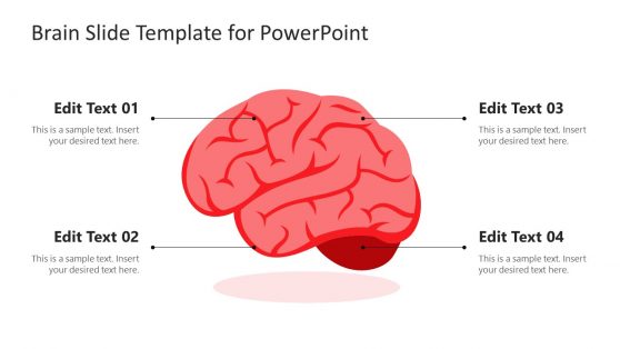
Brain Slide Template for PowerPoint
Nervous System PowerPoint Template
Statistical Bias PowerPoint Templates
Brain Artificial Intelligence PowerPoint Template

6 Step Brain Machinery Circular Diagram for PowerPoint

Smart Head Design for PowerPoint

Group Thinking Phenomenon PowerPoint Template

Artificial Intelligence PowerPoint Template

Kid Head Silhouette Illustration PowerPoint Template

Human Head Puzzle PowerPoint Template
Brain Concept Diagram PowerPoint Templates
Presenters often use the picture of the brain as a metaphor for intelligence. It can also be used to prepare presentations on brainstorming ideas.
Working with brain presentation templates will save you the stress of creating a brain slide from scratch – you can easily download any templates and edit them to suit your presentation preference. For example, our Brain Slide Template for PowerPoint presents a customizable brain diagram characterized by distinct lobes and sections, cleverly illustrated with shadow effects and PowerPoint shapes. The curves of the brain are delineated by dark lines, accompanied by four label lines pinpointing crucial brain divisions: the forebrain (comprising the occipital lobe and cerebrum), midbrain, and brain stem.
In this section, you can download brain illustrations, shapes, and brain slides with pictures of the brain.
What is a Brain Presentation?
A Brain Presentation is a PowerPoint presentation focusing on topics related to the human brain. Such presentations cover brain anatomy, cognitive processes, mental health, neurological disorders, learning theories, and more. These presentations can be educational, informative, or persuasive, depending on the context and audience.
What is the Purpose of a Brain Slide Template?
These templates are intended to visually depict and explain concepts related to the brain, neuroscience, cognitive functions, or related topics in presentations. It provides a consistent and engaging framework to communicate complex ideas, making it easier for your audience to understand and remember the information you presented.
Can I Personalize the Brain PowerPoint Template to Suit my Presentation Needs?
Yes. Our brain PowerPoint template is designed to be fully customizable. You can easily modify text, images, colors, and fonts to align with your content and preferences. Click on the placeholders and replace them with your information.
Can I use this Template for my Classroom Presentation?
Certainly! Our brain PowerPoint template is well-suited for educational purposes. Whether you are teaching students about brain anatomy, cognitive processes, or neuroscience principles, this template provides a visually engaging format.
Download Unlimited Content
Our annual unlimited plan let you download unlimited content from slidemodel. save hours of manual work and use awesome slide designs in your next presentation..
Pete’s PowerPoint Station
- Science Index
- Math/Maths Index
- Language Arts/Literature Index
- Social Studies Index
- Holidays Index
- Art, Music, and Many More, A-Z
- Meteorology
- Four Seasons
- Pre-Algebra
- Trigonometry
- Pre-Calculus & Calculus
- Language Arts
- Punctuation
- Social Studies
- World Religions
- US Government
- Criminal Justice
- Famous People
- American History
- World History
- Ancient History
- The Middle Ages
- Architecture
- All Topics, A–Z
- Privacy & Cookie Policy
- Presentations
The Human Brain
Free Presentations in PowerPoint format
Methods for Studying Brain Behavior
Study of Normal Brain Development
Studying Brain Function and Development
Studying the Brain – Implications for Educators
Anatomy of Mild to Moderate Brain Injury
Anatomy of the Brain
Anatomic Landmarks in the Brain
Brain Evolution
Anatomy of the Human Brain
Parts of the Human Brain
Gross Anatomy of the Brain
Brain Power
Brain Anatomy
See Also: Human Body Systems , The Human Body , The Nervous System
Free Brain Games & Activities for Kids
Brain Games
Brain Games – Neuroscience for Kids
FLASH Human Body Presentations
For Teachers
Lots of Lessons – Human Body
Free Video Clips
Free Online Science Games for Kids
Free Clipart for Science

An official website of the United States government
The .gov means it's official. Federal government websites often end in .gov or .mil. Before sharing sensitive information, make sure you're on a federal government site.
The site is secure. The https:// ensures that you are connecting to the official website and that any information you provide is encrypted and transmitted securely.
- Publications
- Account settings
- Browse Titles
NCBI Bookshelf. A service of the National Library of Medicine, National Institutes of Health.
StatPearls [Internet]. Treasure Island (FL): StatPearls Publishing; 2024 Jan-.

StatPearls [Internet].
Physiology, brain.
Kenia A. Maldonado ; Khalid Alsayouri .
Affiliations
Last Update: March 17, 2023 .
- Introduction
The human brain is perhaps the most complex of all biological systems, with the mature brain composed of more than 100 billion information-processing cells called neurons. [1] The brain is an organ composed of nervous tissue that commands task-evoked responses, movement, senses, emotions, language, communication, thinking, and memory. The three main parts of the human brain are the cerebrum, cerebellum, and brainstem.
The cerebrum is divided into the right and left hemispheres and is the largest part of the brain. It contains folds and convolutions on its surface, with the ridges found between the convolutions called gyri and the valleys between the gyri called sulci (plural of sulcus). If the sulci are deep, they are called fissures. Both cerebral hemispheres have an outer layer of gray matter called the cerebral cortex and inner subcortical white matter.
Located in the posterior cranial fossa, above the foramen magnum, the cerebellum's primary function is to modulate motor coordination, posture, and balance. It is comprised of the cerebellar cortex and deep cerebellar nuclei, with the cerebellar cortex being made up of three layers; the molecular, Purkinje, and granular layers. The cerebellum connects to the brainstem via cerebellar peduncles.
The brainstem contains the midbrain, pons, and medulla. It is located anterior to the cerebellum, between the base of the cerebrum and the spinal cord.
- Issues of Concern
Studies of brain function have focused on analyzing the variations of the electrical activity produced by the application of sensory stimuli. However, it is also essential to study additional features and functions of the brain, including information processing and responding to environmental demands. [2]
The brain works precisely, making connections, and is a deeply divided structure that has remained not entirely explained or examined. [3] Although researchers have made significant progress in experimental techniques, the human cognitive function that emerges from neuronal structure and dynamics is not entirely understood. [4]
- Cellular Level
At the beginning of the forebrain formation, the neuroepithelial cells undergo divisions at the inner surface of the neural tube to generate new progenitors. These dividing neuroepithelial cells transform and diversify, leading to radial glial cells (RGCs).
RGCs also work as progenitors with the capacity to regenerate themselves and produce other types of progenitors, neurons, and glial cells. [5] RGCs have long processes that connect with the neuroepithelium and function as a guide for the migration of neuron cells to ensure that neurons find their resting place, mature, and send out axons and dendrites to participate directly in synapses and electrical signaling. Neurons get produced along with glial cells; glial cells bring support and create an enclosed environment in which neurons can perform their functions.
Glial cells (astrocytes, oligodendrocytes, microglial cells) have well-known roles, which include: keeping the ionic medium of neurons, controlling the rate of nerve signal propagation and synaptic action by regulating the uptake of neurotransmitters, providing a platform for some aspects of neural development, and aiding in recovery from neural damage.
Gray matter is the main component of the central nervous system (CNS) and consists of neuronal cell bodies, dendrites, myelinated and unmyelinated axons, glial cells, synapses, and capillaries. The cerebral cortex is made up of layers of neurons that constitute the gray matter of the brain. The subcortical (beneath the cortex) area is primarily white matter composed of myelinated axons with fewer quantities of cell bodies when compared to gray matter.
Although neurons can have different morphologies, they all contain four common regions: the cell body, the dendrites, the axon, and the axon terminals, each with its respective functions.
The cell body contains a nucleus where proteins and membranes are synthesized. These proteins travel through microtubules down to the axons and terminals via a mechanism known as anterograde transport. In retrograde transport, damaged membranes and organelles travel from the axon toward the cell body along axonal microtubules. Lysosomes are only present in the cell body and are responsible for containing and degrading damaged material. The axon is a thin continuation of a neuron that allows electrical impulses to be sent from neuron to neuron.
Astrocytes occupy 25% of the total brain volume and are the most abundant glial cells. [6] They are classified into two main groups: protoplasmic and fibrous. Protoplasmic astrocytes appear in gray matter and have several branches that contact both synapses and blood vessels. Fibrous astrocytes are present in the white matter and have long fiber-like processes that contact the nodes of Ranvier and the blood vessels. Astrocytes use their connections to vessels to titrate blood flow in synaptic activity responses. Astrocytic endfeet, which forms tight junctions between endothelial cells and the basal lamina, gives rise to the formation of the blood-brain barrier (BBB). [7]
The primary function of oligodendrocytes is to make myelin, a proteolipid critical in maintaining electrical impulse conduction and maximizing velocity. Myelin is located in segments separated by nodes of Ranvier, and their function is equivalent to those of Schwan cells in the peripheral nervous system.
The macrophage populations of the CNS include microglia, perivascular macrophages, meningeal macrophages, macrophages of the circumventricular organs (CVO), and the microglia of the choroid plexus. Microglia are phagocytic cells representing the immune and support system of the CNS and are the most abundant cells of the choroid plexus. [8]
- Development
Human brain development starts with the neurulation process from the ectodermic layer of the embryo and takes, on average, 20 to 25 years to mature. [9] It occurs in a sequential and organized manner beginning with the neural tube formation at the third or fourth week of gestation. This is followed by cell migration and proliferation that leads to the folding of the cerebral cortex to increase its size and surface area, creating a more complex structure. Failure of this migration and proliferation leads to a smooth brain without sulci or gyri, termed lissencephaly. [10] At birth, the general architecture of the brain is mostly complete, and by the age of 5 years, the total brain volume is about 95% of its adult size. Generally, the white matter increases with age, while the gray matter decreases with age.
The most prominent white matter structure of the brain, the corpus callosum, increases by approximately 1.8% per year between the ages of 3 and 18 years. [11] The corpus callosum conjugates the activity of the right and left hemispheres and allows for the progress of higher-order cognitive abilities.
Gray matter in the frontal lobe undergoes continued structural development reaching its maximal volume at 11 to 12 years of age before slowing down during adolescence and early adulthood. The gray matter in the temporal lobe follows a similar development pattern, reaching its maximum size at 16 to 17 years of age with a slight decline afterward. [12]
Below is a list of the brain vesicles and the areas of the brain which develop from them. [13]
Prosencephalon (Forebrain)
- Cerebral cortex
- Basal ganglia (caudate nucleus, putamen, and globus pallidus)
- Hippocampus
- Lateral ventricles
- Hypothalamus
- Epithalamus (pineal gland)
- Subthalamus
- Posterior pituitary
- Optic nerve
- Third ventricle
Mesencephalon (midbrain)
- Cerebral aqueduct
Rhombencephalon (hindbrain)
- Fourth ventricle (rostral)
- Fourth ventricle (caudal)
- Organ Systems Involved
The brain and the spinal cord comprise the central nervous system (CNS). The peripheral nervous system (PNS) subdivides into the somatic nervous system (SNS) and the autonomic nervous system (ANS). The SNS consists of peripheral nerve fibers that collect sensory information to the CNS and motor fibers that send information from the CNS to skeletal muscle. The ANS functions to control the smooth muscle of the viscera and glands and consists of the sympathetic nervous system (SNS), the parasympathetic nervous system (PaNS), and the enteric nervous system (ENS).
Nerves from the brain connect with multiple parts of the head and body, leading to various voluntary and involuntary functions. The ANS drives basic functions that control unconscious activities such as breathing, digestion, sweating, and trembling.
The ENS provides the intrinsic innervation of the gastrointestinal system and is the most neurochemically diverse branch of the PNS. [14] Neurotransmitters such as norepinephrine, epinephrine, dopamine, and serotonin have recently been a topic of interest due to their roles in gut physiology and CNS pathophysiology, as they aid in regulating gut blood flow, motility, and absorption. [15]
The cerebrum controls motor and sensory information, conscious and unconscious behaviors, feelings, intelligence, and memory. The left hemisphere controls speech and abstract thinking (the ability to think about things that are not present). In contrast, the right hemisphere controls spatial thinking (thinking that finds meaning in the shape, size, orientation, location, and phenomena).
The motor and sensory neurons descending from the brain cross to the opposite side in the brainstem. This crossing means that the right side of the brain controls the motor and sensory functions of the left side of the body, and the left side of the brain controls the motor and sensory functions of the right side of the body. Hence, a stroke affecting the left brain hemisphere, for example, will exhibit motor and sensory deficits on the right side of the body.
Sensory neurons bring sensory input from the body to the thalamus, which then relays this sensory information to the cerebrum. For example, hunger, thirst, and sleep are under the control of the hypothalamus.
The cerebrum is composed of four lobes:
- Frontal lobe: Responsible for motor function, language, and cognitive processes, such as executive function, attention, memory, affect, mood, personality, self-awareness, and social and moral reasoning. [16] The Broca area is located in the left frontal lobe and is responsible for the production and articulation of speech.
- Parietal lobe: Responsible for interpreting vision, hearing, motor, sensory, and memory functions.
- Temporal lobe: In the left temporal lobe, the Wernicke area is responsible for understanding spoken and written language. The temporal lobe is also an essential part of the social brain, as it processes sensory information to retain memories, language, and emotions. [17] The temporal lobe also plays a significant role in hearing and spatial and visual perception.
- Occipital lobe: The visual cortex is located in the occipital lobe and is responsible for interpreting visual information.
The cerebellum controls the coordination of voluntary movement and receives sensory information from the brain and spinal cord to fine-tune the precision and accuracy of motor activity. The cerebellum also aids in various cognitive functions such as attention, language, pleasure response, and fear memory. [18]
The brainstem acts as a bridge that connects the cerebrum and cerebellum to the spinal cord. The brainstem houses the principal centers which perform autonomic functions such as breathing, temperature regulation, respiration, heart rate, wake-sleep cycles, coughing, sneezing, digestion, vomiting, and swallowing. The brainstem contains both white and gray matter. The white matter consists of fiber tracts (neuronal cell axons) traveling down from the cerebral cortex for voluntary motor function and up from the spinal cord and peripheral nerves, allowing somatosensory information to travel to the highest parts of the brain. [19]
The brain represents 2% of the human body weight and consumes 15% of the cardiac output and 20% of total body oxygen. The resting brain consumes 20% of the body's energy supply. When the brain performs a task, the energy consumption increases by an additional 5%, proving that most of the brain's energy consumption gets used for intrinsic functions.
The brain uses glucose as its principal source of energy. During low glucose states, the brain utilizes ketone bodies as its primary energy source. During exercise, the brain can use lactate as a source of energy.
In the developing brain, neurons follow molecular signals from regulatory cells like astrocytes to determine their location, the type of neurotransmitter they will secrete, and with which neurons they will communicate, leading to the formation of a circuit between neurons that will be in place during adulthood. In the adult brain, developed neurons fit in their corresponding place and develop axons and dendrites to connect with the neighboring neurons. [20]
Neurons communicate via neurotransmitters released into the synaptic space, a 20 to 50-nanometer area between neurons. The neuron that releases the neurotransmitter into the synaptic space is called the presynaptic neuron, and the neuron that receives the neurotransmitter is called the postsynaptic neuron. An action potential in the presynaptic neuron leads to calcium influx and the subsequent release of neurotransmitters from their storage vesicle into the synaptic space. The neurotransmitter then travels to the postsynaptic neuron and binds to receptors to influence its activity. Neurotransmitters are rapidly removed from the synaptic space by enzymes. [21]
The oligodendrocytes in the CNS produce myelin. Myelin forms insulating sheaths around axons to allow the swift travel of electrical impulses through the axons. The nodes of Ranvier are gaps in the myelin sheath of axons, allowing sodium influx into the axon to help maintain the speed of the electrical impulse traveling through the axon. This transmission is called saltatory nerve conduction, the "jumping" of electrical impulses from one node to another. It ensures that electrical signals do not lose their velocity and can propagate long distances without signal deterioration. [22]
- Related Testing
Functional magnetic resonance imaging (fMRI) can track the effects of neural activity and the energy that the brain consumes by measuring components of the metabolic chain. Other techniques, such as single-photon emission computed tomography (SPECT), study cerebral blood flow and neuroreceptors. Positron emission tomography (PET) assesses the glucose metabolism of the brain. [23] Electroencephalography (EEG) records the brain's electrical activity and is very useful for detecting various brain disorders. Advancements in these techniques have enabled a broader vision and objective perceptions of mental disorders, leading to improved diagnosis, treatment, and prognosis.
- Pathophysiology
Injury to the brain stimulates the proliferation of astrocytes, an immunological response to neurodegenerative disorders called "reactive gliosis." [24] Damage to neural tissue promotes molecular and morphological changes and is essential in the upregulation of the glial fibrillary acidic protein (GFAP). On the other hand, epidermal growth factor receptor (EGFR) allows the transition from non-reactive to reactive astrocytes, and its inhibition improves axonal regeneration and rapid recovery. This means that when astrocytes are reactive, they proliferate and hypertrophy, leading to glial scar formation.
The microglia represent the immune and support system of the CNS. They are neuroprotective in the young brain but can react abnormally to stimuli in the aged brain and become neurotoxic and destructive, leading to neurodegeneration. [25] As the brain ages, microglia acquire an increasingly inflammatory and cytotoxic phenotype, generating a hazardous environment for neurons. [26] Hence, aging is the most critical risk factor for developing neurodegenerative diseases.
The brain is surrounded by cerebrospinal fluid and is isolated from the bloodstream by the blood-brain barrier (BBB). In cases like infectious meningitis and meningoencephalitis, acute inflammation causes a breakdown of the BBB, leading to the influx of blood-borne immune cells into the CNS. In other inflammatory brain disorders such as Alzheimer disease (AD), Parkinson disease (PD), Huntington disease (HD), or X-linked adrenoleukodystrophy, the primary insult is due to degenerative or metabolic processes, and there is no breakdown of the BBB. [27]
Oligodendrocyte loss can occur due to the production of reactive oxygen species or the activation of inflammatory cytokines, causing decreased myelin production and leading to conditions such as multiple sclerosis (MS). [22]
Disturbances in the neurotransmitter systems are related to these substances' production, release, reuptake, or receptor impairments and can cause neurologic or psychiatric disorders. Glutamate is the brain's most abundant excitatory neurotransmitter, while GABA is the primary inhibitory transmitter. Glycine has a similar inhibitory action in the posterior parts of the brain. Acetylcholine aids in processes such as muscle stimulation at the neuromuscular junction (NMJ), digestion, arousal, salivation, and level of attention. Dopamine is involved in the reward and motivational component, motor control, and the regulation of prolactin release. Serotonin influences mood, feelings of happiness, and anxiety. Norepinephrine is involved in arousal, alertness, vigilance, and attention.
Cerebral oxygen delivery and consumption rates are ten times higher than global body values. [28] Blood glucose represents the primary energy source for the brain, and the BBB is highly permeable to it. During low glucose states, the body has developed multiple ways to keep blood glucose within the normal range. As the level drops below 80 mg/dL, pancreatic beta-cells decrease insulin secretion to avoid further glucose decrease. If glucose drops further, pancreatic alpha-cells secrete glucagon, and the adrenal medulla releases epinephrine. Glucagon and epinephrine increase blood glucose levels. Cortisol and growth hormone also act to increase glucose, but they depend on the presence of glucagon and epinephrine to work.
- Clinical Significance
Damage to the Cerebrum
- Frontal lobe - Damage to the frontal lobe causes interruption of the higher functioning brain processes, including social behavior, planning, motivation, and speech production. Individuals with frontal lobe damage may be unable to regulate their emotions, have meaningful or appropriate social interactions, maintain their past personality traits, or make difficult decisions. [29]
- Temporal lobe - The Wernicke area is located in the superior temporal gyrus in an individual's dominant hemisphere, which is the left hemisphere for 95% of people. Damage to the left (dominant) temporal lobe can lead to Wernicke aphasia. This is typically referred to as "word salad" speech, where the patient will speak fluently, but their words and sentences will lack meaning. [30] Damage to the right (non-dominant) temporal lobe may lead to persistent talking and deficits in nonverbal memory, processing certain aspects of sound or music (tone, rhythm, pitch), and facial recognition (prosopagnosia).
- Parietal lobe - Damage to the frontal aspect of the parietal lobe may lead to impaired sensation and numbness on the contralateral side of the body. An individual may have difficulty recognizing texture and shape and may be unable to identify a sensation and its location on their body. Damage to the middle aspect of the parietal lobe can lead to right-left disorientation and difficulty with proprioception. Damage to the non-dominant (right) parietal lobe may lead to apraxia (difficulty with performing purposeful motions such as combing hair or brushing teeth) and difficulty with spatial orientation and navigation (they may get lost in a once familiar area). Patients with non-dominant parietal lobe damage, usually from a middle cerebral artery stroke, may neglect the side opposite of the brain damage (usually the left side), which may manifest as only shaving the right side of their face or drawing a clock with all of the numbers on the right side of the circle. [31]
- Occipital lobe - Damage to the occipital lobe may lead to visual defects, color agnosia (inability to identify colors), movement agnosia (difficulty recognizing object movements), hallucinations, illusions, and the inability to recognize written words (word blindness).
Damage to the Cerebellum
Damage to the cerebellum can lead to ataxia, dysmetria, dysarthria, scanning speech, dysdiadochokinesis, tremor, nystagmus, and hypotonia. To test for possible cerebellar dysfunction, a bedside neurologic exam is commonly the first step. This exam may include the Romberg test, heel-to-shin test, finger-to-nose test, and rapid alternating movement test. [32]
Damage to the Brainstem
Damage to the brainstem may present as muscle weakness, visual changes, dysphagia, vertigo, speech impairment, pupil abnormalities, insomnia, respiratory depression, or death.
Neurodegenerative Diseases
Neuronal degeneration worsens with age and can affect different areas of the brain leading to movement, memory, and cognition problems.
Parkinson disease (PD) occurs due to the degeneration of the neurons that synthesize dopamine, leading to motor function deficits. Alzheimer disease (AD) occurs due to abnormally folded protein deposition in the brain leading to neuronal degeneration. Huntington disease occurs due to a genetic mutation that increases the production of the neurotransmitter glutamate. Excessive amounts of glutamate lead to the death of neurons in the basal ganglia producing movement, cognitive, and psychiatric deficits. Vascular dementia occurs due to the death of neurons resulting from the interruption of blood supply.
Although neurodegenerative diseases are not classically caused by disturbed metabolism, research has shown that there is a reduction in glucose metabolism in Alzheimer disease. [33]
Demyelinating Diseases
Demyelinating diseases result from damage to the myelin sheath that covers the nerve cells in the white matter of the brain, spinal cord, and optic nerves. For example, multiple sclerosis and leukodystrophies are a consequence of oligodendrocyte damage.
A stroke is caused by an interruption in the blood supply to the brain, which may ultimately lead to neuronal death. This condition can result in one of several neurological problems depending on the affected region.
Brain Death
Neurologic evaluation of brain death is a complicated process that non-specialists and families might misunderstand. [34] Brain death is the complete and irreversible loss of brain activity, including the brainstem. It requires verification through well-established clinical protocols and the support of specialized tests.
Hypoglycemia
Glucose is the primary energy source responsible for maintaining brain metabolism and function. The most significant amount of glucose is used for information processing by neurons. [35] The brain requires a continuous supply of glucose as it has limited glucose reserves. CNS symptoms and signs of hypoglycemia include focal neurological deficits, confusion, stupor, seizure, cognitive impairment, or death.
- Review Questions
- Access free multiple choice questions on this topic.
- Comment on this article.
Brain, Encephalon, Connections of the several parts of the brain, Cerebrum, Cerebellum, Pons, Cerebral; Superior; Middle; Inferior Peduncle, Medulla oblongata Henry Vandyke Carter, Public Domain, via Wikimedia Commons
The Fore-brain or Prosencephalon, Mesal aspect of a brain sectioned in the median sagittal plane, Foramen of Monro, Middle commissure, Taenia thalami, Habenular commissure, Genu, Callosum, Fornix, Septum Lucidum, Plenum, Pons, Oblongata, Thalamus Henry (more...)
Areas of localization on lateral surface of hemisphere. Motor area in red, Area of general sensations in blue, Auditory area in green, Visual area in yellow, Brain, Neurology Henry Vandyke Carter, Public Domain, via Wikimedia Commons
Pathways from the Brain to the Spinal Cord, The motor tract, Anterior nerve roots, Anterior and Lateral cerebrospinal Fasciculus, Decussation of pyramids, Geniculate fibers, Internal capsule, Motor area of cortex Henry Vandyke Carter, Public Domain, via (more...)
Homonculus: sensory & motor Image courtesy S. Bhimji MD
Disclosure: Kenia Maldonado declares no relevant financial relationships with ineligible companies.
Disclosure: Khalid Alsayouri declares no relevant financial relationships with ineligible companies.
This book is distributed under the terms of the Creative Commons Attribution-NonCommercial-NoDerivatives 4.0 International (CC BY-NC-ND 4.0) ( http://creativecommons.org/licenses/by-nc-nd/4.0/ ), which permits others to distribute the work, provided that the article is not altered or used commercially. You are not required to obtain permission to distribute this article, provided that you credit the author and journal.
- Cite this Page Maldonado KA, Alsayouri K. Physiology, Brain. [Updated 2023 Mar 17]. In: StatPearls [Internet]. Treasure Island (FL): StatPearls Publishing; 2024 Jan-.
In this Page
Bulk download.
- Bulk download StatPearls data from FTP
Related information
- PMC PubMed Central citations
- PubMed Links to PubMed
Similar articles in PubMed
- Brainstem Stroke. [StatPearls. 2024] Brainstem Stroke. Gowda SN, Munakomi S, De Jesus O. StatPearls. 2024 Jan
- Neuroanatomy, Medulla Oblongata. [StatPearls. 2024] Neuroanatomy, Medulla Oblongata. Iordanova R, Reddivari AKR. StatPearls. 2024 Jan
- Lower Genitourinary Trauma. [StatPearls. 2024] Lower Genitourinary Trauma. Tullington JE, Blecker N. StatPearls. 2024 Jan
- Review Anatomy of the brainstem: a gaze into the stem of life. [Semin Ultrasound CT MR. 2010] Review Anatomy of the brainstem: a gaze into the stem of life. Angeles Fernández-Gil M, Palacios-Bote R, Leo-Barahona M, Mora-Encinas JP. Semin Ultrasound CT MR. 2010 Jun; 31(3):196-219.
- Review Physiology of the cerebellum. [Handb Clin Neurol. 2018] Review Physiology of the cerebellum. D'Angelo E. Handb Clin Neurol. 2018; 154:85-108.
Recent Activity
- Physiology, Brain - StatPearls Physiology, Brain - StatPearls
Your browsing activity is empty.
Activity recording is turned off.
Turn recording back on
Connect with NLM
National Library of Medicine 8600 Rockville Pike Bethesda, MD 20894
Web Policies FOIA HHS Vulnerability Disclosure
Help Accessibility Careers
Ch. 7 – The Nervous System
- Overview & Organization of the Nervous System
Functions of the Nervous System
The master controlling & communicating system of the body…
- Sensory input —gathering information
- To monitor changes occurring inside and outside the body
- Changes = stimuli
- Integration
- To process and interpret sensory input and decide if action is needed
- Motor output
- A response to integrated stimuli
- The response activates muscles or glands
- Structural Classification �of the Nervous System
- Central nervous system (CNS) – dorsal body cavity; integrating and command centers; interpret sensory information & give out instructions
Spinal cord
- Peripheral nervous system (PNS) – outside of CNS
- Nerves outside the brain and spinal cord
- Spinal nerves – carry impulses to and from spinal cord
- Cranial nerves – carry impulses to and from brain
- Functional Classification of �the Peripheral Nervous System
- Sensory (afferent) division
- Nerve fibers that carry information to the CNS
- Somatic sensory fibers – deliver impulses from skin, skeletal muscle, and joints
- Visceral sensory fibers (afferents) – deliver impulses from viscera
- Motor (efferent) division
- Nerve fibers that carry impulses away from the CNS
- Somatic (voluntary) NS – voluntary control of skeletal muscles
- Autonomic (involuntary NS – involuntary control of smooth & cardiac muscle and glands
- Divided into sympathetic and parasympathetic NS
Answer Did You Get It? #1
- Structure & Function of Nervous Tissue
- Support Cells
- Support cells in the CNS are grouped together as neuroglia (AKA glia or glial cells ) = “nerve glue”
- Functions: support, insulate, and protect neurons
- Cannot transmit nerve impulses (as can neurons)
- Never lose their ability to divide (as neurons do)
- Most brain tumors are gliomas
- Glia of the Central Nervous System:
- Ependymal cells
- Oligodendrocytes
- Glia of the Peripheral Nervous System:
- Schwann cells
- Satellite cells
Support Cells, continued…
- Abundant (~1/2 of neural tissue)
- Star-shaped cells
- Brace & anchor neurons to capillaries
- Form living barrier between capillaries and neurons (exchange) (blood-brain barrier)
- Control brain’s chemical environment
- Absorb leaked K + ions
- Absorb released neurotransmitters
- Spiderlike phagocytes
- Protect from infection
- Dispose of debris
- Dead brain cells & bacteria
- Line cavities of the brain and spinal cord
- Beating cilia circulate cerebrospinal fluid (CSF)
- CSF fills brain & spinal cord cavities & serves as cushion
- Wrap around nerve fibers in the CNS
- Produce fatty insulating coverings = myelin sheaths
- Protect neuron cell bodies
- Form myelin sheath around nerve fibers in the PNS
Answer Did You Get It? #’s 2-3
- Neurons = nerve cells
- Cells specialized to transmit nerve impulses from one part of body to another
- Two major regions of neurons:
- Metabolic center: contains nucleus, large nucleolus
- No centrioles = no mitosis
- Nissl substance = specialized RER
- Neurofibrils (intermediate cytoskeleton)
- Maintain cell shape
Neurons, continued…
- Processes outside the cell body
- Microscopic to 3-4 ft in length
- Longest = from lumbar region of spine to great toe
- Dendrites —conduct impulses toward the cell body
- A neuron may have hundreds
- Axons —conduct impulses away from the cell body
- Arises from cone-like region of cell body called axon hillock
- Collateral branches
- End in highly branched axon terminals
- Axon terminals contain vesicles with neurotransmitters
- Axonal terminals are separated from the next neuron by a synaptic cleft
- Synapse —junction between nerves ( syn = clasp/join)
Neuron processes, continued…
- Myelin sheath —whitish, fatty material covering axons
- Protects & insulates fibers
- Increases rate of nerve impulse transmission
- Schwann cells —produce myelin sheaths in jelly roll–like fashion
- Schwann cells in the PNS; oligodendrocytes in the CNS
- Neurilemma – portion of cell membrane on outer layer of coil where most of its cytoplasm resides
- Nodes of Ranvier —gaps in myelin sheath along the axon
- Aid in speeding up nerve impulses – saltatory conduction
- Homeostatic imbalance – multiple sclerosis = gradual destruction of myelin sheaths (become hardened = sclerosis), autoimmune disease (sheath protein)
- Visual & speech disturbances, loss of muscle control, increasingly disabled
- Interferon injections provide relief; no cure
- Terminology of Neurons
- Most neuron cell bodies are found in the CNS
- Nuclei —clusters of cell bodies within the white matter of the CNS (protected within the brain case and vertebral column)
- Ganglia —small collections of cell bodies in the PNS
- Tracts = bundles of nerve fibers in CNS
- White matter – myelinated tracts in CNS
- Gray matter —cell bodies and unmyelinated tracts in CNS
- Nerves = bundles of nerve fibers in PNS
- Functional Classification of Neurons
Direction of nerve impulse with respect to CNS
- Sensory (afferent) neurons
- Carry impulses from the sensory receptors to the CNS
- Ganglion outside of CNS
- Dendrite endings associate with receptors
- Cutaneous sense organs in muscles and tendons
- Proprioceptors —detect stretch or tension
Naked nerve ending; pain/temp
Meissner’s corpuscule: touch
Pacinian corpuscule: deep pressure
Golgi tendon organ & muscle spindle;: proprioception
Functional Classification of Neurons, continued…
- Motor (efferent) neurons
- Carry impulses from the central nervous system to viscera, muscles, or glands
- Cell bodies always in CNS
- Interneurons (association neurons)
- Connect sensory and motor neurons in neural pathways
- Structural Classification of Neurons
- Multipolar neurons—many extensions from the cell body
- most common
- Bipolar neurons—one axon and one dendrite
- Rare in adults
- Act in sensory processing – eye, nose
- Unipolar neurons—have a short single process leaving the cell body
- Divides into proximal (central) and distal (peripheral) processes
- Dendrites only at peripheral end
- Conducts action potentials both ways
- Found in sensory neurons of PNS ganglia
Answer Did You Get It? #’s 4-7
- Physiology of the Nervous System
- Functional Properties of Neurons
- Irritability - ability to respond to stimuli and convert to nerve impulses
- Conductivity - ability to transmit an impulse to other neurons, muscles, or glands
- Nerve Impulses
- Electrical conditions of a resting neuron’s membrane
- Polarized – resting/inactive neuron
- Fewer positive ions on inner face of plasma membrane than on outer face
- Depolarized – stimulated neuron
- More positive ions inside the cell than outside
Nerve Impulses, continued…
- Action Potential Initiation and Generation
- Stimuli excite neurons: light, sound, pressure, mostly neurotransmitters released by other neurons
- Cause a temporary change in the cell membrane’s permeability
- Stimulus causes sodium channel gates to open, and sodium to rush in
- Causes depolarization of the neuron’s membrane
- Inside more positive, outside less positive = graded/local potential
- If stimulus is strong enough, a long distance signal called an action potential or nerve impulse occurs
- Nerve impulses are all-or-nothing responses – they are either propagated over the entire axon or not at all
- Repolarization
- Membrane immediately becomes impermeable to sodium, but permeable to potassium ions
- K + ions rush out of the neuron, restoring electrical conditions to polarized = repolarization
- Repolarization must occur before another impulse can be conducted
- The sodium-potassium pump, using ATP, restores the original concentrations of Na + and K + .
- Saltatory conduction = In myelinated fibers, propagation occurs more quickly since the nerve impulse jumps from node to node.
- Homeostatic imbalance: factors that impair impulse conduction:
- Sedatives & anesthetics block sodium entry
- Cold & continuous pressure interrupt blood circulation (nutrients & O 2 ) – e.g. ice creates numbness, foot “goes to sleep”; prickly feeling caused by impulse transmission starting back up
- Transmission of the Signal at Synapses
- Neurotransmitter is released from vesicles within the axon terminal
- Neurotransmitter molecules diffuse across the synapse
- Neurotransmitters bind to receptors in the membrane of the next neuron
- If enough neurotransmitters are released, another nerve impulse will be generated in this neuron
- Enzymes remove the neurotransmitters from the receptors
- Impulse transmission is an electrochemical event – electrical along the neuron’s membrane; chemical within the synapses
Axon�terminal
Synaptic�cleft
Action�potential�arrives
Axon of�transmitting�neuron
Receiving�neuron
Neurotrans-�mitter is re-�leased into�synaptic cleft
Neurotrans-�mitter binds�to receptor�on receiving�neuron’s�membrane
Vesicle�fuses with�plasma�membrane
Synaptic cleft
Neurotransmitter�molecules
Ion channels
Receiving neuron
Transmitting neuron
Neurotransmitter
Neurotransmitter�broken down�and released
Ion channel opens
Ion channel closes
- Reflex — rapid, predictable, and involuntary response to a stimulus
- Always travel in one direction
- Occurs over pathways called reflex arcs
- Reflex arc — direct route from a sensory neuron, to an interneuron, to an effector
- Neural pathway involving the CNS and PNS
Stimulus at distal�end of neuron
(in cross section)
Interneuron
Sensory neuron
Motor neuron
Integration�center
Reflexes, continued…
- Types of Reflexes
- Somatic reflexes
- Reflexes which stimulate the skeletal muscles
- Example: moving hand away from a hot stove
- Autonomic reflexes
- Regulate the activity of smooth muscles, heart, and glands
- Examples: salivary reflex, pupillary reflex
- Regulate: digestion, elimination, blood pressure, and sweating
- Parts of a reflex arc
- Sensory receptor – reacts to a stimulus
- Integration center
- Effector organ – muscle or gland which is stimulated
- Patellar (knee-jerk) reflex is an example of a two-neuron reflex arc
Figure 7.11d
Figure 7.11b–c
Sensory (afferent)�neuron
Motor�(efferent)�neuron
Sensory receptors�(stretch receptors�in the quadriceps�muscle)
Effector�(quadriceps�muscle of�thigh)
Synapse in�ventral horn�gray matter
Inter-�neuron
Sensory receptors�(pain receptors in�the skin)
Effector�(biceps�brachii�muscle)
- Flexor (withdrawal) reflex is an example of a three-neuron reflex arc
- Withdrawal reflex arc has an interneuron
- The more neurons involved, the slower the communication because of the time it takes for neurotransmitters to diffuse
- Many spinal reflexes do not involve the brain
- Other reflexes require the brain to evaluate different types of information
- Reflex testing evaluates condition of the nervous system
- Exaggerated, distorted, and absent reflexes indicate nervous system disorders
Answer Did You Get It? #’s 8-11
- Central Nervous System (CNS)
- CNS develops from the embryonic neural tube
- Runs along the dorsal median plane
- 4 th week – anterior end expands = brain formation
- Rest of tube = spinal cord
- The central canal of the neural tube enlarges into 4 chambers = ventricles
- Filled with cerebrospinal fluid
- Functional Anatomy of the Brain
- ~3 lbs, wrinkled, texture similar to cold oatmeal
- 4 major regions:
- Cerebral hemispheres (cerebrum)
- Diencephalon
Regions of the Brain: Cerebrum
- Cerebrum (cerebral hemispheres)
- Paired, superior parts of the brain
- Includes more than half of the brain mass; obscures most of the brain stem
- The surface is made of ridges ( gyri = “twisters”) and grooves ( sulci = “furrows”)
- Fissures (deep grooves) divide the cerebrum into lobes
- Occipital lobe
- Temporal lobe
Figure 7.13b
- Cerebral Cortex
- Functions : speech, memory, logic, emotion, consciousness, sensation interpretation, & voluntary movement
- Cell bodies of neurons in cerebral cortex in outermost gray matter
- Primary somatic sensory area
- In parietal lobe posterior to central sulcus
- Receives & interprets impulses from the body’s sensory receptors
- Detects: pain, cold, light touch
Sensory & motor homunculus – the more neurons there are for a function, the larger the area represented by that body region
Figure 7.14
- Visual area in occipital lobe
- Auditory area in temporal lobe
- Olfactory area deep in temporal lobe
- Primary motor area in frontal lobe
- Conscious movement of skeletal muscle
- Axons of these motor neurons form the corticospinal or pyramidal tract
- Descends to spinal cord
- Broca’s area at base of precentral gyrus
- Involved in our ability to speak
- Only located in one (usually left) hemisphere
- Damage here can cause inability to speak – conscious of what you want to say, but unable to do it
- Frontal association areas – higher intellectual reasoning & socially acceptable behavior
- Complex memories stored in temporal and frontal lobes
- Speech/language (Wernicke’s) area – junction of temporal, parietal, & occipital lobes
- Allows us to sound out words
- Usually in just one hemisphere
- Damage: Wernicke’s aphasia – lack of language comprehension; clear speaking though
- Frontal lobes – language comprehension (word meaning)
- Gustatory area – taste – base of primary somatic sensory area (parietal)
- General interpretation area – temporal & parietal
- Cerebral White Matter
- White matter—fiber tracts carrying impulses to, from, and within the cortex
- Corpus callosum – large tract connecting hemispheres; allows hemispheres to communicate with one another
- Called commisures
- Association fiber tracts connect areas within hemispheres ; projection fiber tracts connect cerebrum to lower CNS centers
- Basal nuclei (basal ganglia ) — islands of gray matter buried within the white matter
- Regulate voluntary
motor activities
- Homeostatic Imbalance:
- Problems with basal
nuclei cause difficulty in
walking or other voluntary
movements: Huntington’s
disease & Parkinson’s
Answer Did You Get It? #’s 12-13
- Regions of the Brain: Diencephalon (Interbrain)
- Sits on top of brain stem; enclosed by the cerebral hemispheres
- Made of three parts: Thalamus, Hypothalamus, Epithalamus
- Thalamus – relay station for sensory impulses traveling up to sensory cortex
- Crude awareness of a pending sensation being pleasant or not
- Hypothalamus – floor of diencephalon
- Autonomic NS center: helps body temp, water balance, & metabolism
- Limbic system – “emotional-visceral brain” where thirst, appetite, sex, pain, and pleasure centers are
- Regulates the pituitary gland ; secretes hormones
- Mammillary bodies – reflex centers involved in olfaction
Regions of the Brain: Diencephalon
- Epithalamus
- Forms the roof of the third ventricle
- Houses the pineal body (an endocrine gland)
- Includes the choroid plexus —complex of capillaries which form cerebrospinal fluid
Regions of the Brain: Brain Stem
- Small: ~thumb in diameter & ~3” long
- 3 regions: midbrain, pons, & medulla oblongata
- Provides a pathway for ascending & descending tracts
- Contains nuclei with rigidly programmed autonomic behaviors necessary for survival
- Some connected to cranial nerves controlling breathing & blood pressure
- From mammilary bodies to pons
- Cerebral aqueduct – canal connecting 3 rd ventricle of diencephalon to 4 th ventricle
- Has two bulging fiber tracts — cerebral peduncles : convey ascending & descending impulses
- Mostly composed of tracts of nerve fibers
- Has four rounded protrusions— corpora quadrigemina (“gemini” = twins)
- Reflex centers for vision and hearing
- Pons (“bridge”)
- Rounded part of brain stem just below midbrain
- Mostly composed of fiber tracts
- Includes nuclei involved in the control of breathing
- Medulla Oblongata
- Most inferior part of the brain stem
- Merges into the spinal cord
- Includes important fiber tracts
- Contains nuclei which control:
- Blood pressure
- Fourth ventricle
- Reticular Formation
- Diffuse mass of gray matter along the length of the brain stem
- Involved in motor control of visceral organs
- Reticular activating system (RAS) plays a role in awake/sleep cycles and consciousness
- Damage here can cause a coma (permanent unconsciousness)
- Regions of the Brain: Cerebellum
- Cauliflower-like, dorsally projecting from under the occipital lobe
- Two hemispheres with convoluted surfaces
- Outer cortex composed of gray matter; inner region composed of white matter
- Provides precise timing for skeletal muscle activity and controls balance & equilibrium
- “Automatic pilot” – compares brain’s intentions with body’s actual performance; initiates appropriate corrective measures
- Ataxia – damage to cerebellum can result in clumsy & disorganized movements; appear to be drunk
Answer Did You Get It? #’s 14-16
- Protection of the Central Nervous System
- Nervous tissue is soft and delicate; neurons injured easily
- Brain and spinal cord protected by
- Scalp and skin
- Skull and vertebral column
- Meninges (membranes)
- Cerebrospinal fluid (watery cushion)
- Blood-brain barrier – protection from harmful substances in the blood
Figure 7.17b
- Connective tissue membranes which cover & protect the CNS
- Double-layered, outermost layer; leathery
- Periosteal layer (periosteum)—attached to inner surface of the skull
- Meningeal layer —outer covering of the brain; fuses with the dura mater of the spinal cord
- Layers are fused except in dural venous sinuses where venous blood is collected
- Inward folds attach brain to cranial cavity
- Falx cerebri & tantorium cerebelli
- Arachnoid mater (“spider”)
- Middle layer
- Attached to the pia mater, forming the subarachnoid space
- Filled with cerebrospinal fluid (CSF)
- Arachnoid villi – projections of arachnoid mater; protrude through dura mater
- CSF passes into dural sinuses through these structures
- Pia mater (“gentle mother”)
- Innermost membrane
- Clings tightly brain and spinal cord surfaces
- Epidural injections – “upon the dura”
- Homeostatic Imbalance :
- Meningitis – inflammation of the meninges
- Bacterial or vial infections
- Serious threat to brain if spreads into CNS
- Encephalitis – inflammation of the brain
- Diagnosed by sampling CSF
Cerebrospinal Fluid (CSF)
- Similar to blood plasma composition
- Less protein, more vitamin C, different ion composition
- Formed from blood by choroid plexuses
- Clusters of capillaries hanging from each of brain’s ventricles
- Forms a watery cushion to protect the brain from trauma
- Circulated in arachnoid space, ventricles, and central canal of the spinal cord
- CSF continually circulates in brain
- From two lateral ventricles, to 3 rd ventricle, through cerebral aqueduct, to 4 th ventricle
- Some CSF continues to spinal cord
- Normally circulates at a constant rate
- Changes to CSF composition may indicate meningitis, tumors, or MS
- Lumbar/spinal tap – sample the CSF
- Remain lying down for 12 hrs or “spinal headache”
- Homeostatic Imbalance - Hydrocephalus
- If something obstructs CSF drainage, it accumulates and exerts pressure on the brain
- “Water on the brain”
- Results in enlarged head in newborns with increasing brain size
- Would cause brain damage in adults
- Treated by surgically inserting a shunt (plastic drain); drains excess fluid into a vein
- Blood-Brain Barrier
- Brain is super sensitive to having a constant internal environment
- Neurons kept separated from bloodborne substances by the blood-brain barrier
- Composed of least permeable capillaries of the body
- Bound by tight junctions
- Allowed to enter:
- Water, glucose, and essential amino acids pass easily through
- Metabolic wastes (urea, toxins, proteins, most drugs), nonessential amino acids, K +
- Useless as a barrier against some substances
- Fats and fat soluble molecules
- Respiratory gases
Answer Did You Get It? #’s 17-19
- Traumatic Brain Injuries
- Head injuries are leading cause of accidental death in US; caused by damaging blow to head
- Further damage caused by brain ricocheting on opposite end of skull
- Slight brain injury
- Dizzy/”see stars,” briefly lose consciousness
- No permanent brain damage
- Marked tissue destruction occurs
- May remain conscious if cerebral cortex injury; may be in coma if brain stem is injured severely (especially RAS)
- Nervous tissue does not regenerate
- Intracranial hemorrhage
- Bleeding from ruptured vessels
- May cause death
- Cerebral edema
- Brain swelling from the inflammatory response
- May compress and kill brain tissue – neurological deterioration
- Cerebrovascular Accident (CVA/Stroke)
- 3 rd leading cause of death in US
- Blood circulation to brain is obstructed by a blood clot or ruptured blood vessel
- Brain tissue supplied with oxygen from that blood source dies
- Loss of some functions or death may result; undamaged neurons can spread into damaged areas and take over some lost functions (= neuroplasticity )
- Hemiplegia – one-sided paralysis ( e.g. right-sided paralysis = damage to left motor cortex)
- Apahsia – damage to language areas
- Motor/Broca’s aphasia – loss of ability to speak
- Sensory/Wernicke’s aphasia – loss of ability to understand written & spoken language
- Transient ischemic attack (ITA) – temporary restriction of blood flow (ischemia) to brain
- Last 5-50 min; numbness, temporary paralysis; impaired speech
- Warning of impending, more serious CVA
Answer Did You Get It #20
- The Terrible Three
- Alzheimer’s Disease
- Progressive degenerative brain disease, results in dementia (mental deterioration)
- Mostly seen in the elderly, but may begin in middle age
- Victims experience: memory loss, short attention span, disorientation, eventual loss of language, irritability, moodiness, confusion, sometimes violent, and ultimately, hallucinations.
- Structural changes in the brain include: low Ach, shrinking gyri, brain atrophy (especially in areas of thought and memory), abnormal protein (senile plaque – beta amyloid peptide ) deposits, and twisted tau fibers within neurons
- Treat with acetylcholinesterase inhibitors
- Parkinson’s Disease
- Problem associated with basal nuclei; cause not known
- Typically affects people in 50’s-60’s
- Degeneration of dopamine-releasing neurons in the substantia nigra, causing basal nuclei to become overactive
- Symptoms: persistent tremor (even at rest), head nodding, “pill-rolling” of fingers, forward-bent walking posture, shuffling gait, stiff facial expressions, difficulty in initiating movements
- Treatments: L-dopa for some symptoms (bad side effects); deprenyl to slow degeneration; thalamic stimulation via electrodes alleviates tremors; implants of embryonic tissue promising
- Huntington’s Disease
- Genetic disorder (dominant) – typically occurs at middle-age
- Massive degeneration of basal nuclei and later of the cerebral cortex
- Progressive symptoms: wild, jerky movements ( chorea ), later marked mental deterioration
- Typically fatal within 15 years
- Overstimulation of motor cortex
- Treat with drugs that block dopamine; fetal tissue implants are promising
- Spinal Cord
- 2-way conduction pathway to and from the brain
- Major reflex center (spinal reflexes)
- Extends from the foramen magnum of the skull to the first or second lumbar vertebra
- Cushioned & protected by meninges
- 31 pairs of spinal nerves arise from the spinal cord
- Cervical & lumbar enlargements – origin of upper & lower limb nerves
- Cauda equina (horse’s tail) is a collection of spinal nerves at the inferior end
Spinal Cord Anatomy
- Gray matter of Spinal Cord and Spinal Roots
- Gray matter surrounds the central canal (filled with CSF)
- Dorsal (posterior) horns – project posteriorly
- Contain interneurons
- Sensory neuron cell bodies in dorsal root ganglia ; enter spinal cord through dorsal root
- Anterior (ventral) horns – project anteriorly
- Motor neuron cell bodies in ventral horns; axons exit spinal cord through ventral root
- Homoeostatic imbalance – flaccid paralysis – damage to ventral root = no stimulation of muscles
- Spinal nerves – fusion of dorsal and ventral roots
- White matter of the Spinal Cord
- Myelinated fiber tracts (see 7.22)
- Dorsal, lateral, ventral columns
- Sensory/afferent tracts – conduct sensory impulses to brain
- Motor/efferent tracts – conduct impulses from brain to skeletal muscles
- Dorsal column tracts are all ascending carrying sensory input to brain
- Lateral & ventral tracts contain both ascending & descending tracts
- Homeostatic imbalance – spastic paralysis : transected (cut crosswise) or crushed spinal cord – affected muscles stay healthy b/c still stimulated, but moments become spastic; loss of feeling below injury
- Quadriplegic = 4 limbs affected
- Paraplegic = legs only
Answer Did You Get It? #’s 21-23
- Peripheral Nervous System (PNS)
- Nerves and ganglia outside CNS
- Structure of a Nerve
- Nerve = bundle of neuron fibers outside the CNS
- Neuron fibers are bundled by connective tissue
- Delicate endoneurium surrounds each fiber
- Groups of fibers are bound into fascicles by coarser perineurium
- Fascicles are bound together by tough, fibrous epineurium
- Forms cordlike nerve
Structure of a Nerve, continued…
- Nerves are classified according to the direction in which they transmit impulses:
- Mixed nerves – nerves with both sensory and motor fibers
- Sensory (afferent) nerves – nerves carrying impulses toward the CNS
- Motor (efferent) nerves – nerves carrying impulses away from the CNS
- Cranial Nerves
- 12 pairs of nerves that mostly serve the head and neck
- Only the pair of vagus nerves extend to thoracic and abdominal cavities
- Numbered in order; names typically match the structures they control
- Most are mixed nerves, but three are sensory only (optic, olfactory, & vestibulocochlear)
Cranial Nerves, continued…
- Olfactory nerve — sensory for smell
- Optic nerve — sensory for vision
- Oculomotor nerve — motor fibers to eye muscles (most movements, lens shape, & pupil size)
- Trochlear nerve — motor fiber to eye muscle (superior oblique)
- Trigeminal nerve — sensory for the face, nose, & mouth; motor fibers to chewing muscles
- Abducens nerve — motor fibers to eye muscles (lateral movement)
- Facial nerve — sensory for anterior taste buds; motor fibers for facial expression and lacrimal & salivary glands
- Vestibulocochlear nerve — sensory for balance and hearing
- Glossopharyngeal nerve — sensory for posterior taste buds; motor fibers to the pharynx (swallowing & saliva production); carotid artery pressure sensors
- Vagus nerves — sensory and motor fibers for pharynx, larynx, and thoracic & abdominal viscera (mostly parasympathetic = promote digestion & regulate heart activity)
- Accessory nerve — motor fibers to sternocleidomastoid & trapezius
- Hypoglossal nerve — motor fibers for tongue movements; sensory impulses from tongue
- O h O nce O ne T akes T he A natomy F inal V ery G ood V acations A re H eavenly.
- O nly O wls O bserve T hem T raveling A nd F inding V oldemort G uarding V ery S ecret H orcruxes
- Spinal Nerves & Nerve Plexuses
- There are 31 pairs formed by the combination of the ventral and dorsal roots of the spinal cord
- Named for the region from which they arise
- Spinal nerves divide after leaving the spinal cord
- Dorsal rami — serve the skin and muscles of the posterior trunk
- Ventral rami — for nerves T 1 -T 12 forms intercostal nerves (muscles between ribs & skin and muscles of anterior trunk); for rest of nerves forms a nerve networks ( plexus ) for limb sensory & motor
Answer Did You Get It? #’s 24-27
Spinal Nerves & Nerve Plexuses, continued…
- Cervical plexus – from C 1 –C 5 ventral rami
- Phrenic nerve – diaphragm; shoulder/neck muscles
- Brachial plexus – from C 5 –C 8 and T 1 ventral rami
- Axillary nerve – deltoid muscle, shoulder skin; superior thorax muscles & skin
- Radial nerve – triceps & extensor muscles; upper limb posterior skin
- Median nerve – flexor muscles; forearm skin; some hand muscles
- Musculocutaneous nerve – arm flexor muscles; lateral forearm skin
- Ulnar nerve – some forearm flexor muscles; wrist & hand muscles; hand skin
- Lumbar plexus – from L 1 –L 4 ventral rami
- Femoral nerve – lower abdomen , hip flexors & knee extensors; leg & thigh anteromedial skin
- Obturator nerve – adductor & small hip muscles; medial thigh & hip joint skin
- Sacral plexus – from L 4 –L 5 and S 1 –S 4 ventral rami
- Sciatic nerve – largest nerve in body; splits into two nerves; lower trunk & posterior thigh surface (hip extensors & knee flexors)
- Common fibular nerve – lateral leg & foot
- Tibial nerve – posterior leg & foot
- Superior & inferior gluteal nerves – gluteal muscles
Distribution of Major Peripheral Nerves of the �Upper and Lower Limbs
Spinal Nerve Plexuses
Autonomic Nervous System (AKA Involuntary NS)
- Motor subdivision of the PNS
- Controls body activities automatically
- Special neurons that regulate cardiac muscle, smooth muscle (visceral organs & blood vessels), and glands
- Helps to maintain homeostasis – constantly makes adjustments to keep internal conditions stable
- Consists only of motor nerves
Note the differences between ANS & SNS
Autonomic Nervous System, continued…
- Somatic vs. Autonomic nervous systems (both PNS)
- Different effector organs and neurotransmitters
- Somatic NS has cell bodies in CNS and an axon that extends to the effector organ
- Autonomic NS has a chain of two motor neurons
- Preganglionic axon – 1 st neuron; in the CNS (“before the ganglion”)
- Postganglionic axon – 2 nd neuron; outside of CNS; goes to organ
- Two divisions of ANS
- Sympathetic & parasympathetic division
- Regulate the same organs, but with opposite effects (counterbalance one another)
- Sympathetic division – mobilizes body during extreme situations (“fight vs. flight”)
- Parasympathetic division – rest and digest; unwind & conserve
Brain & Spinal Cord Cranial & Spinal Nerves
Sensory Division Motor Division
(Periphery → CNS) (CNS → Periphery)
Afferent/Incoming Efferent/Outgoing
Cranial Spinal Somatic Motor NS Autonomic NS
Nerves Nerves Voluntary Involuntary
Sympathetic Parasympathetic Enteric
Stimulatory Inhibitory GI
- Anatomy of the Parasympathetic Division
- Originates from brain nuclei of cranial nerves (III, VII, IX, & X) and S 2 -S 4
- AKA craniosacral division
- Cranial neurons synapse with ganglionic motor neuron in terminal ganglia (basically are at the effector organs)
- Sacral preganglionic neurons form pelvic splanchnic nerves (pelvic nerves) – pelvic cavity
- Always uses acetylcholine as a neurotransmitter
- Anatomy of the Sympathetic Division
- Originates from gray matter in spinal cord from T 1 through L 2
- AKA thoracolumbar division
- Ganglia are at the sympathetic trunk (near the spinal cord)
- Short pre-ganglionic neuron and long post-ganglionic neuron transmit impulse from CNS to the effector
- Norepinephrine and epinephrine are neurotransmitters to the effector organs
- Sympathetic Functioning —“fight or flight”
- Response to unusual stimulus
- Takes over to increase activities
- Remember as the “E” division
- Exercise, excitement, emergency, and embarrassment
- Homeostatic Imbalance – excessive sympathetic NS stimulation
- Type A personality – never slows down; may be susceptible to heart disease, high blood pressure, ulcers
- Parasympathetic Functioning —“housekeeping” activites
- Conserves energy (rest & digest)
- Maintains daily necessary body functions
- Remember as the “D” division
- digestion, defecation, and diuresis
Answer Did You Get It? #’s 28-30
- Tracking Down CNS Problems
- EEG – electroencephalography
- Recording of brain neuron’s electrical impulse transmission
- Attach electrodes on scalp
- Record speed of brain waves (unique to each individual)
- Alpha = awake, relaxed state
- Beta = awake, alert state
- Theta = common in children, not normal adults
- Delta = deep sleep
Tracking Down CNS Problems, continued…
- CT, MRI & PET scans
- CT (computed axial tomography) & MRI (magnetic resonance imaging) – easily identify tumors, intracranial lesions, MS plaques & areas of dead brain tissue (infarcts)
- PET scans – localize lesions that cause epileptic sezures; used for Alzheimer’s diagnosis, and in cancer tumor activity
CT Scan: normal vs. tumor
PET Scan: normal vs. Alzheimer’s disease
- Cerebral angiography
- Used to visualize arteries in brain
- Used to guide a catheter carrying clot-busting drugs (tPA)
Cerebral angiogram showing an aneurism
87-year-old man with acute onset left hemiplegia. . The image on the left (A) obtained preoperatively. The image on the right (B) was obtained after intra-arterial thrombolysis.
- Development Aspects of the Nervous System
- The nervous system is formed during the first month of embryonic development; therefore, any maternal infection can have extremely harmful effects
- Maternal measles (rubella) = deafness
- Lack of O 2 for minutes can cause neuron death
- Smoking decreases amount of O 2 in blood; less O 2 to developing fetus’s brain (potentially brain damage)
- Radiation & drugs (alcohol, opiates, cocaine, etc.) can all damage fetal nervous system development
- Homeostatic imbalances :
- Cerebral palsy – poor control and spastic movements of voluntary muscles, seizures, mental retardation, impaired hearing & vision
- Can be caused by lack of O 2 during difficult delivery
- Anencephaly – failure of the cerebrum to develop; cannot hear, see, or process sensory inputs
- Spina bifida – “forked spine”; vertebra fail to completely form; can result in varying degrees of paralysis & loss of bowel and bladder control
Development Aspects of the Nervous System, cont’d
- The hypothalamus is one of the last areas of the brain to develop (regulates body temperature_
- Premature babies can’t thermoregulate well
- Continued growth & maturation of nervous system through childhood
- Myelination: cranial to caudal; proximal to distal
- Brain is maximum weight as young adult
- Neurons then continue to get damaged and die
- Steady decline of brain weight and volume
- Can still learn throughout life; unlimited neural pathways available
- Sympathetic NS becomes less efficient (especially in constricting blood vessels)
- Orthostatic hypotension – pooling of blood in the feet due to lack of activation of vasoconstrictor fibers and lightheadness; common in elderly when they stand up quickly
- Arteriosclerosis (plaque build up in arteries) and high blood pressure result in less O 2 supply to brain
- Can causes senility – forgetfulness, irritability, confusion, and difficulty in concentrating and thinking clearly
- Some drugs, low blood pressure, constipation, poor nutrition, depression, dehydration, and hormone imbalances can cause “reversible senility”
- Professional boxers (& other high impact sports) and chronic alcoholics hasten the effects of aging on the brain
- “Punch drunk” – slurred speech, tremors, abnormal gait, dementia in retired boxers
- Reduced brain size in both
Answer Did You Get It? #’s 31-32

Brain Anatomy and Function
Mar 09, 2019
510 likes | 967 Views
Brain Anatomy and Function. Anatomy of the Brain. Separated into right and left halves by the Interhemispheric Fissure. The Central Sulcus runs down & forward The Lateral Fissure runs backward & up. Frontal and Temporal Lobes. Thought Voluntary movement Speech motor
Share Presentation
- brain function
- brain anatomy
- glucose metabolism
- normal brain anatomy
- brain glucose metabolism normal

Presentation Transcript
Anatomy of the Brain • Separated into right and left halves by the Interhemispheric Fissure • The Central Sulcus • runs down & forward • The Lateral Fissure runs backward & up
Frontal and Temporal Lobes • Thought • Voluntary movement • Speech motor • Covers 1/3rd of area of the brain Frontal Temporal • Memory • Auditory function
Parietal and Occipital Lobes • Sensation • Touch • Pressure • Pain • Temperature • Texture • Position/spatial orientation Parietal Occipital • Vision • Visual processes • Reading
Medulla Oblongata, Cerebellum, and Pons • Large Muscle Coordination • Balance • Walking, Writing Medulla Oblongata Cerebellum • Respiration • Heart rate • Continuous with the spinal cord (2.5 cm) Pons • Relay between the cerebral hemispheres and the cerebellum
Basal Ganglia and Thalamus “The Brakes” • Modifies movement on a minute-to-minute basis • Inhibits Movement • Coordination • Cortical relay
Limbic System • Attention • Sensory gateway • Memory processing • Rage • Aggression • Sexuality • Appetite/Thirst
The Nerve Cell Synaptic junction
Neurotransmitters • Serotonin – major – emotions, judgment, eating and sleep disorders (associated with frontotemporal disorder) • Glutamate/GABA - Widespread, anxiety, sleep, (Valium targets this) • Dopamine – memory, mood, movement, Parkinson's Disease, psychiatric problems • Endorphins – relief of pain, (Morphine targets this) Lichtman, J., et al Washington University 2002
Normal functions Emotions Judgment Sleep Imbalances Depression Suicidal behavior Anxiety Impulsive behavior Eating disorders Serotonin Glutamate/GABA • Normal functions • Involved in most facets of brain function • Imbalances • Memory disturbances • Sleep disturbances • Anxiety
Normal functions Mood Movement Memory Imbalances Movement disorders Schizophrenia Addiction Dopamine Endorphins • Normal functions • Relieve pain • Induce euphoria
Normal Aging Brain • Brain weight and volume decrease • Grooves widen • Surface smoothes • Neurofibrillary tangles increase • Understanding normal variation is key to interpretation
Brain Glucose Metabolism –Normal • Normal brain tissue actively metabolizes glucose and its analogue (F-18 FDG) • Glucose metabolism provides 95% of the energy required for brain function • FDG is irreversibly trapped within brain cells in proportion to its use because it cannot be broken down or stored unlike glucose
FDG-PET Normal Brain Metabolism
FDG-PET Abnormal Brain Imaging • Dementia • Memory loss • Cognitive Decline • Epilepsy • Localization of a seizure focus • Tumor Assessment • Radiation Necrosis vs Tumor • Grade • Objective Imaging Diagnosis of Movement Disorders • Huntington’s Disease • Parkinson’s Disease
Dementia Diagnosis:Current Methods • History and physical examination • Neurologist (Sens. = 50-80%) • Neuropsychologist / Neuropsychiatrist • Neuropsychological testing • MRI / CT • Blood testing • Functional Neuroimaging (SPECT/ PET/MR) • Sens.=80-90%
Summary • Normal Brain Anatomy • Normal Brain Function • Current PET Brain Applications: • Diagnosis of Dementia • Seizure Localization • Tumor Assessment • Objective Imaging Diagnosis of Movement Disorders (not CMS approved)
Contributors • Rebecca Trunnell Hyman • Coordinator of PET Services • Clinical PET of West County - Creve Coeur, MO • Kevin L. Berger, M.D. • Assistant Professor of Radiology • Director of PET Imaging • Michigan State University – East Lansing, MI
- More by User

Brain Death Anatomy and Physiology
Brain Death Anatomy and Physiology. Joel S. Cohen, M.D. Associate Professor of Clinical Neurology Albert Einstein College of Medicine. Historical Perspective. Prior to the advent of mechanical respiration, death was defined as the cessation of circulation and breathing .
991 views • 42 slides

Muscle Function and Anatomy
Muscle Function and Anatomy. Chapter 2. Muscle Architecture. Muscle Architecture. Sections Deepest section contains two proteins Myosin (thick) Actin (thin) Myosin is surrounded by actin. Muscle Architecture. Myofibrils Bundles of actin and myosin. Muscle Architecture. Muscle fiber
785 views • 27 slides

Brain Pathology Review and Anatomy
Brain Pathology Review and Anatomy. Week 2. BRAIN ANATOMY REVIEW. Pathology Review. Anterior of corpus callosum Anterior horn of of LT lateral ventricle Caudate nucleus Thalamus Third ventricle Pineal gland Posterior horn of LT lateral ventricle. Anterior corpus callosum
798 views • 53 slides

Overview of Brain Anatomy and function
Overview of Brain Anatomy and function . Wei-Ching Lee, M.D. INTRODUCTION. Lobes Frontal Parietal Temporal Occipital Brainstem. Anatomy. Anatomy. Homunculus Man. Circle of Willis. Gold: ACA Pink: MCA Blue: PCA. Frontal Lobe. Conscientiousness Judgments
543 views • 36 slides

Neural Anatomy and Function
Neural Anatomy and Function. NERVOUS SYSTEMS. Central nervous system Peripheral nervous system. CENTRAL NERVOUS SYSTEM p. 33. Brain Cerebral Cortex/Cerebrum Motor cortex Basal Ganglia/Diencephalon – sensory input Cerebellum – motor control Brain stem – sensory input Spinal Cord.
661 views • 43 slides

Vestibular Function and Anatomy
. System of balanceMembranous and bony labyrinth embedded in petrous bone5 distinct end organs3 semicircular canals: superior, lateral, posterior2 otolith organs: utricle and saccule. . Semicircular canals sense angular accelerationOtolithic organs (utricle and saccule) sense linear acceleration.
962 views • 43 slides

. System of balanceMembranous and bony labyrinth embedded in petrous bone5 distinct end organs3 semicircular canals: superior, lateral, posterior2 otolith organs: utricle and saccule. . Semicircular canals sense angular accelerationOtolithic organs (utricle and saccule) sense linear acceleratio
581 views • 43 slides

Brain Structure and Function
Brain Structure and Function. Overview. Lobes of the brain (Forebrain) Midbrain/Hindbrain Protection and Blood supply Structure and Function of a neuron Synaptic Transmission Neurotransmitters. The brain. Most complex organ of the body Only weighs 1,300 grams
1.2k views • 29 slides

Brain Structure and Function. “If the human brain were so simple that we could understand it, we would be so simple that we couldn’t” -Emerson Pugh, The Biological Origin of Human Values (1977). The Brain. Brainstem responsible for automatic survival functions Medulla
392 views • 16 slides

Brain Anatomy
Brain Anatomy. Brain. Divided into Forebrain, Midbrain, Hindbrain, Spinal cord. Cerebrum. Largest part Forebrain/Midbrain Left and right sides Parts control senses ( eg . parital lobe = sight ). Cerebral Cortex. Sheet of tissue covering the outermost layer of the cerebrum
2.22k views • 23 slides

Brain Function
Brain Function. By Anna Paul and Natalie Maes. The Three Parts of the Brain. The adult brain is composed of three major divisions: the Cerebrum, Cerebellum and the Brainstem. The Cerebrum . Making up most of (80-85%) of the brain are the right and left hemispheres of the cerebrum.
359 views • 13 slides

Plants: Anatomy, Growth and Function
Plants: Anatomy, Growth and Function. 12.5: Transport in Vascular Plants. Overview of transport in plants. Water and minerals transported in phloem Sugars transported in phloem. Transport of water and minerals.
447 views • 29 slides

Brain Structure and Function. “If the human brain were so simple that we could understand it, we would be so simple that we couldn’t” -Emerson Pugh, The Biological Origin of Human Values (1977). Phineas Gage. September 13 th , 1848 Phineas 25 years old
643 views • 44 slides

Plant Anatomy, Growth and Function
Plant Anatomy, Growth and Function. Candice, Sarah, Stratton, Megan. Unit Plan. Major Unit Concepts. Structure of plants Plants as resources Plants organs and Function Transport in plants Plants growth and development Reproduction in Plants Succession and Sustainability.
323 views • 8 slides

Plants: Anatomy, Growth and Function. 12.5: Transport in Vascular Plants. Overview of transport in plants. Water and minerals transported in xylem Sugars transported in phloem. Transport of water and minerals.
456 views • 28 slides

Brain Anatomy. Meninges - 3 layers. Dura Mater Superficial Fuses brain to skull Arachnoid Reduces friction Filled with CSF; shock absorber Pia Mater Very Vascular; needs a lot of oxygen due to high metabolic rate of neurons. Cerebrum Diencephalon Midbrain Pons Medulla Oblongata
1.5k views • 30 slides

Brain Anatomy Neurotransmission & Brain Neurotransmitters
Brain Anatomy Neurotransmission & Brain Neurotransmitters. Brain Structures. Cerebral Hemisphere - 1/2 of Cerebrum. Cerebral Cortex. Cerebral Cortex (Grey Matter). The cerebrum’s surface—the cerebral cortex—is convoluted into hundreds of folds.
1.13k views • 41 slides

Anatomy of Brain
Anatomy of Brain. Dr. Rima Pathak. Gross Anatomy. Main Sulci & Gyri. Central. Cingulate. Parieto Occipital. Precentral gyrus. Premotor area 6. Postcentral gyrus. Cingulate. Superior frontal gyrus. Thalamus. Caudate. Infundibular stalk. Pineal gland. Mid brain. 4 th ventricle.
1.33k views • 78 slides

Have you ever wondered how much simpler life would be if you could just find a way to increase your brain power?
79 views • 7 slides

Brain Structure and Function. LET II. Evolution of the Human Brain. The ancestors of modern man had a brain weighing only about a pound, which is roughly a third of the weight of our current brain. Most of this increased weight is because of a much larger cerebral cortex. The Triune Brain.
189 views • 14 slides

PLANTS: Anatomy, Growth and Function
PLANTS: Anatomy, Growth and Function. Plant Characteristics. Plants need energy, nutrients, water, gas exchange protection from herbivores and disease and to reproduce. Photosynthesis: Carbon dioxide + water + sunlight -> glucose + oxygen
77 views • 6 slides
Got any suggestions?
We want to hear from you! Send us a message and help improve Slidesgo
Top searches
Trending searches

frankenstein
22 templates

el salvador
32 templates

summer vacation
19 templates

44 templates

17 templates

pediatrician
27 templates
Brain Presentation templates - Page 2
The brain is the chief organ of the nervous system. thanks to it, we can perform all our daily actions, from the simplest ones like eating or talking, to the most fun ones, like discovering these google slides and powerpoint templates about the brain itself..

Aneurysm Disease
Download the "Aneurysm Disease" presentation for PowerPoint or Google Slides. Taking care of yourself and of those around you is key! By learning about various illnesses and how they are spread, people can get a better understanding of them and make informed decisions about eating, exercise, and seeking medical attention....

Mental Health and Well-being - Health - 10th Grade
Mental health and well-being are crucial for leading a fulfilling life. In today's fast-paced world, it is all too easy to sideline our mental health, leading to various issues such as anxiety, depression, and burnout. But it doesn't have to be that way. With the right tools and guidance, we...

Premium template
Unlock this template and gain unlimited access
Download the "Brain Tech" presentation for PowerPoint or Google Slides. The education sector constantly demands dynamic and effective ways to present information. This template is created with that very purpose in mind. Offering the best resources, it allows educators or students to efficiently manage their presentations and engage audiences. With...

Problem-Solving and Reasoning - Math - 5th Grade
Download the "Problem-Solving and Reasoning - Math - 5th Grade" presentation for PowerPoint or Google Slides and easily edit it to fit your own lesson plan! Designed specifically for elementary school education, this design features vibrant colors, engaging graphics, and age-appropriate fonts; elements that capture the students' attention and make...

Neuronal Synapse Case Report
A neuronal synapse case report is a valuable tool for neuroscientists to understand how neurons communicate and form the basis of our brain's activity. Are you ready to share your latest neuronal synapse research? If so, our Google Slides theme and PowerPoint template is your best bet! Create an eye-catching...

Download the "Brain Tech" presentation for PowerPoint or Google Slides. Healthcare goes beyond curing patients and combating illnesses. Raising awareness about diseases, informing people about prevention methods, discussing some good practices, or even talking about a balanced diet—there are many topics related to medicine that you could be sharing with...

Adrenoleukodystrophy Disease
Adrenoleukodystrophy is a rare genetic disorder that affects approximately 1 in between 18,000 and 50,000 people. Although it's rare, it's a disease that can have a devastating impact on the lives of those who are affected by it. It has no cure so far, and being linked to the X...

Encephalon Research Center
At the forefront of neuroscience research lies your research center. Its state-of-the-art facilities are equipped with the latest technology, enabling its team of experts to investigate the intricacies of the brain and nervous system with unprecedented precision. Are we wrong? Of course not! To present your most significant discoveries or...

Mental Health Awareness and Self-Care - Health - 10th Grade
Mental health awareness and self-care are essential topics for any 10th-grade student to understand and prioritize. It's vital to recognize that mental health issues are common and can impact anyone, including teenagers. Start teaching right now! With the help of this editable template and its multiple resources, you can give...

Lewy Body Dementia
Lewy body dementia is a disease that progressively decreases mental abilities. If you need to inform about it we propose you this sober style template, thanks to its gray color. It includes infographics with which you can explain which parts of the brain are affected, tables to discuss the different...

Childhood Anxiety Disorder
This creative lesson is an excellent educational resource for teachers and parents. The five sections provide a comprehensive overview of anxiety in childhood, covering the types of anxiety, how to recognize it, educational measures, and guidelines for managing it in the classroom. Get this presentation today and give teachers and...

Brain Infographics
Download the "Brain Infographics" template for PowerPoint or Google Slides. When it's time to make a medical-themed presentation, a concern arises. "Will I manage to convey a complicated medical concept?" Thanks to this template with infographic resources for medical presentations, that concern will cease to exist. Use your favorite infographics...

Cranioencephalic Trauma
Cranioencephalic trauma sounds like something rare that doesn’t happen unless severe accidents, but it’s actually the third leading cause of death in cities like Mexico. Even though or skulls are extremely resistant, accidents or injuries in our heads are very dangerous to our health. What happens when we get hit...

Brain Technology
Download the "Brain Technology" presentation for PowerPoint or Google Slides. The world of business encompasses a lot of things! From reports to customer profiles, from brainstorming sessions to sales—there's always something to do or something to analyze. This customizable design, available for Google Slides and PowerPoint, is what you were...

Brain Injury Due To Hypoxia Case Report
We all need oxygen to live, every little cell inside us needs it. That’s why, if you don’t breathe for long, or you breathe air that doesn’t include enough oxygen, you can get seriously harmed. The first and most important organ that will miss the oxygen is your brain, and...

Download the "Brain Technology" presentation for PowerPoint or Google Slides. Healthcare goes beyond curing patients and combating illnesses. Raising awareness about diseases, informing people about prevention methods, discussing some good practices, or even talking about a balanced diet—there are many topics related to medicine that you could be sharing with...

Employee Mental Health Care Grants Meeting
There are grants for employee mental health care, and your company would be well served to win one of them. To help you plan the meeting on this important topic, we have designed this wonderful template with evocative illustrations with which you will be able to delve into what mental...

Spinal Muscular Atrophy Breakthrough Infographics
Download the "Spinal Muscular Atrophy Breakthrough Infographics" template for PowerPoint or Google Slides and discover the power of infographics. An infographic resource gives you the ability to showcase your content in a more visual way, which will make it easier for your audience to understand your topic. Slidesgo infographics like...
Previous page
- Page 2 of 7
New! Make quick presentations with AI
Slidesgo AI presentation maker puts the power of design and creativity in your hands, so you can effortlessly craft stunning slideshows in minutes.
IMAGES
VIDEO
COMMENTS
The cerebellum ("little brain") is a fist-sized portion of the brain located at the back of the head, below the temporal and occipital lobes and above the brainstem. Like the cerebral cortex, it has two hemispheres. The outer portion contains neurons, and the inner area communicates with the cerebral cortex.
Brain Basics: Know Your Brain. The brain is the most complex part of the human body. This three-pound organ is the seat of intelligence, interpreter of the senses, initiator of body movement, and controller of behavior. Lying in its bony shell and washed by protective fluid, the brain is the source of all the qualities that define our humanity.
Brain Presentation templates The brain is the chief organ of the nervous system. Thanks to it, we can perform all our daily actions, from the simplest ones like eating or talking, to the most fun ones, like discovering these Google Slides and PowerPoint templates about the brain itself. Filter by ...
Lesson 1: Introduction to the Brain. The brain is a dense organ with various functional units. Understanding the anatomy of the brain can be aided by looking at it from different organizational layers. In this lesson, we'll discuss the principle brain regions, layers of the brain, and lobes of the brain, as well as common terms used to orient ...
Hind Brain. 3. FORE BRAIN Position: It is the anterior part of the brain It has 3 parts 1. Cerebrum 2. Thalamus 3. Limbic System. 4. THALAMUS Position: It is located above the brain stem and between the cerebral cortex and mid-brain Function: It carries sensory information from the body to Cerebrum and The Limbic System. 5.
The human brain is a complex organ, made up of several distinct parts, each responsible for different functions. The cerebrum, the largest part, is responsible for sensory interpretation, thought processing, and voluntary muscle activity. Beneath it is the cerebellum, which controls balance and coordination. The brainstem connects the brain to the spinal cord and oversees automatic processes ...
It is responsible for functions like thought, memory, and controlling bodily systems. 2) The brain is comprised of the cerebrum, cerebellum, and brainstem. The cerebrum controls functions like cognition and movement. The cerebellum regulates balance and motor coordination. The brainstem regulates vital functions like breathing and heart rate.
143 likes • 137,379 views. AI-enhanced title. Silvia Borba. I.F.D "Ercilia Guidali de Pisano",Paysandú, Uruguay. Student's presentation. Education. 1 of 25. Download now. The Structure and Functions of the Human Brain - Download as a PDF or view online for free.
Cranial nervesThe brain communicates with the body through the spinal cord and twelve pairs of cranial nerves (Fig. 6). Ten of the twelve pairs of cranial nerves that control hearing, eye movement, facial sensations, taste, swallowing and movement of the face, neck, shoulder and tongue muscles originate in the brainstem.
Emotions are an extremely complex brain function. The emotional core of the brain is the limbic system. This is where senses and awareness are first processed in the brain. Mood and personality are mediated through the prefrontal cortex. This part of the brain is the center of higher cognitive and emotional functions.
The brain's hemispheres have four lobes. The frontal lobes help control thinking, planning, organizing, problem-solving, short-term memory and movement.; The parietal lobes help interpret feeling, known as sensory information. The lobes process taste, texture and temperature. The occipital lobes process images from your eyes and connect them to the images stored in your memory.
brain, the mass of nerve tissue in the anterior end of an organism. The brain integrates sensory information and directs motor responses; in higher vertebrates it is also the centre of learning.The human brain weighs approximately 1.4 kg (3 pounds) and is made up of billions of cells called neurons.Junctions between neurons, known as synapses, enable electrical and chemical messages to be ...
A Brain Presentation is a PowerPoint presentation focusing on topics related to the human brain. Such presentations cover brain anatomy, cognitive processes, mental health, neurological disorders, learning theories, and more. These presentations can be educational, informative, or persuasive, depending on the context and audience.
Your brain is an essential organ. All of your emotions, sensations, aspirations and everything that makes you uniquely individual come from your brain. This complex organ has many functions. It receives, processes and interprets information. Your brain also stores memories and controls your movements.
Free Presentations in PowerPoint format. The Human Brain. Methods for Studying Brain Behavior. Study of Normal Brain Development. Studying Brain Function and Development. Studying the Brain - Implications for Educators. Anatomy of Mild to Moderate Brain Injury. Anatomy of the Brain. Anatomic Landmarks in the Brain.
The human brain is perhaps the most complex of all biological systems, with the mature brain composed of more than 100 billion information-processing cells called neurons.[1] The brain is an organ composed of nervous tissue that commands task-evoked responses, movement, senses, emotions, language, communication, thinking, and memory. The three main parts of the human brain are the cerebrum ...
The Human Brain Presentation Template. The human brain is arguably one of the most complex entities in the entire universe. Even though it sits right behind our eyes, we know only a fraction of what this incredible machine is capable of. Recent advancements in science, medicine and technology have offered us a slight understanding of our brain.
Cerebrum (cerebral hemispheres) Paired, superior parts of the brain. Includes more than half of the brain mass; obscures most of the brain stem. The surface is made of ridges (gyri = "twisters") and grooves (sulci = "furrows") Fissures (deep grooves) divide the cerebrum into lobes. Occipital lobe. Temporal lobe.
Neurology Presentation templates The nervous system is quite complex. The brain, the skin, the muscles, there's not a single part of our body that doesn't play a part! Let's see what the reaction of your audience is when exposed to an stimulus such as that from witnessing these Google Slides themes & PowerPoint templates about neurology.
Presentation Transcript. Brain Anatomy and Function. Anatomy of the Brain • Separated into right and left halves by the Interhemispheric Fissure • The Central Sulcus • runs down & forward • The Lateral Fissure runs backward & up. Frontal and Temporal Lobes • Thought • Voluntary movement • Speech motor • Covers 1/3rd of area of ...
Brain Infographics. Free Google Slides theme, PowerPoint template, and Canva presentation template. This presentation contains many brain infographics. They are great to express ideas, metaphorically speaking. You can add steps, express the sections of a brainstorming session…. They are great in different areas in which creativity is key ...
Brain Presentation templates - Page 2. The brain is the chief organ of the nervous system. Thanks to it, we can perform all our daily actions, from the simplest ones like eating or talking, to the most fun ones, like discovering these Google Slides and PowerPoint templates about the brain itself. Filters.
The cerebellum has two main functions; 1) Receive input from all sensory sites and project this information to other parts of the brain such as the brainstem and thalamus. 2) Act as part of the motor system regulating equilibrium, muscle tone, postural control, and coordination of voluntary movement. cerebellum is the part of the brain which ...
The company's goal is to connect human brains to computers to help tackle complex neurological conditions. "The surgery was super easy," Mr Arbaugh said during the presentation.
Non-contrast head CT and 1.5T brain MRI with gadolinium revealed symmetric frontal-predominant atrophy but no focal lesions, with normal basal ganglia structure and volume. A comprehensive metabolic panel, complete blood count, blood glucose, and serum inflammatory markers were normal. ... Her rash during initial presentation was likely ...
Cut a tomato in half and then in quarters. Have some deviled eggs ready (hard-boiled eggs cut in half). Arrange the tomato wedges and eggs between each slice of avocado. Have some extra-large ...