Want to create or adapt books like this? Learn more about how Pressbooks supports open publishing practices.
Chapter 10. Tubes and Attachments

10.4 Urinary Catheters
Urinary elimination is a basic human function that can be compromised by illness, surgery, and other conditions. Urinary catheterization may be used to support urinary elimination in patients who are unable to void naturally. Urinary catheterization may be required:
- In cases of acute urinary retention
- When intake and output are being monitored
- For preoperative management
- To enhance healing in incontinent patients with open sacral and perineal wounds
- For patients on prolonged bedrest
- For patients needing end-of-life care
Catheter-Associated Urinary Tract Infections
Catheter-associated urinary tract infections (CAUTI) are a common complication of indwelling urinary catheters and have been associated with increased morbidity, mortality, hospital cost, and length of stay (Gould et al., 2009). Urinary drainage systems are often reservoirs for multidrug-resistant organisms (MDROs) and a source of the transmission of microorganisms to other patients (Gould et al., 2009). The most important risk factor for developing a CAUTI, a health care associated infection (HAI), is the prolonged use of a urinary catheter (Centers for Disease Control and Prevention [CDC], 2015). Urinary tract infections (UTIs) are the most commonly reported HAIs in acute care hospitals and account for more than 30% of all reported infections (Gould et al., 2009). Catheters in place for more than a few days place the patient at risk for a CAUTI. A health care provider must assess patients for signs and symptoms of CAUTIs and report immediately to the primary health care provider. Signs and symptoms of a CAUTI include:
- Fever, chills
- Lower abdominal pain
- Back or flank pain
- Urgency, frequency of urination
- Painful urination
- Change in mental status (confusion, delirium, or agitation), most commonly seen in older adults
The following are practices for preventing CAUTIs (Perry et al., 2014):
- Insert urinary catheters using sterile technique.
- Only insert indwelling catheters when essential, and remove as soon as possible.
- Use the narrowest tube size (gauge) possible.
- Provide daily cleansing of the urethral meatus with soap and water or perineal cleanser, following agency policy.
- Ensure a closed drainage system.
- Ensure that no kinks or blockages occur in the tubing.
- Secure the catheter tube to prevent urethral damage.
- Avoid use of antiseptic solutions on the urethral meatus and/or in the urinary bag.
Urinary Catheterization
Urinary catheterization refers to the insertion of a catheter tube through the urethra and into the bladder to drain urine. Although not a particularly complex skill, urethral catheterization can be difficult to master. Both male and female catheterizations present unique challenges.
Having adequate lighting and visualization is helpful, but does not ensure entrance of the catheter into the female urethra. It is not uncommon for the catheter to enter the vagina. Leaving the catheter in the vagina can assist in the correct insertion of a new catheter into the urethra, but you must remember to remove the one in the vagina.
For some women, the supine lithotomy position can be very uncomfortable or even dangerous. For example, patients in the last trimester of pregnancy may faint with decreased blood supply to the fetus in this position. Patients with arthritis of the knees and hips may also find this position extremely uncomfortable. Catheterization may also be accomplished with the patient in the lateral to Sims position (three-quarters prone).
The male urinary sphincter may also be difficult to pass, particularly for older men with prostatic hypertrophy.
There are two types of urethral catheterization: intermittent and indwelling.
Intermittent catheterization (single-lumen catheter) is used for:
- Immediate relief of urinary retention
- Long-term management of incompetent bladder
- Obtaining a sterile urine specimen
- Assessing residual urine in the bladder after voiding (if a bladder scanner is not available)
Indwelling catheterization (double- or triple-lumen catheter) is used for:
- Promoting urinary elimination
- Measuring accurate urine output
- Preventing skin breakdown
- Facilitating wound management
- Allowing surgical repair of urethra, bladder, or surrounding structures
- Instilling irrigation fluids or medications
- Assessing abdominal/pelvic pain
- Investigating conditions of the genitourinary system
The steps for inserting an intermittent or an indwelling catheter are the same, except that the indwelling catheter requires a closed drainage system and inflation of a balloon to keep the catheter in place. Indwelling catheters may have two or three lumens (double or triple lumens). Double-lumen catheters comprise one lumen for draining the urine and a second lumen for inflating a balloon that keeps the catheter in place. Triple-lumen catheters are used for continuous bladder irrigation and for instilling medications into the bladder; the additional lumen delivers the irrigation fluid into the bladder.
Indwelling urinary catheters are made of latex or silicone. Intermittent catheters may be made of rubber or polyvinyl chloride (PVC), making them softer and more flexible than indwelling catheters (Perry et al., 2014). The size of a urinary catheter is based on the French (Fr) scale, which reflects the internal diameter of the tube. Recommended catheter size is 12 to 16 Fr for females, and 14 to 16 Fr for males. Smaller sizes are used for infants and children. The balloon size also varies with catheters: smaller for children (3 ml) and larger for continuous bladder irrigation (30 ml). The size of the catheter is usually printed on the side of the catheter port.
An indwelling catheter is attached to a drainage bag to allow for unrestricted flow of urine. Make sure that the urinary bag hangs below the level of the patient’s bladder so that urine flows out of the bladder. The bag should not touch the floor, and the patient should carry the bag below the level of the bladder when ambulating. To review how to insert an indwelling catheter, see Checklist 80.
Removing a Urinary Catheter
Patients require an order to have an indwelling catheter removed. Although an order is required, it remains the responsibility of the health care provider to evaluate if the indwelling catheter is necessary for the patient’s recovery.
A urinary catheter should be removed as soon as possible when it is no longer needed. For post-operative patients who require an indwelling catheter, the catheter should be removed preferably within 24 hours. The following are appropriate uses of an indwelling catheter (Gould et al., 2009):
- Improved comfort for end-of-life care
- Assisting in the healing process of an open sacral or perineal pressure ulcer
- Patients requiring prolonged immobilization (unstable thoracic or lumbar fractures, multiple traumatic injuries)
- Select surgical procedures (prolonged procedures, urological surgeries, etc.)
- Intra-operative monitoring of urinary output
- Patients receiving large-volume infusions or diuretic intra-operatively
When a urinary catheter is removed, the health care provider must assess if normal bladder function has returned. The health care provider should report any hematuria, inability or difficulty voiding, or any new incontinence after catheter removal. Prior to removing a urinary catheter, the patient requires education on the process of removal, and on expected and unexpected outcomes (e.g., a mild burning sensation with the first void) (VCH Professional Practice, 2014). The health care provider should instruct patients to
- Increase or maintain fluid intake (unless contraindicated)
- Void when able and within six to eight hours after removal of the catheter
- Inform the health care provider when he or she has voided, and measure the amount, colour, and any abnormal findings; ensure first void (urine output) is measured as per agency policy
- Report any burning, pain, discomfort, or small amount of urine volume
- Report an inability to void, bladder tenderness, or distension
- Report any signs of a CAUTI
Review the steps in Checklist 81 on how to remove an indwelling catheter.
If a patient is unable to void after six to eight hours of removing a urinary catheter, or has the sensation of not emptying the bladder, or is experiencing small voiding amounts with increased frequency, a bladder scan may be performed. A bladder scan can assess if excessive urine is being retained. Notify the health care provider if patient is unable to void within six to eight hours of removal of a urinary catheter. If a patient is found to have retained urine in the bladder and is unable to void, an intermittent/straight catheterization should be performed (Perry et al., 2014).
Critical Thinking Exercises
- Describe the different techniques for cleansing a female and a male patient prior to catheterization.
- Your male patient complains of pain while you are inserting a urinary catheter. Describe your next steps.
Clinical Procedures for Safer Patient Care Copyright © 2015 by Glynda Rees Doyle and Jodie Anita McCutcheon is licensed under a Creative Commons Attribution 4.0 International License , except where otherwise noted.
Share This Book
How to Master Urinary Catheterization: A Step-by-Step Guide
Urinary Catheterization: The Quick Guide
- Purpose: To drain the bladder when it can't empty on its own.
- Types of Catheters:
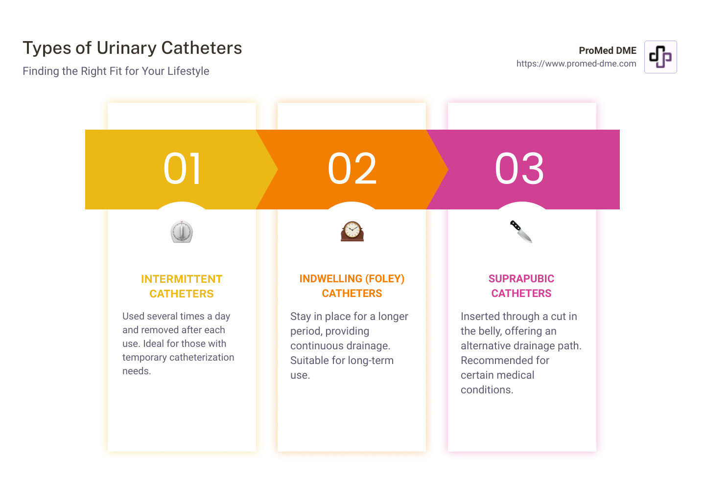
- Intermittent Catheters: Used several times a day and removed after each use.
- Indwelling (Foley) Catheters: Stay in place for a longer period.
- Suprapubic Catheters: Inserted through a cut in the belly.
Urinary catheterization is a procedure that might sound complicated, but it's all about helping your bladder when it needs it the most. Whether you're facing surgery, dealing with a condition that makes going to the bathroom tough, or need help emptying your bladder for another reason, catheters are there to help out.
There are different types of catheters based on how long they'll be used and where they'll be placed. Some are for quick visits to the doctor, while others might stay with you for a little while, helping out 24/7.
Understanding these options can make a big difference in managing your health and finding the right fit for your lifestyle. We'll dive into the specifics and guide you through each step of using and living with a catheter.
Understanding Urinary Catheterization
Urinary catheterization is a medical procedure that might seem complex at first glance, but it's really about helping your body do something it can't do on its own at the moment—release urine.
The basic idea is pretty straightforward: 1. Prepare the area and the materials needed. 2. Insert the catheter—a thin, flexible tube—into the bladder through the urethra (the tube that carries urine out of your body). 3. Allow urine to drain through the catheter into a collection bag.
It's a procedure performed with care, using sterile equipment to minimize infection risks. Pain is minimal, especially with the use of anesthetic gels.
Indications
Why would someone need this? Reasons include: - Difficulty urinating naturally due to medical conditions. - Needing to empty the bladder before, during, or after surgery. - Monitoring urine output in critically ill patients.
Simply put, if the bladder isn't doing its job adequately, catheterization steps in as a temporary or sometimes long-term solution.
Urinary Catheters
There are a few different types of catheters, each suited for different needs: - Intermittent Catheters : These are inserted several times a day, just long enough to empty the bladder, and then removed. - Indwelling Catheters : Often referred to as Foley catheters, these stay in place for longer periods, held by a small balloon filled with water to keep it from slipping out.
Foley Catheter
Speaking of Foley catheters, they're a common choice for long-term use. They can stay in for weeks or months, providing continuous urine drainage. This type of catheter is especially useful for patients who need constant bladder management but can't manage intermittent catheterization on their own.
Understanding urinary catheterization and drainage is key to demystifying the process and realizing it's a helpful, not scary, medical tool. It's about giving control back to individuals when their bodies need a little help.
This knowledge sets the stage for learning the step-by-step guide to catheterization , where we'll dive even deeper into how to safely insert, maintain, and eventually remove a catheter.
Types of Urinary Catheters
When it comes to urinary catheterization and drainage, it's crucial to know that not all catheters are created equal. Different situations call for different types of catheters. Let's break down the main types: Intermittent catheters , Indwelling catheters , Suprapubic catheters , and Foley catheters .
Intermittent Catheters
Intermittent catheters are used several times a day to drain the bladder and then removed. They are a go-to solution for people who can retain some control over their bladder functions. This type of catheter is usually pre-lubricated to make insertion smoother and reduce discomfort. The key here is hygiene and learning the proper technique to minimize the risk of infection. For a closer look at how intermittent catheters work, check out this detailed guide .
Indwelling Catheters
Also known as Foley catheters, these are left inside the bladder. A small balloon filled with water keeps them in place. They're suitable for those who need long-term catheterization, providing continuous urine drainage into a collection bag. Indwelling catheters need regular changing, usually every 3 months, to prevent infections and other complications. For more information on indwelling catheters, including how to manage them, click here .
Suprapubic Catheters
These are similar to indwelling catheters but are inserted through a small incision in the abdomen rather than the urethra. Suprapubic catheters are an option when urethral catheterization is not possible or advisable. They require surgical placement under anesthesia but offer the advantage of bypassing the urethra, which can be beneficial in certain conditions. The catheter is usually changed every 4 to 12 weeks.
Foley Catheters
A subset of indwelling catheters, Foley catheters are specifically designed with a balloon at one end that is inflated to keep the catheter in place. They are made from materials like silicone or natural rubber, tailored to minimize the risk of allergies or sensitivities. Foley catheters are a reliable choice for long-term urinary drainage, ensuring continuous urine flow to a drainage bag.

Understanding the specific needs and medical conditions of each individual is crucial in selecting the right type of catheter. Whether it’s for short-term use following surgery or for long-term management of chronic conditions, the right catheter can significantly improve the quality of life.
With this knowledge, we're ready to move on to the step-by-step guide to catheterization , where we'll cover preparation, insertion, maintenance, and removal, ensuring you're equipped to handle urinary catheterization and drainage confidently and safely.
Step-by-Step Guide to Catheterization
Mastering urinary catheterization and drainage is essential for ensuring comfort, preventing complications, and maintaining a healthy urinary system. Whether it's for temporary relief or long-term management, understanding the process step by step can make all the difference. Let's dive into the essentials of preparation, insertion, maintenance, and removal.
Preparation
1. Gather Your Supplies: Before starting, ensure you have all necessary items within reach. This includes the catheter, lubricant, sterile gloves, a collection bag, and antiseptic wipes. Using the right supplies is crucial for a successful catheterization process.
2. Hygiene is Key: Wash your hands thoroughly with soap and water. If you're assisting someone else, wear sterile gloves to prevent the spread of bacteria.
3. Prepare the Area: Clean the urinary opening and surrounding area using antiseptic wipes. For females, wipe from front to back to avoid introducing bacteria into the urinary tract.
4. Get Comfortable: Find a comfortable position. For self-catheterization, sitting on the toilet or standing with one leg up might work best. If you're assisting, ensure the patient is lying down with legs properly positioned.
5. Lubricate: Apply a generous amount of lubricant to the tip and first few inches of the catheter to ensure a smooth insertion.
6. Gentle Insertion: Carefully insert the catheter into the urinary opening. For males, insert until you reach the bladder and urine starts to flow. For females, insert approximately 2-3 inches until urine flows. It's important to proceed gently to avoid discomfort.
7. Let it Flow: Once the catheter is in place, allow urine to drain completely into the collection bag. Be patient and give it time to ensure the bladder is fully emptied.
Maintenance
8. Secure the Catheter: If using an indwelling catheter, make sure it's securely attached to the leg or abdomen to prevent pulling or movement that could cause injury.
9. Regular Cleaning: Clean the area around the catheter and the catheter itself with mild soap and water at least once a day to reduce the risk of infection.
10. Stay Hydrated: Drinking plenty of fluids helps maintain urine flow and prevents urinary tract infections (UTIs).
11. Wash Your Hands: Just like in the preparation stage, ensure your hands are clean before removing the catheter.
12. Careful Removal: Gently withdraw the catheter, stopping if you encounter any resistance or discomfort. For indwelling catheters, ensure the balloon is fully deflated before attempting to remove.
13. Dispose Properly: After removal, dispose of the catheter and gloves appropriately. For reusable catheters, follow the cleaning instructions provided by the manufacturer.
14. Post-Care: Clean the urinary opening and surrounding area once more. Apply a barrier cream if recommended by your healthcare provider to protect the skin.

Following these steps can help you master urinary catheterization and drainage, ensuring the process is as smooth and comfortable as possible. If you're unsure about any step or encounter difficulties, it's crucial to seek guidance from a healthcare professional. ProMed DME is committed to providing support and high-quality care products to assist in your catheterization process, ensuring you feel confident and well-cared for at every stage.
Managing and Preventing Complications
When it comes to urinary catheterization and drainage, being proactive is key to preventing and managing complications. Here's how you can stay ahead of common issues like infections, bladder spasms, leakages, blockages, UTIs, and kidney damage.
Infections & UTIs
- Keep it Clean: Wash your hands and the catheter area with soap and water before and after handling your catheter. This simple step is crucial in preventing infections.
- Stay Hydrated: Drinking plenty of fluids helps flush bacteria from your urinary system, reducing the risk of UTIs.
- Regular Catheter Care: Follow the cleaning instructions provided by your healthcare provider or the manufacturer. A clean catheter means a lower risk of infection.
Bladder Spasms
- Medication: Some medications can help manage bladder spasms. If you're experiencing discomfort, talk to your doctor about your options.
- Adjustments: Sometimes, the way a catheter is positioned can trigger spasms. If you suspect this is the case, consult your healthcare provider for an adjustment.
Leakages & Blockages
- Check the Position: Ensure your catheter is not kinked or twisted, as this can cause leakages or blockages.
- Monitor Output: Keep an eye on the amount of urine in your drainage bag. If it decreases significantly, it might indicate a blockage.
- Proper Bag Emptying: Empty your drainage bag regularly to avoid overfilling, which can lead to backflow and infections.
Kidney Damage
- Regular Check-ups: Routine visits to your healthcare provider can help monitor the health of your kidneys and catch any issues early.
- Watch for Symptoms: Symptoms like back pain, fever, or changes in urine appearance can indicate kidney problems. If you notice these, contact your healthcare provider immediately.
Anecdotes from the community, such as those shared on Reddit , emphasize the importance of not forcing catheter insertion, as this can lead to complications like false passages or trauma. Instead, patience and proper technique are advised.
In addition, recent developments mentioned on Wikipedia highlight the exploration of alternatives like temporary prostatic stents to reduce the risk of infections associated with long-term catheter use.
Remember: If you experience any issues with your catheter, such as pain, unusual leakage, or signs of infection, it's crucial to seek medical advice promptly. Early intervention can prevent more serious complications.
Next, we'll discuss how to integrate living with a catheter into your daily activities, from exercise to swimming and more, ensuring you can maintain a high quality of life.
Living with a Catheter
Living with a urinary catheter might seem daunting at first, but with the right knowledge and adjustments, it can become a manageable part of your routine. Here’s how to navigate daily activities, maintain hygiene, and care for your catheter.
Daily Activities
You can continue most of your regular activities with a urinary catheter. Whether you're going to work, running errands, or just relaxing at home, your catheter shouldn't significantly disrupt your lifestyle. However, it’s important to secure the catheter and drainage bag properly to prevent discomfort or leaks.
Staying active is important, and yes, you can still exercise with a catheter. Just be mindful of the type of physical activity you choose. Avoid exercises that put direct pressure on the catheter or its insertion site. For many, walking, cycling on a stationary bike, and gentle stretching are good options. Always secure the catheter and bag before starting.
Swimming is possible with a urinary catheter, but it requires some preparation to reduce infection risk. Use a waterproof cover for the catheter's entry point and ensure the drainage bag is securely attached and covered. After swimming, clean the area around the catheter thoroughly.
Sexual Activity
Having a catheter doesn’t mean you have to abstain from sexual activity, but you’ll need to take some precautions. Discuss with your healthcare provider for personalized advice. Generally, ensuring the catheter is securely positioned and being gentle can help prevent discomfort or injury.
Hygiene and Catheter Care
Good hygiene is crucial when living with a catheter to prevent infections. Wash your hands thoroughly before and after touching the catheter or drainage bag. Clean the area around the catheter insertion point daily with mild soap and water, patting it dry gently afterward.
Changing Drainage Bags
Drainage bags need to be changed regularly to maintain hygiene and functionality. Leg bags are typically used during the day and should be emptied when half to three-quarters full to avoid leaks. At night, switch to a larger night bag which can collect more urine while you sleep. Both types of bags should be cleaned regularly with a mixture of vinegar and water or a prescribed solution by your healthcare provider.
Living with a catheter is a significant adjustment, but it doesn’t have to limit your life. Many individuals lead full, active lives with a catheter in place. For more detailed guidance on managing life with a catheter, exploring resources like ProMed DME can be invaluable. They offer support and high-quality supplies that can make managing your catheter easier.
Additionally, a community member’s experience on Reddit highlights the adaptability of individuals living with catheters, showing that with the right approach and attitude, you can continue enjoying many of the activities you love.
In the next section, we'll address some frequently asked questions about urinary catheterization to clarify any uncertainties and provide you with a deeper understanding of how to live comfortably with a catheter.
Frequently Asked Questions about Urinary Catheterization
What is the purpose of urinary catheterization.
The main goal of urinary catheterization is to help drain urine from the bladder when a person cannot do it naturally. This could be due to various reasons like nerve damage, surgery, or conditions that block the flow of urine. Essentially, it's like providing an alternate route for urine to leave the body when the usual path isn’t working right.
Which type of catheter is used for drainage?
There are several types of catheters used for drainage, but the most common ones include:
Intermittent Catheters : These are inserted several times a day, just long enough to empty the bladder, and then removed. It's a go-to for short-term drainage.
Indwelling Catheters (Foley Catheters) : These stay in place for a longer period. They have a small balloon filled with water to keep them from falling out. Perfect for continuous drainage.
Suprapubic Catheters : These are inserted through a small cut in the belly, directly into the bladder. They are used when the urethral route is not possible or advisable.
Each type has its own specific uses and benefits, depending on the patient's condition and needs.
Does a catheter constantly drain urine?
Yes, most catheters are designed to constantly drain urine. Intermittent catheters are an exception since they are removed after each use. But indwelling catheters, like the Foley catheter , and suprapubic catheters are connected to a drainage bag that collects urine continuously. This ensures that the bladder remains empty and reduces the risk of infection.
Managing a catheter and its drainage system correctly is crucial for preventing complications and infections. Proper hygiene, regular check-ups, and following your healthcare provider's instructions will help you maintain a healthy and comfortable life with a catheter.
Urinary catheterization and drainage are essential for individuals who have difficulty emptying their bladder naturally. This procedure, while seemingly daunting at first, can significantly improve the quality of life by preventing uncomfortable and potentially dangerous urinary retention and infections. The key to mastering urinary catheterization lies in understanding the process, choosing the right type of catheter, and following proper maintenance and hygiene protocols.
The benefits of urinary catheterization cannot be overstated. For many, it provides a sense of independence and relief from the symptoms of urinary retention or incontinence. However, the importance of proper care in this context cannot be emphasized enough. Ensuring that the catheter and the area around it are clean reduces the risk of urinary tract infections (UTIs) and other complications, such as bladder spasms and leakages. Regularly emptying the drainage bag, using sterile techniques for insertion and removal, and staying hydrated are all critical steps in catheter care.
At ProMed DME, we are dedicated to supporting you throughout your journey with urinary catheterization. We offer a wide range of catheters and related supplies to meet your unique needs. Our team understands the challenges that come with managing a catheter, and we are here to provide you with the products, information, and support you need to live comfortably and confidently.
You're not alone. With the right resources and a bit of practice, managing your urinary catheter will become a routine part of your day. Whether you're new to catheterization or seeking to improve your technique and care routine, we're here to help. Explore our comprehensive line of catheters and urological supplies and discover the ProMed DME difference. Your health and comfort are our top priorities.
In conclusion, while urinary catheters are a necessary medical tool for many, they come with responsibilities. Awareness, proper care, and prevention can significantly reduce the risk of complications. By working closely with healthcare professionals and adhering to best practices in catheter care, you can maintain your urinary health and lead a comfortable life.
Related Resources & Articles
Stay informed with our informative blog posts.

Health Care Supplies: A Buyer's Guide

5 Essential Medical Supplies in Florida You Need to Know Before Moving

Everything You Need to Know About Rusch Catheters
Discover the promed advantage & try our products.
We offer free shipping and legendary customer service to ensure you receive the best DME products for your needs.
Want to create or adapt books like this? Learn more about how Pressbooks supports open publishing practices.
21.8 Applying the Nursing Process to Catheterization
When preparing to insert an indwelling urinary catheter, it is important to use the nursing process to plan and provide care to the patient. Begin by assessing the appropriateness of inserting an indwelling catheter according to CDC criteria as discussed in the “ Preventing CAUTI ” section of this chapter. Determine if alternative measures can be used to facilitate elimination and address any concerns with the prescribing provider before proceeding with the provider order.
Subjective Assessment
In addition to verifying the appropriateness of the insertion of an indwelling catheter according to CDC recommendations, it is also important to assess for any conditions that may interfere with the insertion of a urinary catheter when feasible. See suggested interview questions prior to inserting an indwelling catheter and their rationale in Table 21.8a.
Table 21.8a Suggested Interview Questions Prior to Urinary Catheterization
Cultural Considerations
When inserting urinary catheters, be aware of and respect cultural beliefs related to privacy, family involvement, and the request for a same-gender nurse. Inserting a urinary catheter requires visualization and manipulation of anatomical areas that are considered private by most patients. These procedures can cause emotional distress, especially if the patient has experienced any history of abuse or trauma.
Objective Assessment
In addition to performing a subjective assessment, there are several objective assessments to complete prior to insertion. See Table 21.8b for a list of objective assessments and their rationale.
Table 21.8b Objective Assessment
Life Span Considerations
Children It is often helpful to explain the catheterization procedure using a doll or toy. According to agency policy, a parent, caregiver, or other adult should be present in the room during the procedure. Asking a younger child to blow into a straw can help relax the pelvic muscles during catheterization.
Older Adults The urethral meatus of older women may be difficult to identify due to atrophy of the urogenital tissue. The risk of developing a urinary tract infection may also be increased due to chronic disease and incontinence.
Expected Outcomes/Planning Expected patient outcomes following urinary catheterization should be planned and then evaluated and documented after the procedure is completed. See Table 21.8c for sample expected outcomes related to urinary catheterization.
Table 21.8c Expected Outcomes of Urinary Catheterization
Implementation
When inserting an indwelling urinary catheter, the expected finding is that the catheter is inserted accurately and without discomfort, and immediate flow of clear, yellow urine into the collection bag occurs. However, unexpected events and findings can occur. See Table 21.8d for examples of unexpected findings and suggested follow-up actions.
Table 21.8d Unexpected Findings and Follow-Up Actions
Evaluate the success of the expected outcomes established prior to the procedure.
Nursing Skills - 2e Copyright © 2023 by Chippewa Valley Technical College is licensed under a Creative Commons Attribution 4.0 International License , except where otherwise noted.
Share This Book

An official website of the United States government
The .gov means it’s official. Federal government websites often end in .gov or .mil. Before sharing sensitive information, make sure you’re on a federal government site.
The site is secure. The https:// ensures that you are connecting to the official website and that any information you provide is encrypted and transmitted securely.
- Publications
- Account settings
Preview improvements coming to the PMC website in October 2024. Learn More or Try it out now .
- Advanced Search
- Journal List
- HHS Author Manuscripts
Diagnosis, Management, and Prevention of Catheter-Associated Urinary Tract Infections
Carol e. chenoweth.
a Division of Infectious Diseases, Department of Internal Medicine, University of Michigan Health System, 3119 Taubman Center, 1500 East Medical Center Drive, Ann Arbor, MI 48109-5378, USA
Carolyn V. Gould
b Centers for Disease Control and Prevention, 1600 Clifton Road Northeast, Mailstop A-31, Atlanta, GA 30333, USA
Sanjay Saint
c Division of General Medicine, Department of Internal Medicine, University of Michigan Health System and Veterans Affairs Ann Arbor Healthcare System, 2800 Plymouth Road, Building 16, Room 430 West, Ann Arbor, MI 48109-2800, USA
Urinary tract infection (UTI) is one of the most common health care–associated infections (HAIs), representing up to 40% of all HAIs. 1 – 3 Most health care–associated UTIs (70%) are associated with urinary catheters, but as many as 95% of UTIs in intensive care units (ICUs) are associated with catheters. 4 , 5 Approximately 20% of patients have a urinary catheter placed at some time during their hospital stay, 6 , 7 especially in ICUs, in long-term care facilities, and increasingly in home care settings. 3 , 4 , 8 The Centers for Disease Control and Prevention (CDC) estimated that up to 139,000 catheter-associated UTIs (CAUTIs) occurred in US hospitals in 2007. 4
CAUTIs are associated with increased morbidity, mortality, and costs. Hospital-associated bloodstream infection from a urinary source has a case fatality of 32.8%. 9 , 10 Each episode of CAUTI is estimated to cost $600; if associated with a bloodstream infection, costs increase to $2800. 11 Nationally, CAUTIs result in an estimated $131 million annual excess medical costs. 4
Moreover, in October 2008, the Centers for Medicare and Medicaid Services (CMS) included hospital-acquired CAUTI under conditions that are no longer reimbursed for the extra costs of managing a patient. 11 To date, there has been no measurable effect of the CMS policy to reduce payments for CAUTIs on CAUTI rates or preventive practices. 12 – 14 Nevertheless, the prevention of CAUTIs has become a priority for most hospitals because 65% to 70% of CAUTIs may be preventable. 15
EPIDEMIOLOGY OF CAUTIs
Likely as a result of widespread interventions occurring nationwide, rates of CAUTIs in ICUs reporting to the CDC decreased significantly between 1990 and 2007. 4 In 2010, the rates of CAUTIs reported to the CDC’s National Healthcare Safety Network (NHSN) ranged from 4.7 per 1000 catheter-days in burn ICUs to 1.3 per 1000 catheter-days in medical/surgical ICUs. 4 Pediatric ICUs reported similar rates of CAUTI, 2.2 to 3.9 per 1000 catheter-days 16 ; however, CAUTIs are infrequently identified in neonatal ICUs. 17 Inpatient wards reported rates equivalent to ICU settings, with a range from 0.2 to 3.2 per 1000 catheter-days. Among inpatient wards, rehabilitation units had the highest rates of CAUTIs. 5 , 16
Microbial Cause of CAUTIs
Most microorganisms causing CAUTIs are from the endogenous microbiota of the perineum that ascend the urethra to the bladder along the external surface of the catheter. 18 A smaller proportion of microorganisms (34%) are introduced by intraluminal contamination of the collection system from exogenous sources, frequently resulting from cross-transmission of organisms from the hands of health care personnel. 18 , 19 Approximately 15% of episodes of health care–associated bacteriuria occur in clusters from patient-to-patient transmission within a hospital. 2 , 19 Rarely, organisms, such as Staphylococcus aureus , cause UTI from hematogenous spread.
Enterobacteriaceae, especially Escherichia coli and Klebsiella spp, are the most common pathogens associated with CAUTI; but in the ICU setting, Candida spp (18%), Enterococcus spp (10%), and Pseudomonas aeruginosa (9%) are more prevalent ( Table 1 ). 16 , 20 , 21 European hospitals report a similar spectrum of microorganisms associated with nosocomial UTIs, except for Pseudomonas spp, which were isolated in only 7% of urine cultures. 22
Selected microorganisms associated with CAUTIs
Abbreviation: LTACH, long-term acute care hospitals.
Data from Chitnis A, Edwards J, Ricks P, et al. Device-associated infection rates, device utilization, and antimicrobial resistance in long-term acute care hospitals reporting to the National Healthcare Safety Network, 2010. Infect Control Hosp Epidemiol 2012;33(10):993–1000; and Sievert D, Ricks P, Edwards J, et al. Antimicrobial-resistant pathogens associated with healthcare-associated infections: summary of data reported to the National Healthcare Safety Network at the Centers for Disease Control and Prevention, 2009–2010. Infect Control Hosp Epidemiol 2013;34(1):1–14.
Among E coli isolates reported to the NHSN from CAUTIs in ICU and non-ICU settings in 2009 to 2010, 29.1% and 33.5%, respectively, were resistant to fluoroquinolones. 21 Many Enterobacteriaceae produced extended-spectrum beta-lactamases; 26.9% of K pneumonia/oxytoca and 12.3% of E coli isolates from patients with CAUTIs were resistant to extended-spectrum cephalosporins. Alarmingly, during this same time period, 12.5% of Klebsiella spp from patients with CAUTIs were resistant to carbapenems. 21 Although long-term acute care hospitals (LTACHs) had a prevalence of carbapenem-resistant Enterobacteriaceae (CRE) in CAUTI isolates similar to that reported in ICUs, a greater percentage of LTACHs reported a CRE CAUTI compared with ICUs. 20
Enterococci emerged as a commonly reported cause of health care–associated UTIs between 1975 and 1984. Although the clinical significance of enterococci isolated from urine is questionable, urinary drainage devices serve as a reservoir for emergence and spread of vancomycin-resistant strains in short- and long-term acute care settings. 20 , 21 Also rarely associated with complications when isolated from the urine, 23 Candida spp account for 28% of CAUTIs reported from ICUs. 20 S aureus are an infrequent cause of CAUTI but, when identified, should prompt consideration for coinciding bacteremia or endocarditis. 10 , 24 CAUTI associated with long-term catheters are associated with 2 or more organisms in 77% to 95% of episodes, and 10% have more than 5 species of organisms present. 3
Biofilms, composed of clusters of microorganisms and extracellular matrix (primarily polysaccharide materials), form on the internal and external surfaces of urinary catheters shortly after insertion. 19 , 25 Typically, the biofilm is composed of one type of microorganism, although polymicrobial biofilms are possible. Microorganisms within the biofilm ascend the catheter to the bladder in 1 to 3 days. Antimicrobials penetrate into biofilms poorly, and microorganisms grow more slowly in biofilms, decreasing the effects of many antimicrobials. 19 , 25 The microorganisms, resistance patterns, and biofilm factors mentioned earlier have significant implications for the management of CAUTIs.
Risk Factors for CAUTIs
Table 2 outlines major modifiable and nonmodifiable risk factors for CAUTI, which have importance for the design and implementation of interventions for the prevention of CAUTI. The duration of catheterization is the dominant risk factor for CAUTI. 1 , 3 , 26 Women have a higher risk of UTI than men, and heavy bacterial colonization of the perineum increases that risk. Other factors that increase the risk of CAUTI include rapidly fatal underlying illness, more than 50 years of age, nonsurgical disease, hospitalization on an orthopedic or urological service, catheter inserted outside the operating room, diabetes mellitus, and serum creatinine greater than 2 mg/dL at the time of catheterization. Nonadherence to aseptic catheter care recommendations has been associated with an increased risk of bacteriuria; conversely, systemic antibiotics have a protective effect on bacteriuria (relative risk 2.0–3.9). 2 , 3 Independent risk factors for urinary tract–related bloodstream infections in patients with bacteriuria include neutropenia, renal disease, and male sex. 27
Risk factors for CAUTIs
DIAGNOSIS OF CAUTIS
Clinical diagnosis of a CAUTI is challenging because pyuria and bacteriuria are almost uniformly present, but neither are reliable indicators of symptomatic UTI in the setting of catheterization. 28 – 31 Symptomatic UTI is defined by the presence of symptoms or signs referable to the urinary tract associated with significant bacteriuria. 28 Fever or other systemic symptoms may be the only clinical indication of UTI in patients who are critically ill or who have spinal cord injuries. 2 , 3 However, outside these patient populations, additional urinary tract-specific signs and symptoms should be sought for the diagnosis of UTI. 28 , 30
Defining significant bacteriuria is difficult because some level of bacterial colonization is universal in urine from catheterized patients. Colony counts in urine as low as 10 2 colony-forming units (CFU)/mL can be associated with symptoms, and colony counts of this level rapidly increase to more than 10 5 CFU/mL within 24 to 48 hours. 28 , 32 , 33 Therefore, the National Institute on Disability and Rehabilitative Research defined bacteriuria in catheterized patients as growth of 10 2 CFU/mL or more of a predominant microorganism. 33 Other guidelines have defined 10 3 CFU/mL as a more reasonable threshold for significant bacteriuria, balancing the sensitivity of detecting CAUTI with the feasibility of the microbiology laboratory to quantify microorganisms. 28
Asymptomatic bacteriuria is defined as bacteriuria in patients without signs or symptoms referable to the urinary tract. 28 The distinction from symptomatic UTI is clinically important because asymptomatic catheter-associated bacteriuria and funguria rarely result in adverse outcomes (eg, pyelonephritis, perinephric abscess, bacteremia) and generally do not require treatment. 30 Nevertheless, a large proportion of antimicrobials in hospitalized patients are prescribed for the treatment of UTIs, most often asymptomatic bacteriuria. 34 – 36
MANAGEMENT OF CAUTIS
The treatment of asymptomatic catheter-associated bacteriuria or candiduria is not indicated except in patients who are at a high risk for the development of complications, such as pyelonephritis or bloodstream infection. 37 Screening and treating pregnant women for asymptomatic bacteriuria to prevent pyelonephritis are recommended. In addition, patients undergoing genitourinary procedures likely to induce mucosal bleeding should be screened and treated in advance for asymptomatic bacteriuria. 28 , 37 As with asymptomatic bacteriuria, asymptomatic candiduria generally does not require treatment, except in neutropenic patients and other high-risk patients noted earlier. 38 Furthermore, because of poor specificity of fever and frequency of bacteriuria and funguria in hospitalized patients with urinary catheters, a thorough investigation for other sources of fever should be conducted before diagnosing a UTI.
Asymptomatic bacteriuria, persisting for 48 hours after the removal of a urinary catheter, has a high risk of progressing to symptomatic UTI; treatment in hospitalized women has been shown to decrease the risk of subsequent UTIs. 39 Therefore, considering the treatment of women with asymptomatic bacteriuria persisting 48 hours after catheter removal is recommended. 28 , 37 , 39 When indicated, 3 to 7 days of appropriate antimicrobial therapy based on culture results should be adequate for the treatment of asymptomatic bacteriuria. 28 , 37
Repeated antimicrobial treatment of bacteriuria during long-term catheterization is a significant risk for colonization with multidrug-resistant organisms, and most of this use is inappropriate. 34 , 35 A recent study reported that a 1-hour educational session reduced inappropriate use of antibiotic therapy for inpatients with positive urine cultures. 40 In addition, audit and feedback to care providers decreased overdiagnosis of CAUTIs and associated inappropriate antibiotic use in another study. 36 Educational efforts aimed at reducing unnecessary urine cultures (eg, pan-culturing for fever without a thorough clinical assessment) would also prevent the inappropriate treatment of bacteriuria and funguria.
Because of the presence of biofilm, leaving the catheter in place during the treatment of CAUTIs makes eradicating bacteriuria or candiduria difficult and can lead to the development of antimicrobial resistance. The management of symptomatic CAUTIs should include removing or replacing the urinary catheter if it has been in place for at least 2 weeks. 28 , 41 In terms of antimicrobial therapy, symptomatic CAUTIs may be treated with 7 days of appropriate antimicrobials if patients have a prompt resolution of symptoms; therapy should be lengthened to 10 to 14 days for those with a delayed response. 28 Initial empiric therapy should be based on local epidemiologic data regarding causative microorganisms of CAUTIs and antimicrobial resistance patterns. Once culture data become available, antimicrobial therapy should be adjusted as necessary, ideally providing the narrowest spectrum of coverage possible while still providing adequate treatment of the UTI. Symptomatic CAUTIs caused by Candida species should be treated with 14 days of antifungal agents. 38
PREVENTING CAUTIS
General strategies, formulated for the prevention of all HAIs, including strict adherence to hand hygiene, are critical for the prevention of CAUTIs. 42 The urinary tract of hospitalized patients, especially those in an ICU setting, represents a significant reservoir for multidrug-resistant organisms. Therefore, precautions recommended for prevention of transmission of multidrug-resistant organisms should be scrupulously observed in catheterized patients. 43 Limiting unnecessary use of antimicrobials, as part of an overall antimicrobial stewardship program, is another important general strategy to prevent the development of antimicrobial resistance related to urinary catheters. 44
The measurement and feedback of results of interventions to the clinical care team is an essential component of any improvement program. The CDC NHSN CAUTI rate (symptomatic UTI per 1000 urinary catheter-days) is the most widely accepted measure for CAUTI surveillance and is endorsed by the Infectious Diseases Society of America, the Society of Healthcare Epidemiology of America, and the Association for Professionals in Infection Control and Epidemiology. 28 , 45 , 46 In addition, beginning in 2012, the CMS has required as a condition of participation that hospitals, long-term care hospitals, and inpatient rehabilitation facilities submit ICU CAUTI rates to the NHSN. A modified definition of UTI is recommended for surveillance in long-term care facilities. 47 Efforts are currently underway to revise the CDC’s NHSN UTI surveillance definitions to improve specificity and clinical relevance of the measure.
However, a population-based measure, using hospital-days as the denominator, has been suggested as an alternative measure to assess improvement interventions at individual hospitals. 48 Other measures, such as rates of asymptomatic bacteriuria, percentage of patients with indwelling catheters, percentage of catheterization with accepted indications, and duration of catheter use, have been used in improvement studies and collaboratives with good success. 49
Several guidelines with specific recommendations for the prevention of CAUTIs have been developed or recently updated ( Box 1 ). 28 , 45 , 46 , 50 However, in 2005, a nationwide survey identified that one-third of hospitals did not conduct surveillance for UTIs, more than one-half did not monitor urinary catheters, and three-quarters did not monitor the duration of catheterization. 12 , 51 In a follow-up study, after the enactment of the CMS nonpayment rule, still no CAUTI prevention practices had been adopted in more than half of the hospitals, except for the use of bladder ultrasound. 12 Even in ICUs, only a small proportion of surveyed sites had policies supporting bladder ultrasound (26%), catheter removal reminders (12%), or nurse-initiated catheter discontinuation (10%). 52 The systematic adoption of prevention practices has begun to be observed through the use of bundles and collaboratives, as detailed later. 49 , 53
Strategies for prevention of CAUTIs
Avoid insertion of indwelling urinary catheters
- Placement only for appropriate indications (see Box 2 )
- Institutional protocols for placement, including perioperative setting
Early removal of indwelling catheters
- Checklist or daily plan
- Nurse-based interventions
- Electronic reminders
Seek alternatives to indwelling catheterization
- Intermittent catheterization
- Condom catheter
- Portable bladder ultrasound scanner
Aseptic techniques for care of catheters
- Sterile insertion
- Closed drainage system
- Maintain gravity drainage
- Avoid routine bladder irrigation
Data from Refs. 28 , 45 , 46 , 50
A qualitative study of 12 hospitals participating in a statewide program identified barriers to adoption of the key interventions to reduce unnecessary use of urinary catheters. Common barriers included difficulty with nurse and physician engagement, patient and family request for indwelling catheters, and catheter insertion practices and customs in emergency departments. 54 In addition, qualitative studies have revealed that staff variations of the perceived risk and perceived strength of evidence supporting preventive practices should be incorporated into implementation plans. 55 , 56
Limiting Use of Urinary Catheters
The foremost strategy for CAUTI prevention is avoidance of or decreasing the duration of urinary catheterization. Catheter utilization varies by ICU type, with the lowest rate in pediatric medical ICUs (0.16 urinary catheter-days/patient-days) and the highest rates reported in trauma ICUs (0.80 urinary catheter-days/patient-days). 16 Decreasing catheter utilization requires interventions at several stages of the lifecycle of the urinary catheter. 26
The first stage in decreasing catheter utilization is limiting the placement of indwelling urinary catheters. Overall, urinary catheters are overused and the documentation surrounding catheterization is inconsistent 7 , 57 – 59 ; urinary catheters are placed for inappropriate indications in 21% to 50% of catheterized patients. 7 , 60 Written policies and criteria for indwelling urinary catheterization, based on accepted indications, is a first step in limiting the placement of urinary catheters; but tracking indications for catheters with feedback to the care team is also important ( Box 2 ). 45 , 50 Some hospitals have had success by targeting interventions for limiting the placement of urinary catheters in emergency departments and operating rooms, locations where the initial placement often takes place. 61
Appropriate indications for indwelling urinary catheters
Acute urinary retention or bladder outlet obstruction
Need for accurate measurements of urinary output
Perioperative use for selected surgical procedures
- Surgical procedures of anticipated long duration
- Urologic procedures
- Intraoperatively for patients with urinary incontinence
- Need for intraoperative urinary monitoring or expected large volume of intravenous infusions
Urinary incontinence in the setting of open perineal or sacral wounds
Improve comfort for end-of-life care or patient preference
Modified from Gould C, Umscheid C, Agarwal R, et al. Healthcare Infection Control Practices Advisory Committee. Guideline for prevention of catheter-associated infections 2009. Infect Control Hosp Epidemiol 2010;31:319–26.
Once catheters are placed, strategies for early removal become necessary to limit the duration of catheterization. Relying on physicians’ orders alone may be inadequate for the management of catheters because, in one study, 28% of physicians were unaware that their patient had a catheter. 7 Nurse-driven interventions have demonstrated effectiveness in reducing the duration of catheterization. 62 – 64 This type of intervention was implemented in a statewide effort that resulted in a significant decrease in catheter use and an increase in appropriate indications of catheters. 49
Computerized physician order entry systems may offer a more cost-effective and efficient system to reduce both the placement of catheters and the duration of catheterization. 65 A systematic review and meta-analysis found that urinary catheter reminder systems and stop orders seem to reduce the mean duration of catheterization by 37% and CAUTIs by 52%. 66
Hospitals have also shown success in decreasing urinary catheter prevalence and CAUTIs through the multimodal interventions noted earlier. 67 , 68 One institution used a multifaceted intervention, which included education, system redesign, rewards, and feedback managed by a dedicated nurse, resulting in a marked decrease in the daily prevalence of urinary catheter days. 67 Strategies to address barriers to the implementation of urinary catheterization bundles include incorporating planned toileting into other patient safety programs, discussing the risk of indwelling urinary catheters with patients and their families, and engaging emergency department personnel to ensure appropriate indications for catheter use are followed have been promoted. 54
Perioperative Management of Urinary Catheters
Approximately 85% of patients admitted for major surgical procedures have perioperative indwelling catheters. Those patients catheterized longer than 2 days are significantly more likely to develop UTIs and are less likely to be discharged to home. 69 Older surgical patients are at the highest risk for prolonged catheterization; 23% of surgical patients older than 65 years are discharged to skilled nursing facilities with an indwelling catheter in place and have substantially more rehospitalization or deaths within 30 days. 70 Therefore, specific protocols for the management of postoperative urinary catheters are important for reducing urinary catheterization utilization and patient outcomes; the Surgical Care Improvement Project has added the removal of urinary catheters as one of their measures.
In a large prospective trial of patients undergoing orthopedic procedures, patients were entered into the following protocol: (1) limiting catheterization to surgeries of more than 5 hours or for total hip and knee replacements and (2) the removal of urinary catheters on postoperative day 1 after total knee arthroplasty and postoperative day 2 after total hip arthroplasty. This intervention resulted in a two-thirds reduction in the incidence of UTIs. 71
Alternatives to Indwelling Urinary Catheters
A randomized trial demonstrated a decrease in bacteriuria, symptomatic UTI, or death in patients who used condom catheters when compared with those with indwelling catheters; this benefit was seen primarily in men without dementia. 72 Condom catheters have also been reported to be less painful than indwelling catheters in some men. 72 , 73 Therefore, condom catheters may be considered in place of indwelling catheters in appropriately selected male patients without urinary retention or bladder outlet obstruction.
Patients with neurogenic bladder and long-term urinary catheters, in particular, may benefit from intermittent catheterization. 50 Intermittent catheterization may also be beneficial for short-term urinary retention. A recent meta-analysis reported a reduced risk of bacteriuria with the use of intermittent catheterization in patients following hip or knee surgery compared with indwelling catheterization. 74 Combining the use of a portable bladder ultrasound scanner with intermittent catheterization may reduce the need for indwelling catheterization. 45 , 75
Aseptic Techniques for Insertion and Maintenance of Urinary Catheters
When indwelling catheterization is necessary, aseptic catheter insertion and maintenance is recommended for preventing CAUTIs. Urinary catheters should be inserted by a trained health care professional using a sterile technique. 50 Cleaning the meatus before catheter insertion is recommended; but ongoing daily meatal cleaning with an antiseptic has not shown benefit and may increase rates of bacteriuria compared with routine care with soap and water. 50 Sterile lubricant jelly should be used for insertion, but antiseptic lubricants are not necessary. 50
Maintaining a closed urinary catheter collection system is important to reduce the risk of CAUTIs. Opening the closed system should be avoided, especially when sampling urine that may be performed aseptically from a port or from the drainage bag. 50 Prophylactic instillation of antiseptic agents or irrigation of the bladder with antimicrobial or antiseptic agents has shown no benefit in preventing bacteriuria and is not recommended. 50 Finally, routine exchange of urinary catheters is not recommended except for mechanical reasons because bacteriuria and biofilms return quickly. 2
Use of Antiinfective Catheters
Antiseptic or antimicrobial impregnated urinary catheters have been studied extensively as an adjunctive measure for preventing CAUTIs with variable results. 76 , 77 However, almost all previous studies used bacteriuria as the primary end point rather than symptomatic UTIs, thus limiting their clinical relevance. In a Cochrane review, silver alloy catheters were found to significantly reduce the incidence of asymptomatic bacteriuria in adult patients catheterized less than 7 days, but the effect was diminished in those catheterized for greater than 7 days. 77 A recent multicenter randomized controlled trial that did use symptomatic CAUTIs as the end point reported no significant clinical benefit with the use of silver alloy-coated or nitrofural-impregnated catheters during short-term (<14 days) catheterization. 78 Few studies have evaluated antiseptic and antimicrobial catheters in long-term urinary catheterization. 79 Therefore, there is no recommendation for routine use of antiinfective urinary catheters to prevent CAUTIs. 50 Despite these recommendations, a national study in 2009 revealed that 45% of nonfederal and 22% of Department of Veterans Affairs hospitals used antimicrobial catheters; hospitals using antiinfective catheters often based their decisions on hospital-specific pilot studies. 12
IMPLEMENTATION: THE ROLE OF BUNDLES, COLLABORATIVES, AND LEADERSHIP
Recently, bundles of interventions have been used with success for the prevention of HAIs, including CAUTIs. The Bladder Bundle outlined using the mnemonic ABCDE in Box 3 applied was successfully adopted by the Michigan Hospital Association Keystone initiative. 49 , 53 After the implementation of this initiative, Michigan hospitals used more key prevention practices and had a lower rate of CAUTIs when compared with hospitals in the rest of the country. 80 Finally, the important role of local hospital leadership and followership for ensuring effective implementation of preventive initiatives has recently been highlighted. 81 – 83 The Web site www.catheterout.org provides a list of common barriers along with solutions that hospitals may wish to use in their CAUTI prevention programs.
The ABCDE for preventing CAUTIs
- Adherence to general infection control principles (eg, hand hygiene, surveillance and feedback, aseptic insertion, proper maintenance, education) is important.
- Bladder ultrasound may avoid indwelling catheterization.
- Condom catheters or other alternatives to an indwelling catheter, such as intermittent catheterization, should be considered in appropriate patients.
- Do not use the indwelling catheter unless you must.
- Early removal of the catheter using a reminder or nurse-initiated removal protocol seems to be warranted.
From Saint S, Olmsted RN, Fakih MG, et al. Translating health care-associated urinary tract infection prevention research into practice via the bladder bundle. Jt Comm J Qual Patient Saf 2009;35(9):449–55; with permission.
CAUTIs are common, costly, and cause significant patient morbidity. CAUTIs are associated with hospital pathogens with a high propensity toward antimicrobial resistance. The treatment of asymptomatic CAUTIs accounts for excess antimicrobial use in hospitals and should be avoided. The duration of urinary catheterization is the predominant risk factor for CAUTI; preventive measures directed at limiting the placement and early removal of urinary catheters have a significant impact on decreasing CAUTIs. Bladder bundles, collaboratives, and the support of hospital leaders are powerful tools for implementing appropriate preventive measures against CAUTI.
- Catheter-associated urinary tract infection (CAUTI) is often caused by hospital-based pathogens with a propensity toward antimicrobial resistance.
- The diagnosis of CAUTI is problematic because pyuria and bacteriuria are not reliable markers of infection. The treatment of bacteriuria in the absence of symptoms is not indicated, except in patients at risk of developing pyelonephritis or bloodstream infection (ie, pregnancy, urologic procedures with bleeding).
- Indwelling urinary catheters that have been in place for more than 2 weeks should be removed when treating CAUTI.
- The duration of urinary catheterization is the predominant risk for CAUTI; preventive measures directed at limiting the placement and early removal of urinary catheters significantly reduce CAUTI rates.
- Bladder bundles, collaboratives, and certain behaviors of hospital-based leaders are powerful tools for implementing preventive measures for CAUTI.
Disclosures/Conflict of Interest:
C.E. Chenoweth, C.V. Gould: None; S. Saint: Honoraria and speaking fees from academic medical centers, hospitals, specialty societies, group-purchasing organizations (eg, Premier, VHA), state-based hospital associations, and nonprofit foundations (eg, Michigan Health and Hospital Association, Institute for Healthcare Improvement) for lectures about catheter-associated urinary tract infection and implementation science.
Publisher's Disclaimer: Disclaimer: The findings and conclusions in this report are those of the authors and do not necessarily represent the official position of the Centers for Disease Control and Prevention.
- MSD careers
How To Do Urethral Catheterization in a Female
- Indications |
- Contraindications |
- Complications |
- Equipment |
- Additional Considerations |
- Relevant Anatomy |
- Positioning |
- Step-by-Step Description of Procedure |
- Aftercare |
- Warnings and Common Errors |
- Tips and Tricks |
Urethral catheterization is the standard method of accessing the urinary bladder. A flexible catheter is passed retrograde through the urethra into the bladder. Several types of catheters are available. If the urethra is impassable, suprapubic catheterization of the bladder will be necessary.
(See also Bladder Catheterization .)
Indications for Urethral Catheterization in a Female
Relief of acute or chronic urinary retention, such as due to urethral obstruction ( obstructive uropathy ) or neurogenic bladder
Treatment of urinary incontinence
Monitoring of urine output
Measurement of postvoid residual urine volume
Collection of sterile urine for culture
Diagnostic studies of the lower genitourinary tract
Bladder irrigation or instillation of medication
Contraindications to Urethral Catheterization in a Female
Absolute contraindications
Relative contraindications
History of urethral strictures
Current urinary tract infection (UTI)
Prior urethral reconstruction
Suspected urethral injury *
Recent urologic surgery
History of difficult catheter placement
*Urethral injury may be suspected following blunt trauma if patients have blood at the urethral meatus (most important sign), inability to void, or perineal or labial ecchymosis, and/or edema. In such cases, urethral disruption should be ruled out with imaging (eg, by retrograde urethrography and sometimes also cystoscopy ) before doing urethral catheterization.
Complications of Urethral Catheterization in a Female
Complications include
Urethral or bladder trauma with bleeding or microscopic hematuria (common)
UTI (common)
Creation of false passages
Scarring and strictures
Equipment for Urethral Catheterization in a Female
Prepackaged kits are typically used but the individual items needed include
Sterile drapes and gloves
Povidone iodine
Applicator swabs, sterile gauze, or cotton balls
Water-soluble lubricant
Urethral catheter (size 16 French Foley catheter is appropriate for most adult women)*
10-mL syringe with water (for catheter balloon inflation)
Sterile collection device with tubing
* A closed catheter system minimizes catheter-associated UTI .
Additional Considerations for Urethral Catheterization in a Female
Sterile technique is necessary to prevent a UTI .
Relevant Anatomy for Urethral Catheterization in a Female
The female urethral meatus appears as an anterior-posterior slit located anterior to the vaginal opening and about 2.5 cm posterior to the glans clitoris. If the meatus recedes superiorly into the vagina, as can happen in older women, it can often be palpated in the midline as a soft mound surrounded by a firm ring of periurethral tissue.
Positioning for Urethral Catheterization in a Female
To expose the vulva, position the patient supine in either lithotomy or frog position (hips and knees partially flexed, heels on the bed, hips comfortably abducted).
Step-by-Step Description of Urethral Catheterization in a Female
Place all equipment within easy reach on an uncontaminated sterile field on a bedside tray. You may put the box containing the catheter and the drainage system between the patient’s legs, so that it is easily accessible during the procedure.
If not done already, attach the catheter to the collection system and do not break the seal unless a different type or size of catheter is required.
Test the retention balloon for leaks by inflating it with water.
Apply lubricant to the tip of the catheter.
Saturate the applicator swabs, cotton balls or gauze with povidone iodine.
Place the sterile fenestrated drape over the pelvis so that the vulva is exposed.
Gently spread the labia and expose the urethral meatus, using your nondominant hand. This hand is now contaminated and must not be removed from the labia or touch any of the equipment during the rest of the procedure.
Cleanse the area around the meatus with each cotton ball saturated in povidone iodine. Use a circular motion, beginning at the meatus and working your way outward. Discard or set aside the newly contaminated gauze or cotton balls.
Hold the lubricated catheter and gently pass it through the urethra, using your free hand. Urine should flow freely into the collection tubing. If the catheter accidentally passes into the vagina, it should be discarded and a new catheter used.
Inflate the balloon with the recommended volume of water, usually 10 mL. Resistance or pain may indicate that the balloon is in the urethra and not the bladder. If so, deflate the balloon, then insert it all the way before reinflation.
Pull the balloon up snug against the bladder neck, after the balloon has been inflated, by slowly withdrawing the catheter until resistance is felt.

Aftercare for Urethral Catheterization in a Female
Remove the drapes.
Secure the catheter to the thigh with an adhesive bandage or tape.
Hang the bag in a dependent position, so that urine can drain via gravity.
Warnings and Common Errors for Urethral Catheterization in a Female
Be sure to maintain strict sterile technique during the procedure to avoid urinary tract infection .
Tips and Tricks for Urethral Catheterization in a Female
It is often helpful to have an assistant to help expose the meatus in women, especially those who are obese or have pelvic organ prolapse. Gentle retraction of the labia is helpful.

- Cookie Preferences

Copyright © 2024 Merck & Co., Inc., Rahway, NJ, USA and its affiliates. All rights reserved.
Urinary Elimination (Urinary Incontinence & Urinary Retention) Nursing Care Plan & Management

Impairments in urinary elimination can be due to urinary incontinence or urinary retention and all refer to the inability to pass urine effectively. Get to know the nursing care plan and management of clients with urinary elimination problems. Learn about the nursing assessment , nursing diagnosis , goals, and interventions for clients with urinary elimination and urinary retention.
Table of Contents
Functional urinary incontinence, urge urinary incontinence, reflex urinary incontinence, stress urinary incontinence, overflow urinary incontinence, mixed urinary incontinence, what is urinary retention, nursing problem priorities, nursing assessment, nursing diagnosis, nursing goals, 1. assessing urinary patterns and etiology, interventions for functional urinary incontinence, interventions for urge urinary incontinence, interventions for reflex urinary incontinence, 3. initiating interventions for urinary retention, 4. preventing urinary tract infections (utis), 5. maintaining skin integrity, 6. providing client and caregiver education, recommended resources, what is urinary incontinence.
Urinary incontinence , also known as overactive bladder , is the involuntary loss of urine due to difficulties controlling the bladder, frequently seen in older individuals, particularly women. This condition often leads to feelings of embarrassment and a loss of independence, as issues such as wet clothing, urine odor, and the need for assistance with toileting can arise. Over time, the inability to control urination can negatively impact a person’s self-image and social interactions, as well as affect their work performance, resulting in feelings of shame and a diminished sense of self. Age, gender, and the number of vaginal deliveries are established risk factors that explain the increased prevalence in women. For men with urinary incontinence, comorbid conditions are a big risk factor. There are different types of urinary incontinence, but the most common are:
Functional urinary incontinence refers to difficulties in reaching or using the toilet when required, despite having normal neurological control mechanisms for urination and the ability to fill, store, and recognize the urge to void urine. Numerous factors may contribute to functional urinary incontinence, including environmental barriers and physical issues that inhibit swift movements to the bathroom or undressing for toilet use. Such physical issues can stem from musculoskeletal problems like back pain or arthritis , or neurological conditions such as Parkinson disease or multiple sclerosis .
Urge urinary incontinence is characterized by unexpected bladder contractions, often strong enough to overpower the sphincter muscles that control urine flow from the bladder through the urethra. The client is aware of the need to void but cannot reach a toilet in time. This “overactive” bladder condition can arise from spinal cord injuries, pelvic surgery , central nervous system disorders like Alzheimer, multiple sclerosis , and Parkinson disease, or due to conditions like interstitial cystitis , urinary tract infections, or pelvic radiation, and even excessive consumption of alcohol.
Reflex urinary incontinence occurs due to a disruption in the normal neurological mechanisms that control the contractions of the detrusor muscle and the relaxation of the sphincter. This condition is generally linked to issues with the central nervous system, resulting from factors such as stroke , Parkinson disease, brain tumors, spinal cord injuries, or multiple sclerosis. Individuals with reflex incontinence tend to urinate regularly without consciously recognizing the need to do so, with a consistent urine volume each time, both day and night. The amount of residual urine typically measures less than 50 mL. Urodynamic tests show that detrusor muscle contractions occur once the bladder reaches a specific volume.
Stress urinary incontinence occurs when urine leaks when pressure is exerted on the bladder by coughing , sneezing, laughing, exercising, or lifting something heavy. It predominantly affects women who have had vaginal deliveries due to decreasing ligament and pelvic floor support of the urethra and decreasing or absent estrogen levels within the urethral walls and bladder base.
Overflow urinary incontinence is the involuntary release of urine caused by an overfilled bladder, often in people who cannot fully empty their bladders. This is commonly caused by benign prostatic hyperplasia in men. Other factors include spinal cord injuries, multiple sclerosis, diabetes mellitus , and bladder obstruction (Tran & Puckett, 2022).
Mixed urinary incontinence refers to the occurrence of multiple types of incontinence – usually stress and urge incontinence. It is usually characterized by involuntary leakage associated with exertion, effort, sneezing, or coughing (Vasavada & Kim, 2023).
Urinary retention , or ischuria , is the inability to fully empty the bladder, and it may or may not coexist with urinary incontinence. Chronic urine retention, however, can lead to overflow incontinence. It can be caused by factors such as immobility, medical conditions like BPH, disk surgery, or hysterectomy, and side effects of various medications including anesthetics, antihypertensives , and antihistamines . These medications may interfere with nerve signals crucial for relaxing the sphincters that enable urination, potentially leading to bladder distention and occasional incontinence. If left untreated, urinary retention can result in severe complications like bladder damage and chronic kidney failure , hence it needs prompt and appropriate management.
Nursing Care Plans and Management
Nursing care planning goals for managing impairments in urinary elimination focus on promoting optimal urinary function and addressing underlying causes. Interventions may include implementing a regular toileting schedule, providing privacy and comfort during toileting, encouraging adequate fluid intake, assisting with mobility and positioning , monitoring urinary output and bladder function, performing bladder scans or catheterizations as necessary, and educating the client on proper hygiene and techniques to promote urinary elimination.
The following are the nursing priorities for clients with problems in urinary elimination:
- Restoring optimal urine function. Optimal urine function directly impacts the client’s quality of life. Restoring it to its most functional helps alleviate discomfort and embarrassment, allowing the client to maintain their dignity and self-worth.
- Impaired skin integrity . Prolonged exposure to urine can lead to skin irritation and breakdown, especially among older adults or immobile clients. Preventing skin breakdown is crucial to maintain the client’s comfort and avoid infections.
- Preventing infection. Incontinence or retention can increase the risk of urinary tract infections, Nursing interventions should also focus on preventing and monitoring for signs of infection.
- Psychological support. Impaired urinary elimination, especially incontinence, can cause embarrassment, anxiety , and depression . Addressing psychological well-being may improve the client’s quality of life.
Here are the common signs and symptoms for clients with problems with urinary elimination:
Urinary incontinence
- Reports of urine leakage. Unintentional discharge or leakage of urine.
- Leakage of urine during physical activities. Incontinence occurs during actions like coughing, sneezing, or exercising.
- The urgency to urinate cannot be controlled. Strong and sudden urge to urinate that is difficult to postpone or control.
- Frequent urination. Need to urinate more often than usual.
- Dampness or wetness in the underwear or clothing. Presence of moisture or wet spots in the undergarments or clothing.
- Skin irritation or infection around the genital area. Redness, itching, or infection of the skin in the genital region.
Urinary retention
- Difficulty initiating urination. Struggles to start the flow of urine.
- Weak or interrupted urine flow. A urine stream that is weak, intermittent, or stops and starts.
- The sensation of incomplete bladder emptying. Feeling that the bladder is not fully emptied after urination.
- Increased frequency of urination. Need to urinate more frequently than usual.
- The urgency to urinate that cannot be relieved. A strong and immediate urge to urinate that cannot be alleviated.
- Lower abdominal discomfort or pain. Mild to moderate pain or discomfort in the lower abdomen.
- Distended or bloated lower abdomen. Swelling or bloating in the lower abdominal area.
- Urinary tract infection or recurrent urinary tract infections. Inflammation and infection of the urinary tract, often lead to repeated infections.
Following a thorough assessment, a nursing diagnosis is formulated to specifically address the challenges associated with urinary incontinence and urinary retention based on the nurse ’s clinical judgement and understanding of the patient’s unique health condition. While nursing diagnoses serve as a framework for organizing care, their usefulness may vary in different clinical situations. In real-life clinical settings, it is important to note that the use of specific nursing diagnostic labels may not be as prominent or commonly utilized as other components of the care plan. It is ultimately the nurse’s clinical expertise and judgment that shape the care plan to meet the unique needs of each patient, prioritizing their health concerns and priorities.
The following are the common goals and expected outcomes for impaired urinary elimination:
- The client will demonstrate proper bladder emptying techniques, including double voiding and complete emptying of the bladder, to prevent urinary retention and reduce the risk of urinary tract infections, as evidenced by maintaining a post-void residual volume of less than 50 mL and reporting clear, odor-free urine.
- The client will actively participate in the identification and management of factors contributing to urinary incontinence, including keeping a bladder diary, identifying triggers, and seeking appropriate treatment options, as evidenced by documenting potential causes and implementing interventions to manage incontinence.
- The client will adhere to a prescribed fluid intake and voiding schedule, monitoring their input and output to maintain a balanced fluid balance , prevent bladder distension, and minimize urinary leakage, as evidenced by maintaining a voiding frequency of every 2 to 3 hours and reporting no episodes of urinary leakage or bladder distension.
- The client will demonstrate an understanding of the rationale behind prescribed treatments for incontinence and retention, including medications, exercises, and lifestyle modifications, by explaining the purpose and benefits of the recommended interventions to the healthcare provider.
- The client will verbalize understanding of their condition, including the underlying causes and potential consequences of urinary incontinence and retention, as evidenced by accurately explaining the condition and its impact on their daily life during education and counseling sessions with healthcare providers.
Nursing Interventions and Actions
Therapeutic nursing interventions for clients with impaired urinary elimination may include:
A complete assessment of a client’s urinary function includes the nursing history, physical assessment and examination of the urine, and related data from any diagnostic tests and procedures taken.
Assess the voiding pattern (frequency and amount). Compare urine output with fluid intake. Note specific gravity. The nurse identifies the client’s normal voiding pattern and frequency, and appearance of the urine. This identifies characteristics of bladder function (effectiveness of bladder emptying, renal function, and fluid balance). The nurse may ask the client how often they urinate during a 24-hour period, if this pattern has changed recently, and if the client needs to void and get up from bed during the night.
Palpate for bladder distension and observe for overflow. Bladder dysfunction is variable but may include loss of bladder contraction and inability to relax the urinary sphincter, resulting in urine retention and reflux incontinence. Bladder distension can precipitate autonomic dysreflexia. The bladder is only palpable if it is moderately distended, and feels like a smooth, firm, round mass rising out of the abdomen, usually at midline. In urinary retention, the bladder is firm and distended and may be displaced to one side of the midline.
Note reports of urinary frequency, urgency, burning, incontinence, nocturia, and size or force of the urinary stream. This provides information about the degree of interference with elimination or may indicate a bladder infection. Fullness over the bladder following void is indicative of inadequate emptying or retention and requires intervention. Frequency, nocturia, urgency, and dysuria often are manifestations of underlying conditions such as UTI. enuresis, retention, and neurogenic bladder may be either a manifestation or the primary problem affecting urinary elimination.
Review drug regimen, including prescribed, over-the-counter (OTC), and street. A number of medications such as some antispasmodics, antidepressants, and narcotic analgesics; OTC medications with anticholinergic or alpha agonist properties; or recreational drugs such as cannabis may interfere with bladder emptying. Many medications contribute to urinary incontinence, directly or indirectly. Medications must always be considered as the cause of new-onset urinary incontinence, especially in older adults, in whom polypharmacy is often encountered.
Assess the availability of toileting facilities and barriers that affect toileting. The client may need a bedside commode if mobility limitations interfere with getting to the bathroom. Access to public restrooms and sanitary facilities should also be considered when outside the home. At home, some environmental factors that interfere with toileting include the distance to the bathroom from living areas or bedrooms; barriers such as stairways, scatter rugs, clutter, or narrow doorways that interfere with bathroom access; and the lighting, especially night lighting.
Assess the client’s usual pattern of urination and occurrence of incontinence. Many clients are incontinent only in the early morning when the bladder has stored a large urine volume during sleep . The clinical presentation of urinary incontinence can be varied in many respects. The client may have minor, situational, severe, constant, and debilitating complaints. Many older adults experience transient episodes of incontinence that tend to be abrupt in onset.
Assess the client for the following common assessment findings:
- Urgency A strong desire to void may be caused by inflammation or infections in the bladder or urethra. It is common in people who have poor external sphincter control and unstable bladder contractions.
- Dysuria This refers to a painful or difficult voiding. The client may report a burning sensation that accompanies or follows voiding. It can be severe, like a hot poker, or more subdued.
- Frequency Voiding that occurs more than usual when compared with the person’s regular pattern or the generally accepted norm of voiding once every 3 to 6 hours. The client’s total fluid intake and output may be normal.
- Hesitancy This means an undue delay and difficulty in initiating voiding. Often, urinary hesitancy is associated with dysuria.
- Polyuria This is a large volume of urine or output voided at any given time. Polyuria can follow excessive fluid intake or may be associated with diseases such as diabetes mellitus , diabetes insipidus, and chronic nephritis.
- Oliguria A small volume of urine or output between 100 to 500 mL/24 hours or less than 30 mL an hour for an adult. This often indicates impaired blood flow to the kidneys or impending renal failure and must be reported promptly to the healthcare provider.
- Anuria This refers to the lack of urine production. This may necessitate kidney failure and may require the use of renal dialysis.
- Nocturia Excessive urination at night that interrupts sleep . It refers to voiding at night two or more times and is expressed in terms of the number of times the client gets out of bed to void.
- Hematuria This refers to red blood cells (RBCs) in the urine. Hematuria should be evaluated as per the American Urological Association hematuria guidelines.
Observe for cloudy or bloody urine and foul odor. Dipstick urine as indicated. Signs of the urinary tract or kidney infection that can potentiate sepsis . Multistrip dipsticks can provide a quick determination of pH, nitrite, and leukocyte esterase suggesting the presence of infection. A dipstick test for blood detects primarily the peroxidase activity of erythrocytes, but myoglobin and hemoglobin can also catalyze this reaction (Queremel Milani & Jialal, 2023).
Obtain periodic urinalysis and urine culture and sensitivity as indicated. These tests monitor renal status. A colony count over 100,000 indicates the presence of infection requiring treatment. UTIs can cause irritative voiding symptoms and urge incontinence. Local inflammation can serve as a bladder irritant, causing uninhibited bladder contractions. Cultures may show bacterial growth in clients whose urinalysis shows little or no evidence of inflammation.
Monitor BUN, creatinine , and white blood cell (WBC) counts. These reflect renal function and identify complications. BUN and creatinine levels should be checked when poor kidney function, obstructed ureters, or urinary retention is suggested. The creatinine clearance test uses 24-hour urine and serum creatinine levels to determine glomerular filtration rate, a sensitive indicator of renal function.
Measure residual urine via postvoid catheterization or ultrasound. Measuring residual urine via postvoid catheterization or ultrasound is helpful in detecting the presence of urinary retention and the effectiveness of a bladder training program. The use of ultrasound is noninvasive, reducing the risk of colonization of the bladder. The nurse may also catheterize the client after voiding. The amount of urine voided and the amount obtained are measured and recorded.
Perform incontinence screening procedures fo female clients annually. The Women’s Preventive Services Initiative (WPSI), a national coalition of women’s health professional orgnizations and client representatives, recommends screening women for urinary incontinence annually. Screening ideally should assess whether women experience urinary incontinence and whther it affects their activities and quality of life.
Provide validated and reliable questionnaires during assessment. Incontinence histories can be very complex and time consuming. Most centers use some form of incontinence questionnaire as an aid. Sending the questionnaire to clients in advance so that they can give appropriate time and thought to their answers amy be beneficial. Parts of the questionnaire should deal with the client’s quality of life, sexual and lifestyle issues, and the relationship of these factors to the incontinence episodes.
Assess the onset and duration of incontinence symptoms. Many cases of urinary incontinence are seen as gradually progressive. Progression from very mild symptoms to more severe and debilitating urine loss may take years. In other clients, the symptoms may appear suddenly and may or may not be associated with a predisposing event.
Determine the client’s weight and BMI. Obesity is an important contributor to stress incontinence, and the presence of obesity may influence management decisions. The magnitude of weight loss was associated with a reduction in urinary incontinence prevalence.
Perform neurological assessment tests. The sensation of the perineum and perianal area should be tested with a soft touch and light prick. Using a cotton swab, the anal wink pelvic floor reflex can be elicited by stroking laterally to the anal canal. The bulbocavernosus reflex can be elicited by gently tapping the clitoris with a cotton swab in the female client.
Perform a paper towel test for a client with stress incontinence. A paper towel test provides a quick estimate of the degree of stress urine loss. The client is asked to cough repetitively and forcefully with a paper towel held a short distance from the urethra. The area of each visible spread of the liquid on the towel is calculated for each known volume. The area of staining on the paper towel used by the client with incontinence can be measured and the volume of the loss estimated.
2. Establishing Normal Urinary Elimination
Most interventions to establish normal urinary elimination include independent nursing functions such as promoting adequate fluid intake, maintaining normal voiding habits, and assisting with toileting.
Begin bladder retraining per protocol when appropriate. Timing and type of bladder program depend on the type of injury (upper or lower neuron involvement). Bladder retraining requires the client to postpone voiding, resist or inhibit the sensation of urgency, and void according to a timetable rather than the urge to void. The goals are to gradually lengthen the intervals between urination to correct the client’s frequent urination, stabilize the bladder, and diminish urgency.
Start habit training along with bladder training. This refers to scheduled toileting and attempts to keep clients dry by having them void at regular intervals, such as every two to four hours. The goal is to keep the client dry and is a common therapy for frail older adults and those who are bedridden or have cognitive impairment .
Limit the use of Crede’s maneuver as much as possible. Credé’s maneuver should be used with caution because it may precipitate autonomic dysreflexia. The Crede maneuver involves manual compression of the bladder and can be useful in clients with decreased bladder tone or areflexia and low outlet resistance (Carter & Moberg, 2023).
Encourage adequate fluid intake (2 to 4 L per day), avoiding caffeine and use of aspartame, and limiting intake during late evening and at bedtime. Recommend the use of cranberry juice/vitamin C. Sufficient hydration promotes urinary output and aids in preventing infection. When a client is taking sulfa drugs, sufficient fluids are necessary to ensure adequate excretion of the drug, reducing the risk of cumulative effects. Aspartame, a sugar substitute (e.g., Nutrasweet), may cause bladder irritation leading to bladder dysfunction. Clients at risk for UTI or urinary calculi should increase their fluid intake to dilute urine and increase the frequency of their urination, which helps in reducing the risk of UTI as well as stone formation.
Teach Kegel exercises. These exercises improve pelvic floor muscle tone and ureterovesical junction sphincter tone, thereby reducing or eliminating episodes of incontinence. The client can identify the perineal muscles by tightening the anal sphincter as if to control the passage of gas or hold a bowel movement. There are two types of exercises: a quick contraction followed by immediate relaxation and a long contraction followed by relaxation.
- Instruct the client to contract their pelvic floor muscle (PFM) by pulling their rectum, urethra, and vagina up inside, and contracting the PFM. this is followed by relaxation.
- Advise to complete 45 of the quick and 45 of the long contractions exercises daily. The long contractions may be lengthened up to 10 seconds gradually.
- Instruct to perform a PFM contraction when initiating any activity that increases intra-abdominal pressure.
Catheterize as indicated. Catheterization may be necessary as a treatment and for evaluation if the client is unable to empty the bladder or retains urine. This is performed only when absolutely necessary because the danger exists of introducing microorganisms into the bladder.
Teach self-catheterization and instruct in the use and care of indwelling catheters. This method helps clients maintain autonomy and encourages self-care . An indwelling catheter may be required, depending on the client’s abilities and degree of urinary problem. The care of an indwelling catheter is directed toward the prevention of infection and encouraging urinary flow through the drainage system. It includes encouraging large amounts of fluid intake, changing the retention catheter and tubing, maintaining the patency of the drainage system, preventing contamination of the drainage system, and teaching these measures to the client.
Keep the bladder deflated by means of an indwelling catheter initially. Begin intermittent catheterization program when appropriate. An indwelling catheter is used during the acute phase for the prevention of urinary retention and for monitoring output. Intermittent catheterization may be implemented to reduce complications usually associated with the long-term use of indwelling catheters. A suprapubic catheter may also be inserted for long-term management. Clean intermittent self-catheterization can also be performed by the client, especially those who have some form of neurogenic bladder dysfunction. A clean or medical aseptic technique is used.
Administer medications as indicated. The goal of pharmacologic therapy is to improve the symptoms of frequency, nocturia, urgency, and urge incontinence. These include anticholinergics , antispasmodics, tricyclic antidepressants (TCAs), and beta-3-adrenergic receptor agonists.
- Antispasmodics (oxybutynin, flavoxate hydrochloride,) These drugs reduce bladder spasticity and associated symptoms of frequency, urgency, incontinence, and nocturia. Oxybutynin causes direct smooth muscle relaxation of the urinary bladder. Flavoxate is used for symptomatic relief of dysuria, urgency, nocturia, and incontinence.
- Anticholinergics (dicyclomine, darifenacin, propantheline, hyoscyamine sulfate, and tolterodine) These agents represent first-line medicinal therapy in women with urge incontinence. Anticholinergic suppresses involuntary bladder contraction of any etiology and increases the urine volume at which first involuntary bladder contraction occurs.
- Tricyclic antidepressants (imipramine, amitriptyline) TCAs function to increase norepinephrine and serotonin levels and exhibit an anticholinergic and direct muscle relaxant effect on the urinary bladder. However, due to their black box warning, the use of TCAs is often avoided.
- Beta-3 agonists (mirabegron, vibegron) These agents cause relaxation of the detrusor smooth muscle of the urinary bladder and increase bladder capacity.
Refer for further evaluation for bladder and bowel stimulation. Clinical research is being conducted on the technology of electronic bladder control. The implantable device sends electrical signals to the spinal nerves that control the bladder and bowel. Early results look promising. Electrical stimulation is known to elicit a passive contraction of the PFM, thus re-educating these muscles to provide enhanced levels of continence. This modality is often used with biofeedback-assisted pelvic muscle exercise training and voiding schedules.
Refer to a urinary continence specialist as indicated. Collaboration with specialists is helpful for developing an individual plan of care to meet clients’ specific needs using the latest techniques, and continence products. In more complicated cases of urinary incontinence, collaboration among primary care providers and specialists is needed to deliver seamless, quality care to clients.
Instruct the client about vaginal cone retention exercises. Vaginal cone retention exercises are an adjunct to the Kegel exercises. Vaginal cones of varying weight are inserted intravaginally twice a day. The client tries to retain the cone for 15 minutes by contracting the pelvic muscles.
Complete a focused record of the incontinence including duration, frequency, and severity of leakage episodes, and alleviating and aggravating factors. This provides evidence of the causes, the severity of the condition, and its management. The following mnemonic, DIAPPERS, may be helpful in remembering the functional contributors to incontinence:
- D – Delirium
- I – Infection
- A – Atrophic urethritis or vaginitis
- P – Pharmacologic agents
- P – Psychiatric illness
- E – Endocrine disorders
- R – Reduced mobility or dexterity
- S – Stool impaction
Assess the client’s recognition of the need to void. Clients with functional urinary incontinence are incontinent because they are unable to get to an appropriate place to void. In some cases, functional incontinence may result from problems with thinking or communicating. A person with Alzheimer disease or other forms of dementia , for example, may not think clearly enough to plan trips to the restroom, recognize the need to use the restroom or find the restroom. People with severe depression may lose all desire to care for themselves, including using the restroom.
Assess the client for potentially reversible causes of acute/transient urinary incontinence. Transient or acute incontinence can be reduced or eliminated by reversing the underlying cause. Transient urinary incontinence is often seen in both older adults and hospitalized clients. Conditions such as bladder cancer , bladder stones, and foreign bodies can irritate the bladder, resulting in involuntary bladder contractions and incontinence. Stones or neoplasms may also result in incontinence due to obstruction.
Assess the availability of functional toileting facilities (working toilet, bedside commode). A bedside commode is necessary for an immobile client. Environmental factors must be assessed, such as access to toilets, chair or bed height, toilet height, and sufficient space in the toilet to accommodate equipment such as walking aids and wheelchairs, floor surfaces, and unambiguous signage that may affect continence (Yates, 2019).
Assess the client for established/chronic incontinence: stress urinary incontinence, urge urinary incontinence, reflex, or extra urethral (“total”) urinary incontinence. If present, begin treatment for these forms of urine loss. Functional incontinence is often accompanied by another form of urinary leakage, particularly among older adults. Functional incontinence may be accompanied by severe cognitive impairment that makes it difficult for the client to identify the need to void or physical impairments that make it difficult for the client to reach the toilet in time.
Assess the client’s ability to get to a toileting facility, both independently and with help. This information allows the nurse to plan for assistance with transfer to a toilet or bedside commode. Functional continence requires the client to be able to get to a toilet either independently or with assistance. Clients who are weakened by a disease or impaired physically may require assistance with toileting.
Evaluate the home, acute care, or long-term care environment for convenience to toileting facilities, giving special consideration to the following: Functional continence demands access to the toilet; environmental barriers blocking this access can produce functional incontinence. The ability to successfully toilet requires competence in the physical, functional, and cognitive domains, along with the need for a familiar environment to toilet (Yeung et al., 2019).
- Distance of toilet from bed, chair, and living quarters
- Characteristics of the bed, including the presence of side rails and distance of the bed from the floor
- Characteristics of the pathway to the toilet, including barriers such as stairs, loose rugs on the floor, and inadequate lighting
- Characteristics of the bathroom, including patterns of use; lighting; the height of the toilet from the floor; the presence of handrails to assist transfers to the toilet; and breadth of the door and its accessibility for a wheelchair, walker, or other assistive devices
Assess the client’s normal pattern of urination and an episode of incontinence. This information is the source for an individualized toileting program. Many clients are incontinent only in the early morning when the bladder has collected a large urine volume during sleep. Clients with cognitive impairment may require assistance with voiding from nursing personnel or family members.
Assess the client’s need for physical assistive devices such as a cane , walker, or wheelchair. Functional continence requires the ability to gain access to a toilet facility, either independently or with the assistance of devices to increase mobility. If there are physical barriers, toileting aids, easily removed clothing, and wiping aids can help. Removing clutter, ensuring good lighting, and mobility aids can also assist the client during toileting difficulties (Continence Foundation of Australia, 2022).
Assess the client for dexterity, including the strength to manage buttons, hooks, snaps, Velcro, and zippers needed to remove clothing. Consult physical or occupational therapists to promote optimal toilet access as indicated. Functional continence requires the ability to remove clothing to urinate. Skills specific for independent toileting, which includes undressing and getting to the toilet, are unique and should not be combined during assessment with all the other ADL abilities. More specifically, dressing and personal hygiene can be classified as early loss ADL on a hierarchy scale.
Assess cognitive status with cognitive assessment tools as designated. Functional continence needs satisfactory mental acuity to respond to sensory input from a filling urinary bladder by locating the toilet, and moving to it, and emptying the bladder. The Revised Hasegawa’s Dementia Scale (HDS-R) consists of 9-language-related questions regarding orientation, memory retention, and calculation. This scale is used to assess a client’s cognitive function (Koitabashi & Uchida, 2019).
Monitor older adults for dehydration in the long-term care facility, acute care facility, or home. Dehydration can intensify urine loss, produce acute confusion , and increase the risk of morbidity and mortality, especially in frail older adults. Polyuria can cause excessive fluid loss, leading to intense thirst, dehydration , and weight loss.
Set a toileting schedule. A toileting schedule guarantees the client a designated time for voiding and reduces episodes of functional incontinence. Habit training, also known as scheduled toileting, attempts to keep clients dry by having them void at regular intervals, such as every 2 to 4 hours. The goal is to keep the client dry and is a common therapy for frail older adults and those who are bedridden or have Alzheimer disease.
Eliminate environmental barriers to toileting in acute care, long-term care, or home setting. Help the client remove loose rugs from the floor and improve lighting in hallways and bathrooms. Loose rugs and inadequate lighting can be a barrier to functional continence. The toilet should contain an easily accessible call signal to call for help if needed. The client should also be encouraged to use handrails placed near the toilet.
Place an appropriate, safe urinary receptacle such as a 3-in-1 commode, female or male hand-held urinal, no-spill urinal, or containment device when toileting access is limited by immobility or environmental barriers. Provide privacy. The client must take this alternative toileting facility. Some people may be ashamed when using a toilet in a more open area. For clients who are unable to use toileting facilities, the nurse must provide urinary equipment close to the bedside, such as urinals, bedpans, or commodes, and provide necessary assistance to use them as needed.
Assist the person to change their clothing to maximize toileting access. Select loose-fitting clothing with stretch waistbands rather than buttoned or zippered waist; minimize buttons, snaps, and multilayered clothing; and substitute Velcro or other easily loosened systems for buttons, hooks, and zippers in existing clothing. Clothing can be a barrier to functional continence if it takes time to remove before voiding. Women may find skirts or dresses easier to wear while implementing a toileting program. Pants with elastic waistbands may be easier for men and women to remove for toileting. Adaptive clothing designed for clients with specific mobility or health challenges can be helpful because they feature easy-open fasteners or open-back designs, which makes dressing and undressing more convenient.
Start a prompted voiding program or patterned urge response toileting program for older adults with functional incontinence and dementia in the home or long-term care facility: Prompted voiding or patterned urge response toileting has been revealed to considerably lessen or eliminate functional incontinence in selected clients in the long-term care facility and in the community setting. A level A guideline from the American College of Obstetricians and Gynecologists (ACOG) recommends behavioral therapy, including bladder training and prompted voiding, as a non-invasive method for improving symptoms of urge and mixed incontinence in women.
- Ascertain the frequency of current urination using an alarm system or check and change the device.
- Note urinary elimination and incontinent patterns on a bladder log to use as a baseline for assessment and evaluation of treatment efficacy
- Start a prompted toileting program based on the results of this program; toileting frequency may vary from every 1.5 to 2 hours, to every 4 hours
- Praise the client when toileting occurs with prompting
- Refrain from any socialization when incontinent episodes occur; change the client and make them comfortable
Tell the client to limit fluid intake 2 to 3 hours before bedtime and to void just before bedtime. Restricting fluid intake and voiding before bedtime reduces the need to disrupt sleep for voiding. Regulation of fluid intake, particularly during nighttime, can help reduce the need to void during the night. If the client has been prescribed diuretics , instruct that they take this early in the morning to avoid disrupting their sleep if they take the medication at night.
Determine the client’s episodes of incontinence. Urge incontinence happens when the bladder muscle abruptly contracts. The client may report feeling the need suddenly to urinate but being unable to get to the bathroom in time. Urge incontinence is a type of uncontrolled urine loss that cannot be prevented. The entire contents of the client’s bladder are lost rather than a few drops of urine.
Tell the client to keep a daily diary indicating voiding frequency and patterns. This information enables the nurse to recognize patterns in voiding. This information will allow for an individualized treatment plan. The client may be voiding as often as every two hours. Voiding diaries should record the volume and type of fluid intake and the frequency and volume of voids. Episodes of nocturia and incontinence should be recorded, including an estimate of the volume; associated activities such as coughing, straining, and dishwashing; and associated symptoms such as urgency.
Observe the results of cytometry or cystometrography (CMG). Diagnostic testing is used to measure bladder pressures and fluid volume during filling, storage, and urination. The results of this test may show the underlying problem leading to urge incontinence. CMG is used to assess the first sensation of filling, fullness, and urge. Bladder compliance and the presence of uninhibited detrusor contractions can be noted during this filling CMG. water is the most common filling medium.
Promote access to toilet facilities, and instruct the client to make scheduled trips to the bathroom. Scheduled voiding allows for frequent bladder emptying. Timed, frequent voiding can be used to minimize incontinence, especially if the bladder is kept empty before incontinence-producing activities. Another method of bladder training is to maintain the prearranged schedule and disregard the unscheduled voids.
Aid the client with developing a bladder training program that includes voiding at scheduled intervals, and gradually increasing the time between voidings. A bladder training program helps increase bladder capacity through the regulation of fluid intake, pelvic exercises, and scheduled voiding. A regular schedule of voiding helps decrease detrusor overactivity and increase bladder fluid volume capacity. Bladder training generally consists of self-education, scheduled voiding with conscious delay of voiding, and positive reinforcement.
Administer medications as ordered:
- Anticholinergics Anticholinergics lessen or block detrusor contractions, thereby reducing the occurrence of incontinence. Propantheline bromide decreases the rate of urge incontinence by 13 to 17%, according to a study. It must be taken on an empty stomach . Tolterodine has also caused a decrease in urge incontinence by 50% and a decrease in urinary frequency by 17%.
- Tricyclic antidepressants The tricyclics increase serotonin or norepinephrine, which results in the relaxation of the bladder wall and increased bladder capacity. Imipramine is the most widely used TCA for urologic indications. It facilitates urine storage by decreasing bladder contractility and increasing outlet resistance.
Instruct client on pelvic floor exercises. The client can perform pelvic floor muscle exercises by drawing in or lifting up the muscles of the pelvic floor, as if to control urination or defecation with minimal contraction of abdominal, buttock, or inner-thigh muscles. The client can confirm that she is using the correct muscles at home by periodically performing the contractions during voiding with the goal of interrupting the urinary stream.
Promote biofeedback therapy and assist the client in performing them. Biofeedback therapy is a form of pelvic floor muscle rehabilitation using an electronic device for clients having difficulty levator ani muscles. Biofeedback therapy is recommended for treatment of urge incontinence. This therapy uses a computer and electronic instruments to relay auditory or visual information to the client about the status of pelvic muscle activity. These devices allow the client to receive immediate visual feedback on the activity of the pelvic floor muscles.
Ascertain the client’s recognition of the need to urinate. Clients with neurological impairments may have damaged sensory fibers, and may not have the sensation of the need to void. It is hypothesized that impairments in the higher micturition center due to dementia causes a deficit in the inhibition of the voiding reflex. This results in the onset of dysfunction of urine storage, which may be characterized by frequent urination and urinary incontinence.
Measure and record urine volume with each voiding. Urine volumes are usually consistent with reflex incontinence. Urine output can be affected by many factors, including fluid intake, body fluid losses through other routes such as perspiration and breathing or diarrhea , and the cardiovascular and renal status of the client. Urine outputs below 30 mL/hour may indicate low blood volume or kidney malfunction.
Review the results of urodynamic studies. A cystometrogram will measure bladder pressures and fluid volumes during filling, storage, and urination. Electromyography will record detrusor activity during voiding. Test results will indicate the point of coordination between the detrusor muscle and sphincter activity. Urodynamics is a means of evaluating the pressure-flow relationship between the bladder and the urethra for the purpose of defining the functional status of the lower urinary tract.
Ascertain the quantity, frequency, and character of urine, such as color, odor, and specific gravity. Urinary retention, vaginal discharge, and the presence of a catheter predispose the client to infection, especially if the client has perineal sutures. Normal urine consists of 96% water and 4% solutes. Concentrated urine is darker in color. Dilute urine may appear almost clear, or very pale yellow. Some foods and drugs may affect the color of the urine too. White blood cells, bacteria, pus, or contaminants such as prostatic fluid, sperm, or vaginal drainage may cause cloudy urine.
Monitor time intervals between voiding and document the quantity voided. Keeping an hourly record for 48 hours can help in establishing a toileting program and gives a clear picture of the client’s voiding pattern. Instructing the client to keep a voiding diary can help as a pre-therapy diagnostic tool, as well as in measuring pots-therapy outcomes. Estimates of voiding frequency and amounts obtained by history alone can be unreliable.
Ask the client about stress incontinence when moving, sneezing, coughing, laughing, and lifting objects. High urethral pressure can inhibit voiding until abdominal pressure increases enough for urine to be involuntarily lost. Also, hinders bladder emptying. Triggers of stress incontinence are predictable: typically, the client may report involuntary urine loss during coughing, laughing, and sneezing. Incontinence worsens during high-impact sports activities such as golf, tennis, or aerobics.
Allow the client to maintain a “bladder diary.” Data about fluid intake and voiding patterns provide a basis for planning bladder management techniques. A bladder diary includes recording the time of each micturition, voided volume, fluid intake, episodes of urgency and incontinence, and even pad usage. Two or three days of recording generally provide useful clinical data (Hsiao & Lin, 2022).
Tell the client to limit fluid intake 2 to 3 hours prior to bedtime and to void just before going to bed. Restricting fluid intake and voiding before going to bed reduces the need to interrupt sleep for voiding. There is less urine production during the night if fluid intake is reduced at night. This leads to fewer instances of waking up to use the toilet, improving sleep quality and reducing disruptions to rest.
Allow voiding at scheduled intervals before predictable urination. Voiding at regular intervals, based on knowledge of the client’s voiding pattern, lowers the possibility of uncontrolled incontinence. Assist the client who has the urge to void immediately. Delays only increase the difficulty in starting to void, and the desire to void may pass.
For the male client, acknowledge the application of an external catheter. An external catheter attached to a gravity drainage device enables the client to remain dry. The use of a condom appliance is preferable to the insertion of a retention catheter because the risk of UTI is minimal. The nurse also needs to determine when the client experiences incontinence. Some clients may only require a condom appliance at night.
Catheterize the client at regular intervals if spontaneous voiding is not possible. Emptying the bladder at regular intervals will reduce incontinence episodes. The risk for infection is noteworthy with indwelling catheters. The nurse may implement automatic stop orders for 48 to 72 hours after catheter insertion. Continue catheter use only with a documented order from the healthcare provider.
Assess vital signs. Check for changes in mentation, hypertension , and peripheral or dependent edema. Weigh daily. Maintain precise I&O records . Kidney failure results in reduced fluid excretion and builds up of toxic wastes. It may lead to a complete renal shutdown. Urinary retention may result from diabetes, prostatic enlargement, urethral pathology, trauma, pregnancy, or neurologic disorders.
Palpate and percuss suprapubic area. Examine verbalization of discomfort, pain, fullness, and difficulty of voiding. A distended bladder could be felt by the client in the suprapubic area. Perception of bladder fullness and bladder distention above the symphysis pubis implies urinary retention. Physical examination should also include a complete abdominal assessment, with palpation and percussion of the bladder and abdominal/pelvic organs; evaluation for flank tenderness; and digital rectal examination for males to assess prostate size.
Monitor urinalysis, urine culture, and sensitivity. Urinary tract infection can cause retention. Laboratory studies are not required to diagnose urinary retention but may be useful in identifying associated complications. Urinalysis and urine culture should be obtained in clients with urinary retention for the main purpose of evaluating for urinary tract infection.
Monitor blood urea nitrogen (BUN) and creatinine. This laboratory test will differentiate between renal failure and urinary retention. Elevated BUN and creatinine levels can result from bilateral renal obstructive processes or obstruction in a solitary kidney.
Review previous patterns of voiding. There is a wide range of “normal” voiding frequencies. Acute urinary retention requires immediate medical intervention. With chronic urinary retention, one is able to urinate but may have trouble starting the stream or emptying the bladder completely. These clients may be more likely to have overflow incontinence, which can rarely be mistaken for continued spontaneous voiding, and sometimes even frequent urination.
Allow the client to keep a record of the amount and time of each voiding. Take down decreased urinary output. Determine specific gravity as ordered. Retention of urine increases pressure in the kidneys and ureters which may lead to renal insufficiency. Insufficiency of blood circulation to the kidney alters its capability to filter and concentrate substances. A voiding diary can be used to provide a written record of the amount of urine voided and the frequency of voiding.
Use a bladder scan (portable ultrasound instrument) or catheterize the client to measure residual urine if incomplete emptying is presumed. Retention of urine in the bladder predisposes the client to urinary tract infection and may indicate the need for an intermittent catheterization program. Several commercial products exist to quickly estimate bladder volume. These automated point-of-care devices utilize 3-dimensional ultrasound to estimate bladder volume and can be useful both on presentation and after attempted decompression (Billet & Windsor, 2019).
If an indwelling catheter is in place, assess for patency and kinking. An occluded or kinked catheter may lead to urinary retention in the bladder. Ensure that the catheter tubing is securely taped or fastened to the client’s body to prevent unnecessary movement or pulling, which could lead to kinks. The urinary drainage bag must be positioned below the level of the bladder to allow gravity to facilitate drainage.
Promote fluids, if not contraindicated. Unless medically restricted, fluid intake should be at least 1500 mL per 24 hours. Large amounts of fluid ensure a large urine output, which keeps the bladder flushed out and decreases the likelihood of urinary stasis and subsequent infection.
Encourage regular intake of cranberry juice. Cranberry juice keeps the acidity of urine. This aids in preventing infection. Acidifying clients with indwelling catheters may reduce the risk of UTI and calculus formation. Cranberry, plum, or prune juice tend to increase the acidity of the urine.
Place the client in an upright position to facilitate successful voiding. An upright position on a commode or in bed on a bedpan increases the client’s voiding success through the force of gravity. Assist the client to a normal position for voiding; standing for male clients, squatting, or leaning slightly forward when sitting for females. These positions enhance the movement of urine through the tract by gravity.
Provide privacy . Privacy aids in the relaxation of urinary sphincters. Providing privacy also contributes to the client’s sense of comfort and sense of security. Feeling relaxed and at ease is essential for proper urination, especially for clients with urinary retention. Voiding is a sensitive and potentially embarrassing situation especially if they are experiencing difficulties due to urinary retention.
Encourage the client to void at least every four hours. Voiding at frequent intervals empties the bladder and reduces the risk of urinary retention. Encourage the client to void on their regular voiding schedule to help maintain it, whether the client feels the urge or not. The stretching-relaxing sequence of such a schedule tends to increase bladder muscle toner and promote more voluntary control.
Encourage the use of the toilet or a commode instead of a bedpan if applicable. Assist the client with the use of the toilet or bedside commode rather than a bedpan, to provide a more natural setting for voiding. If the client’s condition allows, the male client may stand beside the bed to use the urinal; most men find this position mroe comfortable and natural.
Allow the client to listen to the sound of running water, dip hands in warm water/pour lukewarm water over the perineum. Provide sensory stimuli that may help the client relax. Pour warm water over the perineum of a female or have the client sit in a warm bath to promote muscle relaxation. Applying a hot water bottle to the power abdomen of both men and women may also foster muscle relaxation.
Offer fluids before voiding. Sufficient urine volume is necessary to stimulate the voiding reflex. A normal daily intake averaging 1,500 mL of measurable fluids is adequate for most adult clients. Some clients may have increased fluid requirements, requiring a higher daily fluid intake. These include clients who are perspiring excessively or who have abnormal fluid losses through vomiting , gastric suction, diarrhea , or wound drainage.
Perform Credé’s maneuver. Credé’s method (pressing down over the bladder with the hands) enhances urinary bladder pressure, and this consequently induces relaxation of the sphincter to allow voiding. Clients who have a flaccid bladder may use manual pressure on the bladder to promote bladder emptying. This is not advised without a healthcare provider or nurse’s order and is used only for clients who have lost and are not expected to empty their bladder.
Decompress bladder moderately. Once a huge amount of urine has accumulated, fast urinary bladder decompression produces pressure on pelvic arteries and may cause venous pooling. Prompt bladder decompression is the mainstay of treatment for nearly all etiologies of urinary retention. This can be accomplished by urethral or suprapubic catheterization.
Encourage the client to take bethanechol as indicated. Bethanechol stimulates the parasympathetic nervous system to release acetylcholine at nerve endings and to enhance the tone and amplitude of contractions of smooth muscles of the urinary bladder. This agent is used for selective stimulation of the bladder to produce contraction and thereby initiate micturition and empty the bladder.
If incomplete emptying is presumed, catheterize and measure residual urine. Urinary retention predisposes the client to urinary tract infection and may be a sign of the need for an intermittent catheterization program. Postvoid residual urine (PVR) is normally 50 to 100 mL. However, a bladder obstruction or loss of bladder muscle tone may interfere with the complete emptying of the bladder during urination. PVR is measured to assess the amount of retained urine after voiding and determine the need for interventions.
Secure the catheter of the male client to the abdomen and thigh for the female. This technique prevents urethral fistula and avoids accidental dislodgement. Ensuring that the catheter is securely taped or fastened to the client’s body aids in preventing unnecessary movement and pulling, which could lead to kinks.
Suggest a sitz bath as ordered. A sitz bath supports muscle relaxation, reduces edema, and may improve voiding attempts. Warm water increases blood circulation in the pelvic area, which can improve the blood supply to the bladder and surrounding structures. An increased blood supply may enhance bladder function and make urination more efficient.
Assist in the insertion of suprapubic catheter. A suprapubic catheter is inserted surgically through the abdominal wall above the symphysis pubis into the urinary bladder. The suprapubic catheter may be placed for temporary bladder drainage until the client is able to resume normal voiding or it may become a permanent device, such as in cases with urethral or pelvic trauma.
The rate of UTIs is greater in women than men because of the short urethra and its proximity to the anal and vaginal areas. For women who have experienced a UTI, the nurse needs to provide instructions about ways to prevent recurrence. The most frequent healthcare-associated infection is the catheter-associated urinary tract infection (CAUTI). This occurs while an indwelling catheter is in place or within 48 hours of its removal.
Consider a criteria for appropriate catheter insertion. Catheterization is the only way to treat overflow incontinence. If the underlying cause of the overflow problem can be treated or eliminated, these clients may be able to return to normal voiding, thus the removal of the catheter can occur. If unsuccessful, intermittent catheterization is usually preferred.
Use aseptic technique and sterile equipment during insertion. The open system of the urinary catheter reuires the nurse to be especially vigilant to ensure sterile technique is maintained when inserting and connecting the catheter and drainage tubing. The closed system has a reduced risk of microorganisms netering the system and infecting the urinary tract.
Use the smallest catheter possible that allows proper drainage. Determine the appropriate catheter size by the size of the urethral canal. Men frequently require a larger size than women. Adults may use sizes #14 or #16. The lumen of the silicone catheter is slightly larger than that of a same-sized latex catheter. An appropriate size decreases the risk for trauma that can predispose to an infection.
Promote continued mobility unless contraindicated. Mobility decreases the risk of developing UTIs. Studies suggest that clients with stroke who have indwelling catheters but were able to ambulate have a lower rate of UTI. early ambulation may result in earlier removal of the indwelling catheter which explains the protective effect of ambulation against UTI (Sisante et al., 2015).
Cleanse the perineal area and keep it dry. Provide catheter care as appropriate. Proper perineal hygiene decreases the risk of skin irritation or breakdown and the development of ascending infection. Vigorous cleansing of the urethral meatus while the catheter is still in place is discouraged because the cleansing action can move the catheter back and forth, increasing the risk of infection. To clean the external catheter surface, the nurse may gently wash it with soap and water or wipes during daily baths.
Maintain unobstructed urine flow and avoid kinking of the tubing. If the tubing is kinked or there is an obstruction in the flow of the urine, backflow may occur. Bacteria may enter the urinary drainage bag, multiply rapidly, and then migrate to the bladder. By keeping the drainage bag lower than the client’s bladder and not allowing urine to flow back into the bladder, this risk is reduced.
Recommend good hand washing and proper perineal care. Handwashing and perineal care reduce skin irritation and the risk of ascending infection. No special cleaning other than routine hygienic care is necessary for clients with indwelling catheters, nor is special meatal care recommended. Other studies, however, report that using 2% chlorhexidine gluconate no-rinse wipes during daily baths helps decrease CAUTI rates.
Take a specimen of urine for culture. A bladder infection can result in a strong urge to urinate; successful management of a urinary tract infection may reduce or improve incontinence. If catheterization can be discontinued, the culture can be obtained in a voided midstream urine specimen. If an indwelling catheter has been in place for two weeks at the onset of the UTI and is still indicated, it should be replaced, and the urine culture should be obtained from the freshly placed catheter (Brusch & Stuart, 2021).
Empty the collection bag regularly. The nurse must empty the urine drainage or collection bag routinely with a separate, clean collecting container for each client. When draining, the nurse must prevent contact of the drainage spigot with the nonsterile collecting container to avoid ascending of harmful microorganisms into the tubing to the urethra.
Administer anti-infective agents as necessary, such as nitrofurantoin macrocrystals, co-trimoxazole, ciprofloxacin, and norfloxacin. Bacteriostatic agents inhibit bacterial growth and destroy susceptible bacteria. Prompt treatment of infection is necessary to prevent serious complications of sepsis /shock. Seven days are the recommended duration of antibiotic treatment for clients whose symptoms resolve promptly. For those with a delayed response or with bacteremia, 10 to 14 days of treatment is recommended. If the client is not severely ill, a 5-day regimen of quinolone may be considered.
Remove the catheter as soon as feasible. Catheters should be kept in place only for as long as needed. Indwelling catheters placed in clients undergoing surgery should be removed as soon as possible postoperatively. Cathter use and duration should be minimized in all clients, especially those at higher risk for CAUTI.
Use alerts in chart or computerized charting system. This is to inform the healthcare provider of the presence of a catheter and require an order for continued use. Not all providers know which of their clients has an indwelling catheter. As a result, some facilities have incorporated an alert system that requires the provider to take an action after a specified time frame.
Avoid adding antimicrobials or antiseptics to the urine drainage bags. The Infectious Diseases Society of America (IDSA) guidelines advise against the routine addition of antimicrobials or antiseptics to the drainage bag of clients who are catheterized in an effort to reduce the risk of catheter-associated bacteriuria or CAUTI. This does not have a significant effect on the outcomes of clients with CAUTI.
Protecting the tissue integrity of clients during hospital stays is one of the most important goals in the management of urinary incontinence. When skin integrity is lost, the treatment cost can change dramatically. Additionally, the client may also suffer physical, psychological, social, and economic loss. Nursing interventions and approaches to protecting skin integrity facilitate the prevention of skin integrity deterioration and decrease healthcare costs (Avsar & Karagdag, 2017).
See also: Wound Care and Skin/Tissue Integrity Nursing Care Plan and Management
Assess the skin surrounding the client’s catheter, as well as areas that are continually exposed to urine, such as the buttocks or the client’s back. Skin that is continually moist becomes macerated. Urine that accumulates on the skin is converted to ammonia, which is very irritating to the skin. Because both skin irritation and maceration predispose the client to skin breakdown and ulceration, the client requires meticulous skin care.
Wash, rinse, and dry the client’s perineal area regularly. To maintain skin integrity, the nurse must wash the client’s perineal area with mild soap and water or a commercially prepared no-rinse cleanser after episodes of incontinence. The nurse then rinses the area thoroughly if soap and water were used, and dries it gently and thoroughly.
Avoid rubbing the skin when drying. It is important not to rub the skin dry after washing, in oder to avoid additional friction. Simple patting with an absorbent towel should be sufficient to dry the skin and is less damaging than rubbing.
Use gentle bath products for the client’s skin. Bath oil or shower oil without perfume or other possible allergens, or a pH-neutral cleansing foam applied with a soft cloth are the preferred products for a gentle cleansing of the skin, for both the genital region and the rest of the skin.
Pay attention to skin folds during daily baths. Continuous moisture on the skin must be prevented by meticulous hygiene measures. It is important to pay special attention to skin folds, including areas under the breasts, arms, and groin, and between the toes. Perspiration, urine, stool, and drainage must be removed from the skin promptly.
Provide hydration to the client’s dry skin. It is important to hydrate the dry skin with a hydrating topical product. Ointments are too greasy and tend to be too occlusive; therefore, it is better to use a cream. Creams are emulsions of water and oil. When the skin is very dry, it is better to use a rich cream, whereas an oil in water cream may be sufficient when the skin is not extremely dry. Caution must be used in the amount of cream used, especially in skin folds, in order to avoid softening of the skin and maceration. The cream must be applied in a gentle, patting way to avoid friction.
Manage any existing perineal skin excoriation with a vitamin-enriched cream, followed by a moisture barrier. Moisture barrier ointments are beneficial in protecting perineal skin from urine. The skin may be lubricated with a bland lotion to keep the skin smooth and pliable. Drying agents and powders are avoided. Topical barrier ointments such as petroleum jelly may be helpful in protecting the skin of clients who are incontinent.
Use linens that are effective in absorbing moisture. Specially designed incontinence drawsheets provide significant advantages over standard drawsheets for incontinent clients confined to the bed. These sheets are like a drawsheet but are double-layered, with a quilted nylon or polyester surface and an absorbent viscose rayon layer below. This sheet helps maintain skin integrity; it does not stick to the skin when wet, decreases the risk of bedsores , and reduces odor.
Avoid vigorous and excessive washing. Excessive cleansing must be avoided as this contributes to skin dryness and skin irritation. When older adults are admitted at the hospital or to a nursing home, they are often washed more often and more vigorously than they were used to at home. This might be the reason for the increased risk of incontinence-associated dermatitis (IAD) (Beele et al., 2018).
The client and their caregivers need to understand their condition, potential causes, and management options. The nurse should assess the client’s knowledge and provide appropriate education and communication support.
Explain to the client and caregiver the rationale behind and implementation of a toileting program. Successful functional continence requires consistency in the use of a toileting program. A stndard voiding program may require assistance from family members if the client has cognitive impairment. Explain that the purpose of this program is to empty the bladder before the bladder reaches critical volume that would cause an urge or stress incontinence episode.
Educate caregivers and family members about the importance of responding immediately to the client’s request for assistance with voiding. Functional continence is promoted when caregivers respond promptly to the client’s request for help with voiding. The caregiver should assist the client who have the urge to coid immediately. Delays only increase the difficulty in starting to void, and the desire to void may pass if not acted upon urgently.
Advise the client about the benefits of using disposable or reusable insert pads, pad-pant systems, or replacement briefs specifically designed for urinary incontinence (or double urinary and fecal incontinence ) as indicated. Most absorptive products utilized by community-dwelling older adults are not designed to absorb urine, prevent odor, and protect the perineal skin. Absorbent pads that wick moisture away from the body should be used to absorb drainage. Clients who are incontinent need to be checked regularly and have their wet incontinence pads and linens changes promptly, then their skin needs to be cleansed and dried properly.
Educate the client about the effects of extreme alcohol and caffeine intake. These chemicals are known to be bladder irritants. They can increase detrusor overactivity. Fluids containing caffeine, carbonation, alcohol, or artificial sweeteners should be avoided because they irritate the bladder wall, thus resulting in urinary urgency. Eben chocolate and milk and many over-the-counter medications contain caffeine. These should be slowly decreased if consumed in large amounts to avoid withdrawal responses, such as headache and depression.
Educate the client about Kegel exercises. Kegel exercises are done to strengthen the muscles of the pelvic floor and can be followed with a minimum of exertion. Two types of Kegel exercises can be taught to the client. One is a quick 2-second contraction where the client squeezes the pelvic muscle quickly and hard and then relaxes immediately. The other is a slow 3 to 5-second long contraction, then the client relxaes after the sustained contraction. When the exercise is properly performed, contraction of the muscles of the buttocks and thighs is avoided.
Explain the importance of absorbent pads in social situations. Absorbent pads will preserve clothing when the client is in public. The client needs to learn about replacing the pads at regular intervals to prevent skin irritation from exposure to urine and moisture. Absorbent pads should not be used in place of definitive interventions to decrease or eliminate urinary incontinence. Dependency on absorbent pads may be a deterrent to achieving continence, providing the client a false sense of security.
Demonstrate to the client or caregiver intermittent catheterization. This method drains the bladder at particular periods. The client may perform clean intermittent self-catheterization to retain their independence and gain control of their bladder. Initially, catheterization may be necessary every 2 to 3 hours, increasing 4 to 6 hours.
- Encourage the client to attempt to void first before catheterization.
- If unable to void or to completely empty the bladder, the client may insert the catheter to remove residual urine.
- Instruct the client to gather all necessary supplies and perform hand hygiene .
- The client should cleanse their urinary meatus with a towelette or a soapy washcloth, then rinse with a wet washcloth. Females should clean from front to back.
- The client may assume a semi-reclining position in bed or sitting ina chair. Male clients may prefer to stand over the toilet.
- Instruct the client to apply lubricant to the catheter tip, then insert the catheter until urine flows through.
- For female clients, they may locate their urinary meatus using a mirror or use the “touch” technique by placing their non dominant hand on their clitoris, placing their third and fourth fingers at the vagina, and locating the meatus between the index finger and the third finger.
- For male clients, they should hold their penis with a slight upward tension at a 60 to 90-degree angle to insert the catheter.
- Hold the catheter in place until all urine is drained, then withdraw the catheter slowly.
Work with the client and family to establish a reasonable, manageable voiding program. Participation in the plan of care promotes additional knowledge and appropriate management. Bladder training generally consists of self-education, scheduled voiding with the conscious delay of voiding, and positive reinforcement. Bladder training requires the client to resist or inhibit the sensation of urgency and postpone voiding.
Educate the client on the importance of caring for the urethral meatus. This should be done twice daily with soap and water and dry thoroughly. Meatal care reduces the risk of infection. Instruct the client to wash their perineal area with soap and water at least twice a day amd avoid a to-and-fro motion of the catheter. They should dry the area well but avoid applying powder because it may irritate the perineum.
Discuss the importance of adequate fluid intake. Increased fluid stimulates voiding and decreases the risk of urinary tract infections. The quantity and types of fluids consumed influences urinary voiding symptoms. The recommended amount of fluids consumed in 24 hours totals six to eight glasses for all types of fluid. The benefits of adequate fluid intake include prevention of dehydration , constipation , UTI, and kidney stone formation.
Inform the client and significant other to observe the different signs and symptoms of bladder distention like reduced or lack of urine, urgency, hesitancy, frequency, distention of the lower abdomen, or discomfort. Knowledge of the signs and symptoms allows the client, significant other, or caregiver to recognize them and seek treatment. The retention of urine can lead to chronic infections that if unresolved predispose the client to renal calculi, pyelonephritis, sepsis, or hydronephrosis. Additionally, urine leakage can lead to perineal skin breakdown.
Instruct the client and significant other to observe the different signs and symptoms of urinary tract infection like chills and fever , frequent urination or concentrated urine, and abdominal or back pain. Knowledge of the signs and symptoms allows the client, significant other, or caregiver to recognize them and seek treatment. Symptoms of CAUTI are generally nonspecific; most clients present with fever and leukocytosis. Significant pyuria is characterized by more than 50 white blood cells per high-power field (HPF).
Teach the client to achieve an upright position on the toilet if possible. An upright position is the natural position for voiding and uses the force of gravity. For female clients, squatting or leaning slightly forward while sitting, and for male clients, standing, facilitates the movement of urine through the tract by gravity. If the client is unable to ambulate, they may use a bedside commode for females and a urinal for males while standing at the bedside.
Inform the client about possible surgical treatment as needed. If prostate enlargement is involved, surgery may be required. Women may need surgery to lift a fallen bladder or rectum. A urethral stent may be required to treat urethral stricture. Surgical care for stress incontinence involves procedures that increase urethral outlet resistance. Surgical care for urge incontinence involves procedures that improve bladder compliance, bladder capacity, or both.
Inform the client of the importance of weight loss in incontinence management. The benefits of weight loss in clients who are overweight or obese are numerous and encompass improvements in type 2 diabetes mellitus , hypertension , dyslipidemia, and mood. The results should encourage the client to consider weight loss as a first-line treatment for reducing urinary incontinence before embarking on more invasive medical and surgical therapies.
Advice the client and caregivers on home modification for facilitation of self-care. Teach the client and the family to maintain easy access to toilet facilities, including removing scatter rugs and ensuring that halls and doorways are free of clutter. Suggest graduated lighting for night-time voiding, a dim night light in the bedroom, and low-wattage hallway lighting to help the client reach the toilet even at night safely. Grab bars may be installed in the bathroom and elevated toilet seats as necessary.
Recommended nursing diagnosis and nursing care plan books and resources.
Disclosure: Included below are affiliate links from Amazon at no additional cost from you. We may earn a small commission from your purchase. For more information, check out our privacy policy .
Ackley and Ladwig’s Nursing Diagnosis Handbook: An Evidence-Based Guide to Planning Care We love this book because of its evidence-based approach to nursing interventions. This care plan handbook uses an easy, three-step system to guide you through client assessment, nursing diagnosis, and care planning. Includes step-by-step instructions showing how to implement care and evaluate outcomes, and help you build skills in diagnostic reasoning and critical thinking.

Nursing Care Plans – Nursing Diagnosis & Intervention (10th Edition) Includes over two hundred care plans that reflect the most recent evidence-based guidelines. New to this edition are ICNP diagnoses, care plans on LGBTQ health issues, and on electrolytes and acid-base balance.

Nurse’s Pocket Guide: Diagnoses, Prioritized Interventions, and Rationales Quick-reference tool includes all you need to identify the correct diagnoses for efficient patient care planning. The sixteenth edition includes the most recent nursing diagnoses and interventions and an alphabetized listing of nursing diagnoses covering more than 400 disorders.

Nursing Diagnosis Manual: Planning, Individualizing, and Documenting Client Care Identify interventions to plan, individualize, and document care for more than 800 diseases and disorders. Only in the Nursing Diagnosis Manual will you find for each diagnosis subjectively and objectively – sample clinical applications, prioritized action/interventions with rationales – a documentation section, and much more!

All-in-One Nursing Care Planning Resource – E-Book: Medical-Surgical, Pediatric, Maternity, and Psychiatric-Mental Health Includes over 100 care plans for medical-surgical, maternity/OB, pediatrics, and psychiatric and mental health. Interprofessional “patient problems” focus familiarizes you with how to speak to patients.

Other recommended site resources for this nursing care plan:
- Nursing Care Plans (NCP): Ultimate Guide and Database MUST READ! Over 150+ nursing care plans for different diseases and conditions. Includes our easy-to-follow guide on how to create nursing care plans from scratch.
- Nursing Diagnosis Guide and List: All You Need to Know to Master Diagnosing Our comprehensive guide on how to create and write diagnostic labels. Includes detailed nursing care plan guides for common nursing diagnostic labels.
- Avsar, P., & Karagdag, A. (2017). Efficacy and Cost-Effectiveness Analysis of Evidence-Based Nursing Interventions to Maintain Tissue Integrity to Prevent Pressure Ulcers and Incontinence-Associated Dermatitis . Worldviews on Evidence-Based Nursing , 15 (1).
- Beele, H., Smet, S., Van Damme, N., & Beeckmann, D. (2018). Incontinence-Associated Dermatitis: Pathogenesis, Contributing Factors, Prevention and Management Options . Drugs & Aging , 35 .
- Berman, A., Snyder, S., & Frandsen, G. (2016). Kozier and Erb’s Fundamentals of Nursing . Pearson.
- Billet, M., & Windsor, T. A. (2019). Urinary Retention . Emergency Medicine Clinics of North America , 37 (4).
- Brusch, J. L., & Stuart, M. (2021, April 1). Catheter-Related Urinary Tract Infection (UTI): Transmission and Pathogens, Guidelines for Catheter Use, Diagnosis . Medscape Reference.
- Carter, G. T., & Moberg, E. A. (2023, May 16). Bladder Dysfunction: Practice Essentials, Pathophysiology, Etiology . Medscape Reference.
- Continence Foundation of Australia. (2022, December 7). Functional incontinence | Urinary . Continence Foundation of Australia.
- Fisher, J. S., & Kim, E. D. (2020, June 1). Urinary Tract Obstruction: Practice Essentials, Background, Pathophysiology . Medscape Reference.
- Hinkle, J. L., & Cheever, K. H. (2018). Brunner & Suddarth’s Textbook of Medical-surgical Nursing . Wolters Kluwer.
- Hsiao, S.-M., & Lin, H.-H. (2022). Feasibility and clinical implications of 3-day bladder diary derived classification of female storage lower urinary tract symptoms. Scientific Reports , 12 .
- Koitabashi, R., & Uchida, Y. (2019). Analysing the relationship between cognition and urine storage function . International Journal of Urological Nursing , 13 (2).
- Queremel Milani, D. A., & Jialal, I. (2023, May 1). Urinalysis – StatPearls . NCBI.
- Serlin, D. C., Heidelbaugh, J. J., & Stoffel, J. T. (2018). Urinary Retention in Adults: Evaluation and Initial Management . American Family Physician , 98 (8).
- Sisante, J.-F., Abraham, M., Billinger, S., & Mittal, M. (2015). A Retrospective Cohort Study: Effect of Ambulation on Urinary Tract Infections in Acute Stroke Patients . Stroke , 46 .
- Tran, L. N., & Puckett, Y. (2022, August 8). Urinary Incontinence – StatPearls . NCBI.
- Vasavada, S. P., & Kim, D. (2023, May 26). Urinary Incontinence: Practice Essentials, Background, Anatomy . Medscape Reference.
- Yates, A. (2019). Understanding incontinence in the older person in community settings. British Journal of Community Nursing , 24 (2).
- Yeung, J., Jones, A., Jhangri, G. S., Gibson, W., Hunter, K. F., & Wagg, A. (2019). Toileting Disability in Older People Residing in Long-term Care or Assisted Living Facilities . Journal of Wound, Ostomy and Continence Nursing , 45 (5).
2 thoughts on “Urinary Elimination (Urinary Incontinence & Urinary Retention) Nursing Care Plan & Management”
Brilliant and so informative, I have learnt and consolidated hugely from this resource. Thank you
Hi Deborah, You’re welcome! I’m thrilled to hear that the resource on urinary elimination nursing care plans was both informative and helpful for your learning and consolidation. It’s always great to know when the material we provide makes a real difference. If there’s anything else you’re curious about or need further explanation on, don’t hesitate to reach out. Happy to help!
Leave a Comment Cancel reply
COMMENTS
Urinary Catheterization is the introduction of a catheter through the urethra into the bladder for the purpose of withdrawing urine. Purposes. To relieve urinary retention. To obtain a sterile urine specimen from a woman. To measure the amount of residual urine in the bladder. To obtain a urine specimen when a specimen cannot secure ...
Urinary elimination is a basic human function that can be compromised by illness, surgery, and other conditions. Urinary catheterization may be used to support urinary elimination in patients who are unable to void naturally. Urinary catheterization may be required: In cases of acute urinary retention. When intake and output are being monitored.
Evaluate whether the ED has a high placement rate of unnecessary (inappropriate) urinary catheters. Calculate a one‐day urinary catheter placement rate in the ED. One day placement rate = (Number of urinary catheters placed/Number of patients admitted during 24 hours) x 100. 8.
using Foley urethral catheters. These have a self-retaining balloon that, when filled Nursing Practice Practical procedures Catheter care Author Ann Yates is director of continence services, Cardiff and Vale University Health Board. Abstract This article, the first in a six-part series on urinary catheters, explains the reasons for catheterisation,
Urinary Elimination Devices. This section will focus on the devices used to facilitate urinary elimination. Urinary catheterization is the insertion of a catheter tube into the urethral opening and placing it in the neck of the urinary bladder to drain urine.There are several types of urinary elimination devices, such as indwelling catheters, intermittent catheters, suprapubic catheters, and ...
Procedure. The basic idea is pretty straightforward: 1. Prepare the area and the materials needed. 2. Insert the catheter—a thin, flexible tube—into the bladder through the urethra (the tube that carries urine out of your body). 3. Allow urine to drain through the catheter into a collection bag.
Catheter Characteristics Urinary catheters can be divided into two categories: indwelling (referred to as indwelling urinary catheters [IUCs] or Foley catheters) or inserted as a single catheteri-zation (referred to as "straight" or "in-and-out," or inter-mittent catheterization [IC]) (Newman, 2017; Newman et al., 2018).
Bladder catheterization is a commonly performed procedure in all hospitals. It can be performed by external, urethral, and suprapubic techniques. It is associated with complications including but not limited to urinary tract infection which is the most common hospital-acquired infection. This activity describes in detail the working knowledge ...
Leave the catheter in the vagina as a landmark to avoid incorrect reinsertion. Obtain a new catheter kit and cleanse the urinary meatus again before reinsertion. If reinsertion is successful into the bladder, remove the catheter that is in vagina after the second attempt. Sterile field is broken during the procedure.
Each scenario involves caring for a fictional resident with an indwelling urinary catheter. This resident can either be the CPR mannequin or another staff member who volunteer. Please note that the fifth scenario requires two people, and the sixth, and final scenario should be acted out last because it requires an empty drainage bag.
Urinary tract infection (UTI) is one of the most common health care-associated infections (HAIs), representing up to 40% of all HAIs. 1-3 Most health care-associated UTIs (70%) are associated with urinary catheters, but as many as 95% of UTIs in intensive care units (ICUs) are associated with catheters. 4,5 Approximately 20% of patients have a urinary catheter placed at some time during ...
Urethral catheterization is the standard method of accessing the urinary bladder. A flexible catheter is passed retrograde through the urethra into the bladder. Several types of catheters are available. If the urethra is impassable, suprapubic catheterization of the bladder will be necessary.
Biology class 12 assignment neet level practise chapter wise. ... Definition In urinary catheterization a latex, polyurethane, or silicone tube known as a urinary catheter is inserted into a patient's bladder via the urethra. Catheterization allows the patient's urine to drain freely from the bladder for collection.
Urinary Catheterization - Reading Assignments. Module outline. Lesson 1: Overview of Urinary Catheterization. Introduction - . It is the role of a nurse to support bladder emptying as needed by helping the patient in toileting, which may include use of a commode, urinal, or bedpan - During acute illness a patient may require urinary ...
Short term drainage of the bladder, Urinary retention, incontinence; When would we NOT want to use urinary catheterization? We would not want to use a urinary catherization when the client isn't showing any signs of urinary retention, or incontinence. If you use catheters for unnecessary reasons then it could put the client at a bigger risk.
1. Introduction. Indwelling urinary catheterisation is a common clinical procedure performed by nurses in hospital and community settings (Forde and Barry, 2018; Prinjha et al., 2016).Between 12%-16% of patients require this clinical procedure during the course of their hospitalisation with higher prevalence among those admitted to specialised intensive care units (Dudeck et al., 2013).
Intermittent catheterization is used if you have urinary retention — where you find it difficult to empty your bladder on your own. Urinary retention is divided into acute and chronic urinary ...
Urinary retention predisposes the client to urinary tract infection and may be a sign of the need for an intermittent catheterization program. Postvoid residual urine (PVR) is normally 50 to 100 mL. However, a bladder obstruction or loss of bladder muscle tone may interfere with the complete emptying of the bladder during urination.
Study with Quizlet and memorize flashcards containing terms like A nurse is caring for a group of newly admitted clients. For which of the following clients should the nurse suspect to receive a prescription for urinary catheterization?, A nurse is planning to obtain a urinary specimen from a client's closed urinary system. Identify the correct sequence of steps that the nurse should take., A ...
Study with Quizlet and memorize flashcards containing terms like An hourly output less than _____ (amount) for _____ (time) is cause for concern and requires further assessment. FILL IN THE BLANKS WITH THE CORRECT RESPONSES., Identify valid indications for indwelling urinary catheters. SELECT ALL THAT APPLY., Which statement accurately describes an indwelling (Foley) catheter versus a straight ...
case study clinical kim johnson documentation assignments document your initial focused urinary assessment of ms. johnson. first as always do, introduced. Skip to document. University; ... ,I slowly inserted it into her urethra. The bladder started to empty and the urine flowed into the basin. I removed the catheter, made sure she was ...
#assignment #catheterization #nursingclasses Assignment On Urinary Catheterization || Nursing Assignment || Most Attractive & Readable AssignmentIn This Nurs...