Thank you for visiting nature.com. You are using a browser version with limited support for CSS. To obtain the best experience, we recommend you use a more up to date browser (or turn off compatibility mode in Internet Explorer). In the meantime, to ensure continued support, we are displaying the site without styles and JavaScript.
- View all journals

Ultrasound articles from across Nature Portfolio
Ultrasound is a non-invasive imaging technique that uses the differential reflectance of acoustic waves at ultrasonic frequencies to detect objects and measure distances. It is commonly used for medical imaging of internal organs and developing fetuses during pregnancy.
Latest Research and Reviews
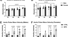
In vivo phenotypic vascular dysfunction extends beyond the aorta in a mouse model for fibrillin-1 ( Fbn1 ) mutation
- M. E. Barrameda
- M. Esfandiarei
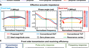
An ultrasensitive and broadband transparent ultrasound transducer for ultrasound and photoacoustic imaging in-vivo
Transparent ultrasound transducers suffer from practical limitations due to acoustic impedance mismatch. By using a transparent adhesive based on silicon dioxide epoxy, the authors demonstrate a broadband, ultrasensitive transparent ultrasound transducer, advancing the possibilities of sensor fusion.
- Seonghee Cho
- Chulhong Kim
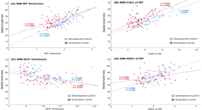
Sarcopenia prediction using shear-wave elastography, grayscale ultrasonography, and clinical information with machine learning fusion techniques: feature-level fusion vs. score-level fusion
- Young Han Lee
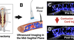
Non-contrast ultrasound image analysis for spatial and temporal distribution of blood flow after spinal cord injury
- Denis Routkevitch
- Amir Manbachi
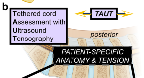
Tethered spinal cord tension assessed via ultrasound elastography in computational and intraoperative human studies
Kerensky et al. quantify tension across human spinal cords in computational simulations, a cadaveric benchtop model, and a neurosurgical case series. Their direct methodology successfully differentiates stretched spinal cords from healthy states in all sub-studies.
- Max J. Kerensky
- Abhijit Paul
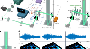
Ultrafast longitudinal imaging of haemodynamics via single-shot volumetric photoacoustic tomography with a single-element detector
Photoacoustic tomography using a single laser pulse and a single element functioning as thousands of virtual detectors allows for the volumetric capture of fast haemodynamic changes in the feet of human volunteers.
- Lihong V. Wang
News and Comment
Mid-ir optoacoustic microscopy.
- Rita Strack
Ultrasound success removes barriers to targeted drug delivery in amyotrophic lateral sclerosis

A sound strategy for gene expression
A new study in Science reports a synthetic biology approach to encode an ultrasound-based gene expression reporter that is applicable to mammalian cells in vitro and in vivo.
- Darren J. Burgess
Unbiased, whole-brain imaging of neural circuits
Functional ultrasound imaging enables unbiased identification of behaviorally relevant brain regions across the whole brain.
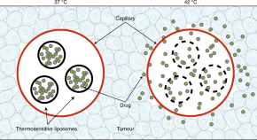
Hyperthermia-induced drug delivery in humans
A clinical study shows the feasibility and safety of the intratumoral release of an anticancer drug encapsulated in thermosensitive liposomes by heating the patient’s tumour via focused ultrasound.
- Kullervo Hynynen
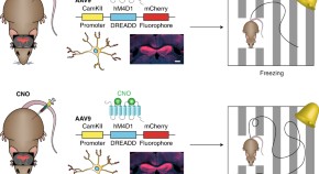
Non-invasive chemogenetics
A technique combining focused ultrasound for opening the blood–brain barrier and virally encoded engineered G-protein-coupled receptors for promoting the expression of a gene targeting excitatory neurons enables the non-invasive stimulation of specific brain regions and cell types in mice.
- Caroline Menard
- Scott J. Russo
Quick links
- Explore articles by subject
- Guide to authors
- Editorial policies
Ultrasound News
Top headlines, latest headlines.
- Biomedical Imaging Technology
- Ultrasound and Oxygen Saturation in Blood
- Ultrasound Imaging: Ultrafast Tech
- Soundwaves Harden 3D-Printed Treatments in Body
- Network of Robots to Monitor Pipes Acoustically
- 2 Droplets Levitated and Mixed
- New Laser Setup Probes Metamaterials
- Medical Imaging Fails Dark Skin: Researchers ...
- Ultrasound May Rid Groundwater of Toxic ...
- Ultra-Sensitive Photoacoustic Microscopy
Earlier Headlines
Friday, july 28, 2023.
- A Wearable Ultrasound Scanner Could Detect Breast Cancer Earlier
Wednesday, July 26, 2023
- A Quick Look Inside a Human Being
Tuesday, June 27, 2023
- Researchers Use Ultrasound to Control Orientation of Small Particles
Thursday, June 22, 2023
- When Soft Spheres Make Porous Media Stiffer
Thursday, June 15, 2023
- A 'spy' In the Belly
Wednesday, June 7, 2023
- Sponge Makes Robotic Device a Soft Touch
Monday, May 22, 2023
- A Giant Leap Forward in Wireless Ultrasound Monitoring for Subjects in Motion
Tuesday, May 2, 2023
- Wearable Ultrasound Patch Provide Non-Invasive Deep Tissue Monitoring
Tuesday, April 4, 2023
- Detecting, Predicting, and Preventing Aortic Ruptures With Computational Modeling
Friday, March 10, 2023
- New Ultrasound Method Could Lead to Easier Disease Diagnosis
Wednesday, March 1, 2023
- The Future of Touch
Tuesday, February 28, 2023
- Ultrasound Device May Offer New Treatment Option for Hypertension
Friday, February 24, 2023
- Faster and Sharper Whole-Body Imaging of Small Animals With Deep Learning
Thursday, February 23, 2023
- Making Engineered Cells Dance to Ultrasound
Wednesday, February 22, 2023
- Study Offers Details on Using Electric Fields to Tune Thermal Properties of Ferroelectric Materials
Monday, February 13, 2023
- Creating 3D Objects With Sound
Tuesday, January 31, 2023
- Focused Ultrasound Technique Leads to Release of Neurodegenerative Disorders Biomarkers
Wednesday, January 25, 2023
- Wearable Sensor Uses Ultrasound to Provide Cardiac Imaging on the Go
Friday, January 13, 2023
- A Precision Arm for Miniature Robots
Tuesday, January 3, 2023
- Tracking Radiation Treatment in Real Time Promises Safer, More Effective Cancer Therapy
- Team Writes Letters With Ultrasonic Beam, Develops Deep Learning Based Real-Time Ultrasonic Hologram Generation Technology
Thursday, December 1, 2022
- An Exotic Interplay of Electrons
Wednesday, September 21, 2022
- The Super-Fast MRI Scan That Could Revolutionize Heart Failure Diagnosis
Friday, August 12, 2022
- Using Sound and Bubbles to Make Bandages Stickier and Longer Lasting
Tuesday, August 9, 2022
- Ultrasound Could Save Racehorses from Bucked Shins
Thursday, July 28, 2022
- Engineers Develop Stickers That Can See Inside the Body
Thursday, July 21, 2022
- Flexible Method for Shaping Laser Beams Extends Depth-of-Focus for OCT Imaging
Wednesday, June 15, 2022
- High-Intensity Focused Ultrasound (HIFU) Can Control Prostate Cancer With Fewer Side Effects
- Moth Wing-Inspired Sound Absorbing Wallpaper in Sight After Breakthrough
Tuesday, May 31, 2022
- Direct Sound Printing Is a Potential Game-Changer in 3D Printing
Monday, May 30, 2022
- Ultrasound-Guided Microbubbles Boost Immunotherapy Efficacy
Thursday, May 5, 2022
- How MRI Could Revolutionize Heart Failure Diagnosis
Wednesday, April 27, 2022
- 3D Bimodal Photoacoustic Ultrasound Imaging to Diagnose Peripheral Vascular Diseases
Monday, April 18, 2022
- Tumors Partially Destroyed With Sound Don't Come Back
Tuesday, April 12, 2022
- Ultrasound Gave Us Our First Baby Pictures Can It Also Help the Blind See?
Monday, April 4, 2022
- Dual-Mode Endoscope Offers Unprecedented Insights Into Uterine Health
Wednesday, March 23, 2022
- Concert Hall Acoustics for Non-Invasive Ultrasound Brain Treatments
Tuesday, March 22, 2022
- Quantum Dots Shine Bright to Help Scientists See Inflammatory Cells in Fat
Friday, March 11, 2022
- Acoustic Propulsion of Nanomachines Depends on Their Orientation
Monday, February 28, 2022
- Ultrasound Scan Can Diagnose Prostate Cancer
Friday, February 25, 2022
- Ultrasounds for Endangered Abalone Mollusks
Thursday, February 24, 2022
- Transparent Ultrasound Chip Improves Cell Stimulation and Imaging
Tuesday, February 22, 2022
- Low-Cost, 3D Printed Device May Broaden Focused Ultrasound Use
Tuesday, February 15, 2022
- Speed of Sound Used to Measure Elasticity of Materials
Tuesday, January 25, 2022
- Ultrasound Technique Predicts Hip Dysplasia in Infants
Wednesday, January 5, 2022
- The First Topological Acoustic Transistor
Friday, December 17, 2021
- New Research Sheds Light on How Ultrasound Could Be Used to Treat Psychiatric Disorders
Wednesday, December 8, 2021
- CRISPR/Cas9 Gene Editing Boosts Effectiveness of Ultrasound Cancer Therapy
Wednesday, November 10, 2021
- A Personalized Exosuit for Real-World Walking
Monday, November 1, 2021
- Noninvasive Imaging Strategy Detects Dangerous Blood Clots in the Body
Friday, September 10, 2021
- Acoustic Illusions
Tuesday, August 24, 2021
- Researchers Developing New Cancer Treatments With High-Intensity Focused Ultrasound
Tuesday, August 17, 2021
- Prediction Models May Reduce False-Positives in MRI Breast Cancer Screening
Tuesday, August 3, 2021
- Does Visual Feedback of Our Tongues Help in Speech Motor Learning?
Tuesday, July 27, 2021
- Researchers Demonstrate Technique for Recycling Nanowires in Electronics
Thursday, July 22, 2021
- Soft Skin Patch Could Provide Early Warning for Strokes, Heart Attacks
Monday, July 12, 2021
- Magnetic Field from MRI Affects Focused-Ultrasound-Mediated Blood-Brain Barrier
Monday, June 21, 2021
- A Tiny Device Incorporates a Compound Made from Starch and Baking Soda to Harvest Energy from Movement
Friday, May 28, 2021
- New Tool Activates Deep Brain Neurons by Combining Ultrasound, Genetics
Monday, May 24, 2021
- Silicon Chips Combine Light and Ultrasound for Better Signal Processing
Tuesday, May 11, 2021
- Tiny, Wireless, Injectable Chips Use Ultrasound to Monitor Body Processes
Wednesday, May 5, 2021
- Release of Drugs from a Supramolecular Cage
- Focused Ultrasound Enables Precise Noninvasive Therapy
Wednesday, April 14, 2021
- Using Sound Waves to Make Patterns That Never Repeat
Monday, March 22, 2021
- Reading Minds With Ultrasound: A Less-Invasive Technique to Decode the Brain's Intentions
- LATEST NEWS
- Top Science
- Top Physical/Tech
- Top Environment
- Top Society/Education
- Health & Medicine
- Mind & Brain
- Living Well
- Space & Time
- Matter & Energy
- Business & Industry
- Automotive and Transportation
- Consumer Electronics
- Energy and Resources
- Engineering and Construction
- Telecommunications
- Textiles and Clothing
- Biochemistry
- Inorganic Chemistry
- Organic Chemistry
- Thermodynamics
- Electricity
- Energy Technology
- Alternative Fuels
- Energy Policy
- Fossil Fuels
- Nuclear Energy
- Solar Energy
- Wind Energy
- Engineering
- 3-D Printing
- Civil Engineering
- Construction
- Electronics
- Forensic Research
- Materials Science
- Medical Technology
- Microarrays
- Nanotechnology
- Robotics Research
- Spintronics
- Sports Science
- Transportation Science
- Virtual Environment
- Weapons Technology
- Wearable Technology
- Albert Einstein
- Nature of Water
- Quantum Computing
- Quantum Physics
- Computers & Math
- Plants & Animals
- Earth & Climate
- Fossils & Ruins
- Science & Society
- Education & Learning
Strange & Offbeat
- Leafhopper Inspires Invisibility Devices
- AI Meet AI: Talk to Each Other
- Managing Anger: Breathe, Don't Vent
- Tanks of the Triassic
- Holographic Message Encoded in Simple Plastic
- Maybe Our Universe Has No Dark Matter
- A New World of 2D Material Is Opening Up
- Best Way to Memorize Stuff? It Depends...
- Chimp: Persistence of Mother-Child Play
- High-Speed Microscale 3D Printing
Trending Topics
Open Access is an initiative that aims to make scientific research freely available to all. To date our community has made over 100 million downloads. It’s based on principles of collaboration, unobstructed discovery, and, most importantly, scientific progression. As PhD students, we found it difficult to access the research we needed, so we decided to create a new Open Access publisher that levels the playing field for scientists across the world. How? By making research easy to access, and puts the academic needs of the researchers before the business interests of publishers.
We are a community of more than 103,000 authors and editors from 3,291 institutions spanning 160 countries, including Nobel Prize winners and some of the world’s most-cited researchers. Publishing on IntechOpen allows authors to earn citations and find new collaborators, meaning more people see your work not only from your own field of study, but from other related fields too.
Brief introduction to this section that descibes Open Access especially from an IntechOpen perspective
Want to get in touch? Contact our London head office or media team here
Our team is growing all the time, so we’re always on the lookout for smart people who want to help us reshape the world of scientific publishing.
Home > Books > Medical Imaging
Ultrasound Imaging - Current Topics

Book metrics overview
3,563 Chapter Downloads
Impact of this book and its chapters
Total Chapter Downloads on intechopen.com
Total Chapter Citations
Academic Editor
Gregory University , Nigeria
Published 11 May 2022
Doi 10.5772/intechopen.95178
ISBN 978-1-78985-186-1
Print ISBN 978-1-78984-877-9
eBook (PDF) ISBN 978-1-78985-331-5
Copyright year 2022
Number of pages 154
Ultrasound Imaging - Current Topics presents complex and current topics in ultrasound imaging in a simplified format. It is easy to read and exemplifies the range of experiences of each contributing author. Chapters address such topics as anatomy and dimensional variations, pediatric gastrointestinal emergencies, musculoskeletal and nerve imaging as well as molecular sonography. The book is...
Ultrasound Imaging - Current Topics presents complex and current topics in ultrasound imaging in a simplified format. It is easy to read and exemplifies the range of experiences of each contributing author. Chapters address such topics as anatomy and dimensional variations, pediatric gastrointestinal emergencies, musculoskeletal and nerve imaging as well as molecular sonography. The book is a useful resource for researchers, students, clinicians, and sonographers looking for additional information on ultrasound imaging beyond the basics.
By submitting the form you agree to IntechOpen using your personal information in order to fulfil your library recommendation. In line with our privacy policy we won’t share your details with any third parties and will discard any personal information provided immediately after the recommended institution details are received. For further information on how we protect and process your personal information, please refer to our privacy policy .
Cite this book
There are two ways to cite this book:
Edited Volume and chapters are indexed in
Table of contents.
By Solomon Demissie, Mulatie Atalay and Yonas Derso
By Ercan Ayaz
By Haithem Zaafouri, Meryam Mesbahi, Nizar Khedhiri, Wassim Riahi, Mouna Cherif, Dhafer Haddad and Anis Ben Maamer
By Felix Okechukwu Erondu
By Stefan Cristian Dinescu, Razvan Adrian Ionescu, Horatiu Valeriu Popoviciu, Claudiu Avram and Florentin Ananu Vreju
By María Eugenia Aponte-Rueda and María Isabel de Abreu
By Jong Hwa Lee, Jae Uk Lee and Seung Wan Yoo
By J.M. López Álvarez, O. Pérez Quevedo, S. Alonso-Graña López-Manteola, J. Naya Esteban, J.F. Loro Ferrer and D.L. Lorenzo Villegas
By Arthur Fleischer and Sai Chennupati
IMPACT OF THIS BOOK AND ITS CHAPTERS
3,563 Total Chapter Downloads
1 Crossref Citations
2 Dimensions Citations
Order a print copy of this book
Available on

Delivered by
£119 (ex. VAT)*
Hardcover | Printed Full Colour
FREE SHIPPING WORLDWIDE
* Residents of European Union countries need to add a Book Value-Added Tax Rate based on their country of residence. Institutions and companies, registered as VAT taxable entities in their own EU member state, will not pay VAT by providing IntechOpen with their VAT registration number. This is made possible by the EU reverse charge method.
As an IntechOpen contributor, you can buy this book for an Exclusive Author price with discounts from 30% to 50% on retail price.
Log in to your Author Panel to purchase a book at the discounted price.
For any assistance during ordering process, contact us at [email protected]
Related books
Medical imaging.
Edited by Felix Okechukwu Erondu
Medical Imaging in Clinical Practice
Medical and biological image analysis.
Edited by Robert Koprowski
Edited by Yongxia Zhou
Ultrasound Elastography
Edited by Monica Lupsor-Platon
New Advances in Magnetic Resonance Imaging
Edited by Denis Larrivee
Frontiers in Neuroimaging
Edited by Xianli Lv
Optical Coherence Tomography
Edited by Giuseppe Lo Giudice
Updates in Endoscopy
Edited by Somchai Amornyotin
Elastography
Edited by Dana Stoian
Call for authors
Submit your work to intechopen.

Volume 5 Supplement 1
Topics in emergency abdominal ultrasonography
Edited by Luca Brunese and Antonio Pinto
Publication of this suppement has been funded by the University of Molise, Universiy of Siena, University of Cagliari, University of Ferrara and University of Turin. The Supplement Editors declare that they have no competing interests.
Sources of error in emergency ultrasonography
To evaluate the common sources of diagnostic errors in emergency ultrasonography.
- View Full Text
Accuracy of ultrasonography in the diagnosis of acute appendicitis in adult patients: review of the literature
Ultrasound is a widely used technique in the diagnosis of acute appendicitis; nevertheless, its utilization still remains controversial.
US detection of renal and ureteral calculi in patients with suspected renal colic
The purpose of this study was to determine whether the color Doppler twinkling sign could be considered as an additional diagnostic feature of small renal lithiasis (_5mm).
Gastrointestinal perforation: ultrasonographic diagnosis
Gastrointestinal tract perforations can occur for various causes such as peptic ulcer, inflammatory disease, blunt or penetrating trauma, iatrogenic factors, foreign body or a neoplasm that require an early re...
Sigmoid diverticulitis: US findings
Acute diverticulitis (AD) results from inflammation of a colonic diverticulum. It is the most common cause of acute left lower-quadrant pain in adults and represents a common reason for acute hospitalization, ...

The role of US examination in the management of acute abdomen
Acute abdomen is a medical emergency, in which there is sudden and severe pain in abdomen of recent onset with accompanying signs and symptoms that focus on an abdominal involvement. It can represent a wide sp...
Intestinal Ischemia: US-CT findings correlations
Intestinal ischemia is an abdominal emergency that accounts for approximately 2% of gastrointestinal illnesses. It represents a complex of diseases caused by impaired blood perfusion to the small and/or large ...
US in the assessment of acute scrotum
The acute scrotum is a medical emergency . The acute scrotum is defined as scrotal pain, swelling, and redness of acute onset. Scrotal abnormalities can be divided into three groups , which are extra-testicula...
Contrast enhanced ultrasound ( CEUS ) in blunt abdominal trauma
In the assessment of polytrauma patient, an accurate diagnostic study protocol with high sensitivity and specificity is necessary. Computed Tomography (CT) is the standard reference in the emergency for evalua...
Abdominal vascular emergencies: US and CT assessment
Acute vascular emergencies can arise from direct traumatic injury to the vessel or be spontaneous (non-traumatic).
Accuracy of ultrasonography in the diagnosis of acute calculous cholecystitis: review of the literature
To evaluate the accuracy of ultrasonography in the diagnosis of acute calculous cholecystitis in comparison with other imaging modalities.
Ultrasonography (US) in the assessment of pediatric non traumatic gastrointestinal emergencies
Non traumatic gastrointestinal emergencies in the children and neonatal patient is a dilemma for the radiologist in the emergencies room and they presenting characteristics ultrasound features on the longitudi...
- Editorial Board
- Sign up for article alerts and news from this journal
- Follow us on Twitter
- Follow us on Facebook
- ISSN: 2524-8987 (electronic)
Disclaimer » Advertising
- HealthyChildren.org

- Previous Article
- Next Article
Benign Focal Parenchymal Lesions
Malignant focal lesions, organ perfusion, vesicoureteral reflux, elastography, 3d and 4d ultrasound, high-frequency ultrasound, conclusions and future directions, advanced ultrasound techniques for pediatric imaging.
POTENTIAL CONFLICT OF INTEREST: The authors have indicated they have no potential conflicts of interest to disclose.
FINANCIAL DISCLOSURE: Dr Hwang has received funding from the National Institutes of Health KL2 award. Dr Piskunowicz has received funding from National Science Center grant DEC-2012/05/B/NZ5/01554. Dr Darge has financial relationships with Bracco Diagnostics Inc, Lantheus Medical Imaging, Philips Healthcare, and Siemens Healthineers.
- Split-Screen
- Article contents
- Figures & tables
- Supplementary Data
- Peer Review
- CME Quiz Close Quiz
- Open the PDF for in another window
- Get Permissions
- Cite Icon Cite
- Search Site
Misun Hwang , Maciej Piskunowicz , Kassa Darge; Advanced Ultrasound Techniques for Pediatric Imaging. Pediatrics March 2019; 143 (3): e20182609. 10.1542/peds.2018-2609
Download citation file:
- Ris (Zotero)
- Reference Manager
Ultrasound has become a useful tool in the workup of pediatric patients because of the highly convenient, cost-effective, and safe nature of the examination. With rapid advancements in anatomic and functional ultrasound techniques over the recent years, the diagnostic and interventional utility of ultrasound has risen tremendously. Advanced ultrasound techniques constitute a suite of new technologies that employ microbubbles to provide contrast and enhance flow visualization, elastography to measure tissue stiffness, ultrafast Doppler to deliver high spatiotemporal resolution of flow, three- and four-dimensional technique to generate accurate spatiotemporal representation of anatomy, and high-frequency imaging to delineate anatomic structures at a resolution down to 30 μm. Application of these techniques can enhance the diagnosis of organ injury, viable tumor, and vascular pathologies at bedside. This has significant clinical implications in pediatric patients who are not easy candidates for lengthy MRI or radiation-requiring examination, and are also in need of a highly sensitive bedside technique for therapeutic guidance. To best use the currently available, advanced ultrasound techniques for pediatric patients, it is necessary to understand the diagnostic utility of each technique. In this review, we will educate the readers of emerging ultrasound techniques and their respective clinical applications.
Ultrasound in which advanced techniques are used can enhance the diagnostic sensitivity of conventional grayscale and color Doppler ultrasound while offering additional functional information. In contrast-enhanced ultrasound (CEUS), intravascular microbubble agents are used to highlight perfusion abnormalities associated with various pathologies, including organ injury and residual tumors, which would otherwise be challenging to identify with conventional ultrasound. 1 , – 3 The enhanced diagnostic sensitivity of CEUS can negate the need for further high-cost, cross-sectional imaging requiring sedation.
Elastography is another functional ultrasound technique that allows for the quantification of tissue stiffness, which reflects tissue composition and/or architecture, edema, injury, and perfusion. The technique provides a quantitative or semiquantitative measure of the tissue evaluated. Two main types of elastography include strain and shear elastography. Strain elastography can produce a semiquantitative measure of tissue stiffness by detecting axial displacements arising from either manual compression or internal physiologic motion (eg, respiration and heartbeat). Shear-wave elastography applies high-intensity pulses to deform the tissue and produce laterally propagating shear waves that can be used to quantify stiffness. Elastography is approved by the Food and Drug Administration (FDA) for the evaluation of all abdominal organs in pediatric patients.
Furthermore, improvements in three-dimensional (3D) ultrasound techniques enable accurate quantification and delineation of anatomic structures for improved diagnosis and surgical guidance. The integration of four-dimensional (4D) ultrasound, with the fourth dimension representing time, allows for 3D depiction of moving objects, such as the heart valves in echocardiography. There is ongoing research to simultaneously scan the whole-organ volume without reconstructive postprocessing, which, if successful, will significantly advance the clinical potential of 3D ultrasound. 4
Ultrafast Doppler (UfD) is an advanced Doppler technique with significantly higher temporal resolution than conventional Doppler (up to 100 000 frames per second as opposed to 50 frames per second), offering real-time blood volume changes shown to correlate with neuronal activation. 5 Moreover, high-resolution imaging in which a frequency of up to 70 MHz (compared with conventional imaging with a frequency of up to 15 MHz) is used can enhance the diagnostic sensitivity of various dermatologic, neuromuscular, vascular, and other superficial pathologies because of the improved resolution in the region close to the transducer.
In this review, we will highlight recent advancements in anatomic and functional ultrasound techniques that have the potential to significantly improve the diagnostic and therapeutic potential of ultrasound. Our goal for the review is to educate the readers of the availability of these emerging ultrasound techniques and their potential clinical applications.
The ultrasound contrast agents are gas-containing, phospholipid-encapsulated microbubbles of 2 to 3 µm in size (compared with 7–8 µm of red blood cells) that generate an increased ultrasound signal because of a high acoustic impedance mismatch. Lumason (Bracco Diagnostics Inc, Monroe, NJ), sulfur hexafluoride gas-filled microbubbles encapsulated by phospholipids, was approved for characterization of focal liver lesions and evaluation of vesicoureteral reflux by the FDA in 2016. 6 The technique requires injection of the manually reconstituted contrast agent into a peripheral vein and detecting the signal reflected from the microbubbles flowing in the vessels. Its safety profile is much superior to computed tomography (CT) and MRI contrast agents. 7 , 8 The contrast agent is primarily exhaled and not renally cleared. Because of the excretion through the lungs, the contrast agent can be safely given to patients with renal insufficiency. In addition, it allows for an ultrasound study with contrast, avoiding the radiation of CT and the sedation needed for MRI. These serve as major advantages in pediatric imaging. CEUS is also lower in cost compared with CT or MRI.
The main benefit of CEUS in small children is the ability to diagnose certain benign focal lesions, such as hemangioma, focal nodular hyperplasia, or fatty infiltration, obviating the need for expensive radiation or sedation requiring, at times, lengthy cross-sectional imaging. Hemangiomas have a characteristic centripetal pattern of enhancement, with avid peripheral enhancement and progressive mild central enhancement, which can be diagnosed with CEUS alone. 9 Grayscale ultrasound and color Doppler findings of these benign focal lesions can be puzzling because the dynamic wash-in and wash-out pattern seen with CEUS cannot be obtained.
We present a challenging case of a large congenital hepatic hemangioma diagnosed with CEUS on initial scanning that would have otherwise been a diagnostic challenge with conventional ultrasound alone ( Fig 1 ). The lesion demonstrated heterogeneous echotexture and an indeterminate color Doppler signal. With a CEUS scan, however, the lesion proved to have avid enhancement in the periphery, to the same degree as the aorta, and partial centripetal filling on day 2 of life ( Supplemental Video 1 ). The diagnosis of hemangioma was made at the time. Subsequent scans obtained monthly until 8 months revealed progressive irregular enhancement of the periphery of the lesion, still enhancing avidly as the aorta, with an increase in the size of the lesion. MRI obtained at 8 months of life revealed the same peripherally avid enhancement pattern seen on CEUS acquired the same month. A biopsy was performed because of the increasing size of the lesion, and the diagnosis of congenital hemangioma was made. Subsequent CEUS of the lesion performed 2 months after the MRI revealed a slight decrease in the size of the lesion and a smoother echotexture and enhancement pattern, most compatible with rapidly involuting congenital hemangioma.

A large liver hemangioma shown with grayscale ultrasound, CEUS, and MRI, and CEUS images demonstrating centripetal enhancement over time. A, Grayscale ultrasound image of a hemangioma on the initial scan 8 months before the CEUS (C–G) and MRI (B) acquisition. B, Coronal, fat-saturated contrast-enhanced MRI image obtained a few days after the CEUS scan showing a large liver hemangioma with irregular peripheral rim enhancement. C, CEUS image of the lesion (white arrowheads) shown in contrast mode (left) and grayscale mode (right) before microbubble administration. The image is dark, as expected, before microbubble administration on contrast mode. Note the irregular streaks of bright signals within the lesion in the absence of microbubbles, likely due to internal calcifications. D, CEUS image of the lesion is shown at 10 seconds after intravenous microbubble administration. E, CEUS image of the lesion is shown at 12 seconds after administration. F, CEUS image of the lesion is shown at 15 seconds after administration. G, CEUS image of the lesion is shown at 95 seconds after administration.
The above case represents an example in which increased CEUS experience and familiarity within the medical community would have prevented a biopsy, which is not without risks, especially because the lesion was seen to regress in size at 10 months of life. Noninvasive monitoring of the lesion with CEUS would have been sufficient. It also highlights the value of CEUS in reducing the number of MRI scans and associated sedation, which has been associated with neurotoxic effects in the developing brain. 10 , 11 Additionally, magnetic resonance contrast administration is posing a challenge because of the gadolinium accumulation in the body. Further benign CEUS enhancement characteristics include well-defined peripheral and/or central vascular pattern (ie, benign lymph nodes, hyperplastic thyroid nodules, focal nodular hyperplasia, and splenic hamartoma), delayed wash out (ie, liver regenerative nodules), and lack of enhancement (ie, hemorrhage, cyst, column of Bertin, and calyceal diverticulum). In summary, CEUS can serve as a diagnostic and/or monitoring tool for many benign lesions without the need for additional imaging and the possibility of a bedside examination.
CEUS permits real-time imaging of dynamic perfusion with high temporal resolution and quantitative analysis of in flow and out flow of the microbubbles through the vessels and within the lesion. Because the vascularization pattern of benign and malignant lesions is different, CEUS can be a useful tool in distinguishing the 2. 12 On CEUS, most malignant lesions reveal fast arrival of the contrast agent (wash in) during the arterial phase followed by rapid wash out. The quick wash-out phenomenon is one of the most characteristic features of a malignant lesion because of a nearly complete arterial blood supply and abnormal arterio-venous shunts. Accurate assessment of tumor vascularity is possible because of retention of microbubbles within the intravascular space in contrary to increased permeability of iodinated and gadolinium-based contrast agents. 13 , – 15
The potential spectrum of the clinical application of CEUS in pediatric oncology is broad. Immediate exclusion of malignancy without the need to wait for further imaging studies substantially reduces the stress for parents and their child. In terms of biopsy guidance, CEUS could identify areas of a necrotic versus viable tumor so as not to result in false-negative results ( Fig 2 ). With the technique, serial quantitative estimation of tumor vasculature is feasible for assessment of response to anticancer treatment ( Fig 3 ). Lastly, CEUS could reduce a number of oncologic follow-up examinations with the use of ionizing radiation and/or general anesthesia. In a study in which the authors evaluated the utility of CEUS in children, it was shown that CEUS substantially decreased the need for further evaluation with MRI or CT. 16

CEUS of pelvic osteosarcoma in a 13-year-old boy was obtained for biopsy guidance. A, Grayscale ultrasound of pelvic osteosarcoma (short white arrows) of heterogeneous echogenicity reveals bone destruction, (long white arrows) as evidenced by a disruption of the echogenic line representing the cortex of the adjacent ilium. B, CEUS of pelvic osteosarcoma (short white arrows) at 14 seconds after contrast administration with a visible large region of necrosis (red circumscribed area) and enhancing tumor tissue on the periphery.

CEUS of the nephroblastoma in a 3-year-old boy extending into the right atrium and inferior vena cava was obtained before and after therapy to assess for treatment response. A, Transverse grayscale ultrasound of the nephroblastoma in the right atrium revealing solid cystic echotexture (arrowheads) and extending into the inferior vena cava (white arrows). B, Sagittal grayscale ultrasound revealing extension of the tumor thrombus from the inferior vena cava into the right atrium (white arrows). C, CEUS of the vascularized tumor thrombus in the inferior vena cava (black arrows) before treatment is shown. D, CEUS of the tumor thrombus in the inferior vena cava after 2 cycles of chemotherapy revealing marked decreased vascularization and diameter (black arrows).
The degree of microbubble enhancement is reflective of organ perfusion and viability, and CEUS may be used for diagnosis of organ ischemia, injury, or death. CEUS can depict varying degrees of brain injury, as can be seen with postinjury hyperperfusion, and the evolution of reperfusion response has important prognostic implications ( Fig 4 , Supplemental Video 2 ). For infants who are critically ill with significant brain injury, timely performance of either 4-vessel cerebral angiography or radionuclide cerebral blood flow studies for diagnosis of brain death is not easy. If further validated against the gold standard examinations, CEUS has the potential to serve as an alternate tool for diagnosis of brain death. 17 Moreover, the bedside use of CEUS for diagnosis of trauma has shown much potential because of the portability and repeatability of the examination combined with superior diagnostic sensitivity compared with conventional ultrasound. 18 , 19 In the case of necrotizing enterocolitis, CEUS can serve as a complementary tool for assessing bowel perfusion and identifying bowel ischemia, the diagnosis of which would otherwise be delayed because of the presence of an oscillator causing a false color Doppler signal or the absence of flow in the bowel wall misconstrued as technical limitations of color Doppler ( Fig 4 , Supplemental Video 3 ).

CEUS images of brain and bowel perfusion are shown. A, Midcoronal brain CEUS image of a normal 1-month-old boy obtained at 15 seconds after microbubble administration revealing relative hyperperfusion to the central gray nuclei compared with the remainder of the brain, which is expected for that age. Normal-sized ventricles and brain volume for age are seen. B, A midcoronal brain CEUS image of a 10-month-old girl after cardiac arrest status post extracorpreal membrane oxygenation. Midcoronal brain CEUS obtained15 seconds after microbubble administration reveals marked increased perfusion to the cortical and subcortical gray and white matter, which is not typically seen in normal neonates and infants. Note also the hyperperfusion to the central gray nuclei, which is slightly higher than expected for that age. C, A midcoronal brain CEUS image of the same 10-month-old girl obtained 1 week after the examination obtained in (B), with the image acquired at 15 seconds after microbubble administration. Compared with the previous brain CEUS examination (B), significant overall decreased perfusion to the brain is noted, likely due to evolving injury, compared with the normal brain CEUS (A). Less avid perfusion to the central gray nuclei and cortex is demonstrated, as evidenced on the time intensity curve analysis (E). D, Midcoronal brain CEUS image of a 6-month-old boy 3 hours after cardiac arrest of unknown etiology reveals a near absent brain perfusion and to-and-fro flow in the avidly enhancing extracranial vessels. Note that the images (A, B, and D) were obtained with an EPIQ scanner (Philips Healthcare, Bothell, WA) and C5-1 transducer with settings of 12 Hz and a mechanical index of 0.06. Image (C) was obtained with Aplio i800 (Toshiba Medical Systems Corp, Tokyo, Japan) and an i8-C1 transducer with settings of 9 Hz and a mechanical index of 0.06. E, Normalized time intensity curve graph of the wash in of microbubbles into the brain in patients (A–D), with y-axes representing changes in signal intensity (decibels) and x-axes representing time in seconds. Note the marked hyperperfusion in the patient (B) and absent perfusion in near brain death (D). F, Bowel CEUS of a 39-day-old, formerly premature girl with necrotizing enterocolitis reveals a hyperemic bowel segment (white arrowheads). G, A bowel CEUS of a 1-day-old, formerly premature girl found to have complete bowel ischemia with no enhancement of the bowel and surrounding mesentery (white arrowheads), likely due to prenatal insult.
In addition, CEUS can be used to assess the degree of inflammation or fibrosis and/or scarring. In Crohn disease, the discernment of fibrotic versus inflammatory bowel strictures has important clinical implications. Inflammatory bowel strictures respond better to anti-inflammatory medications, whereas fibrotic strictures may benefit from surgical resection. Although MRI should be the gold standard for the diagnosis of Crohn disease, its use for routine monitoring of the diseased bowel segment is limited in children because of high cost and sedation needs. In this setting, CEUS may be used for serial monitoring of therapeutic response and evolution of the disease during clinic visits and potentially serve as a comparable tool to MRI by providing a quantifiable dynamic perfusion pattern of the diseased bowel segment. If combined with elastography, an advanced ultrasound technique that can assess tissue stiffness, CEUS may improve our understanding and management of diseased bowel segments with varying degrees of fibrosis and inflammation. 20 , 21
In the 1990s, contrast-enhanced voiding urosonography (ceVUS) was introduced as a safe, radiation-free alternative to voiding cystourethrography (VCUG) in children. The technique consists of administration of microbubbles through a urinary catheter and evaluation for vesicoureteral reflux with ultrasound ( Fig 5 ). Since then, thousands of children have been evaluated with ceVUS for vesicoureteral reflux, with a favorable safety profile. In a study of 1010 children ages 15 days to 17.6 years evaluated with ceVUS, no serious adverse event occurred, and only a few minor events occurred, likely because of catheterization. 22 More recent prospective clinical trials in which the authors compared the diagnostic sensitivity of VCUG and ceVUS revealed comparable 23 or superior 24 diagnostic sensitivity of ceVUS in comparison with the conventional VCUG. The higher sensitivity of ceVUS in the detection of vesicoureteral reflux and the ability to conduct a detailed evaluation of the urethra have enabled ceVUS to replace VCUG.

ceVUS images are shown. A, Side by side sagittal grayscale and corresponding ceVUS image of the right kidney (white arrows) of a 19-month-old boy revealing grade 2 reflux into the renal pelvis (white arrowheads). B, Sagittal ceVUS image of the right kidney (white arrows) with grade 4 reflux into the dilated renal pelvis and calyces (white arrowheads) in a 16-month-old boy. C, Microbubble-filled tortuous megaureter (arrowheads) and bladder (white arrows) in a 3-week-old boy. D, Sagittal ceVUS image of the microbubble-filled urinary bladder (white arrows) and urethra (white arrowheads) in an 8-month-old boy during the voiding phase.
Ultrasound elastography is a functional imaging technique in which tissue stiffness can be inferred and quantified by studying the alterations in speed of traveling ultrasound waves. The stiffer the tissue, the faster a wave travels. Ultrasound elastography was initially developed by the food industry to evaluate the stiffness, or maturity, of cheeses. 25 Since its introduction in 2002 for assessment of liver fibrosis, 26 rapid improvements in the technique has resulted in the ability to accurately identify different stages of liver fibrosis for application in a variety of disorders, including nonalcoholic fatty liver disease and metabolic, autoimmune, and veno-occlusive diseases. 27 , – 36
In brief, there are 2 main types of ultrasound elastographies: shear-wave and strain elastography ( Fig 6 ). The former is used to quantify absolute tissue stiffness by applying high-intensity pulses and detecting laterally propagating waves as a result of tissue deformation. The latter is a semiquantitative technique in which manual compression or internal physiologic motion, such as the heartbeat, results in axial displacements reflective of tissue stiffness. The strain elastography is semiquantitative because the exact force used to generate tissue displacement is unknown, but relative tissue stiffness (ie, stiffness of a lesion compared with the surrounding area) can be obtained in the interrogated region of interest.

Shear-wave and strain elastography images are shown. A, A rectangular cursor is placed in the cortex of the brain of a normal neonate on a sagittal plane for shear-wave elastography measurement, here shown as 1.08 ± 0.26 m/s. B, Similar shear-wave elastography measurement of the periventricular gray-white matter in an infant after anoxic brain injury reveals a markedly elevated elastography measurement of 3.17 ± 0.26 m/s. C, A solid, isoechoic thyroid nodule depicted on grayscale ultrasound (left) and strain elastography (right), with the strain elastography revealing mild increased stiffness as noted by the green color (blue noting soft and red noting hard). D, A normal testis shown in grayscale ultrasound (left) and strain elastography mode (right) reveals slightly stiffer tissue (this time denoted by blue/green) close to the echogenic band of connective tissue across the testis, so-called mediastinum testis, compared with the adjacent softer tissue (red). Note that depending on the system settings, color representation of tissue stiffness may differ.
One of the major benefits of elastography is the reduced need for an invasive liver biopsy, which oftentimes requires moderate sedation to general anesthesia in small children and is associated with a 6% complication risk (up to 0.1% life-threatening). 37 Although a biopsy is accepted as the gold standard, it has its own limitations, including sampling error and substantial variability in the staging of histopathologic fibrosis of up to 33% in previous literature. 38 There is also no validated histologic scoring system of liver fibrosis for the pediatric population.
Although further research is needed, elastography may be used to diagnosis other pathologies, including severe brain injury, 39 fibrotic bowel segment, 21 , 40 and renal fibrosis. 41 Noninvasive inference of the severity of brain injury is of clinical importance because invasive intracranial pressure measurements are not without risks. Severe anoxic brain injury can be quantitatively detected by elastography as an increase in stiffness due to brain edema, although the exact biochemical and physiologic basis of such elasticity change in the affected tissue warrants further research. 39 Close monitoring of brain injury also permits timely institution of neuroprotective and/or adjunct therapies for improved outcome. In the setting of Crohn disease, in which distinction between fibrosis and inflammation is critical to determining a surgical versus anti-inflammatory medical approach, elastography may be of importance in the diagnosis of inflammatory and/or fibrotic bowel strictures. In patients with chronic kidney disease, elastography may be of prognostic value in identifying those at risk for renal function deterioration. 41
UfD is an advanced Doppler technique with a high temporal resolution of >100 000 frames per second compared with the typical 50 frames per second used in conventional color Doppler imaging ( Fig 7 ). 42 The technique sends out a plane wave to simultaneous scan a whole field of view rather than transmitting focused beams, as in traditional Doppler techniques. The high-frame rate permits up to a 50-fold increase in sensitivity for blood flow changes in the human brain, which correlate neuronal activity as assessed by EEG. 5 In preclinical imaging, cerebral blood volume changes seen on UfD highly correlated with neuronal activity, with a high spatiotemporal resolution of ∼100 mm and 1 millisecond. 43 , 44 Improved sensitivity for slow-flow and smaller vessels is particularly important in rheumatology, 42 , 45 brain vascular imaging, 46 , 47 and carotid flow velocity estimation. 48 , – 50

UfD images of the normal neonatal brain are shown. A, Ultrafast power Doppler images of the neonatal brain acquired through the anterior fontanel from left to right: coronal view, tilted parasagittal view, parasagittal view. B, Directional power Doppler images based on the speed information from left to right: medial sagittal view revealing venous network around lateral ventricle, parasagittal view revealing small thalamic and cortical arterioles and venules, and transtemporal transverse view through mastoid fontanel for imaging the circle of Willis and cerebellum. C, Resistivity mapping based on UfD data. The RI in big arteries is between 0.8 and 1 (left and center image), whereas the RI in small and veins are lower at ∼0.6 and <0.3, respectively. Images reprinted from Demené et al, 2018 51 with permission. CBV, cerebral blood volume; RI, resistivity index.
Note that the super high temporal resolution of UfD surpasses that of functional MRI (fMRI). The high temporal resolution of up to 1 millisecond contrasts with that of traditional fMRI, which is at best 1 second. This has important clinical implications in the neonatal and infant population as fMRI is not routinely performed because of the complexity of the examination, including sedation, transportation, and high cost. It is also highly convenient for intraoperative use and for continuous, noninvasive monitoring of cerebral function during surgeries. Close bedside monitoring of perfusion changes associated with brain injury and/or seizure activity for children of all ages may be feasible with this technique, although further research is required in this regard. Further studies will be needed, however, to understand the potential correlation between fMRI and UfD because the former measures oxygenated blood whereas the latter measures both oxygenated and deoxygenated blood, technically different physiologic parameters. UfD on the other hand is low cost, portable, and easily repeatable without the need for sedation.
Although 3D ultrasound suffered from long image-processing times during its infancy, current computer technology allows for high speed of image acquisition and reconstruction. In the 3D technology, positional information gained from the transducer or external positional devices is used to reconstruct 3D images from two-dimensional (2D) images. In the case of hydrocephalus, the traditionally used manual quantification of the ventricular size on a 2D plane is more cumbersome and less accurate than the volumetric reconstruction of the ventricular system ( Fig 8 ). 52 , – 57 The 3D technique is also useful in generating accurate organ or tumor volume. 58 The 4D technology allows for the incorporation of time dimension into 3D imaging, which can be useful in the imaging of moving objects, such as the heart valves. The 3D-4D technique overcomes the limitations of a 2D technique, namely lengthy acquisition times, operator dependence during multiple sweeps, and mental integration of 2D-acquired images. With further developments of 3D-4D technology, simultaneous acquisition of whole-organ perfusion with UfD or CEUS may be possible, which will drastically change the way in which ultrasound is used in the clinical setting. Further improvements will be needed to minimize artifacts associated with 3D-4D technology 59 that may cause diagnostic challenges.

A representative image revealing the 3D reconstruction and quantification of the brain ventricular volume. Dotted lines trace the borders of the mildly enlarged third and fourth ventricles in sagittal (top left), axial (bottom left), and coronal planes (top right) for 3D reconstruction (bottom right). The calculated ventricular volume (red) is shown at the bottom. COR, coronal.
The frequency range of medical ultrasound systems for most scanners is from 2 to 20 MHz. Recently, ultrasound systems of much higher frequency ranges have been introduced to the clinical setting, permitting a significant increase in resolution. VisualSonics ultrahigh-resolution ultrasound systems (FUJIFILM VisualSonics, Inc, Toronto, Canada), used for preclinical studies for the past 10 years, are now FDA approved for clinical applications. Note that the higher frequency peak of up to 70 MHz provides a tremendous increase in resolution compared with that of conventional ultrasound systems, permitting resolution down to 30 μm. Such resolution surpasses that of CT or MRI. Note that an increased frequency results in an increase in ultrasound attenuation in tissues, which means that the use of the system is limited to the first 3 cm of the body. Such resolution can be used to depict single hair follicles growing out of the skin.
The best applications of the high-resolution ultrasound system are neonatology, neuromusculoskeletal, dermatology, lymphatic, vascular, thyroid, and other small-part imaging. 60 , – 66 Further neuromusculoskeletal application of this technique would be helpful for guided superficial nerve blocks or reconstructive surgeries. Higher resolution imaging of the subungual space, moreover, can help assess the integrity of the subungual vessels (terminal branches of the proper digital artery) in the setting of trauma or help characterize various types of benign and malignant tumors affecting the subungual space ( Fig 9 ). A variety of pediatric venous diseases leading to the formation of debris or thrombus in the superficial and deep veins can be evaluated with high-resolution ultrasound. Other applications include improved characterization of nerve fascicles, large- and small-vessel intima-media thickness, internal contents of peripheral veins, and salivary glands ( Fig 10 ). Thyroid nodules, superficial vascular anomalies, and ocular pathology, especially in small children, would also be better evaluated with high-resolution imaging.

The nail plate of the distal phalanx of the second digit was imaged with a high-resolution ultrasound scanner by using a 40 MHz transducer. Shown is the picture diagram of the distal aspect of the second digit (B), with the magnification box noting the region of ultrasound evaluation. Annotated on the corresponding grayscale image (A) are the nail plate of the distal phalanx of the second digit (red arrows), nail bed with germinal matrix (between white arrows), eponychium (cuticle) (black arrow), and vessels supplying the nail unit (inside white box).

Small parts imaged with a high-resolution ultrasound scanner. A, A 50-MHz transducer was used to image the right median nerve in the transverse plane (white arrows) and the numerous fascicles visible within the nerve. B, A 50-MHz transducer was used to image the wall of the superficial palmar arch artery with intima-media (white arrows). C, A 30-MHz transducer was used to image the dilated superficial vein of the lower extremity with the valves (white arrows) and debris in the folds of valves preventing full opening of the valves (red arrows). D, A 50-MHz transducer was used to image the inner oral cavity sublingual glands (white arrows) and the mucous membrane (between red arrows).
It is important to note that for evaluation of superficial soft-tissue abnormalities in small children, ultrasound provides higher spatial resolution than MRI. The spatial resolution of a high-resolution ultrasound scanner is in micrometers, whereas that of MRI is in millimeters. With the availability of FDA-approved ultrasound contrast, ultrasound can now provide dynamic enhancement information similar to multiphase contrast-enhanced MRI. Moreover, sedation required for MRI of most small children is not without risks. Although the diagnostic benefits of MRI should be acknowledged, the awareness that, in some circumstances, advanced ultrasound techniques can provide the necessary diagnostic information is worth noting. In referral of pediatric patients to radiologic exams, such knowledge would be beneficial.
Rapid advancements in ultrasound techniques are changing the role of ultrasound in the management of a pediatric patient. Ultrasound is not merely a screening tool that precedes cross-sectional examinations; it is a critical tool in disease diagnosis and monitoring as well as therapeutic guidance, including as a point-of-care modality. The aforementioned techniques now offer functional information, including dynamic perfusion, tissue stiffness, and hemodynamic changes at the spatiotemporal resolution surpassing that of MRI. Improved 3D-4D and high-resolution techniques additionally serve to enhance the diagnostic potential of ultrasound. Combined with portability, low cost, lack of the need for radiation and sedation, and safety, ultrasound in which advanced techniques are used can serve as a useful clinical tool in the management of pediatric patients.
Further research in validating the emerging ultrasound techniques is ongoing. CEUS, for instance, is increasingly being explored for novel applications besides focal liver lesions and vesicoureteral reflux. In the case of elastography, the exact histopathologic and physiologic mechanisms behind changes in quantitatively measured tissue stiffness warrant further study. The continued work in this regard will improve our understanding of the parameters measured with advanced ultrasound techniques and help generate standardized guidelines to help discern healthy from diseased and/or injured organs.
Dr Hwang conceptualized and wrote the manuscript; Drs Piskunowicz and Darge contributed to the writing and image acquisition; and all authors approved the final manuscript as submitted and agree to be accountable for all aspects of the work.
This trial has been registered at www.clinicaltrials.gov (identifiers NCT03549507 and NCT03549520).
FUNDING: Funded by the National Institutes of Health KL2 award and Radiological Society of North America Research Scholar Grant (Dr Hwang) and National Science Center grant DEC-2012/05/B/NZ5/01554 (Dr Piskunowicz). Funded by the National Institutes of Health (NIH).
two-dimensional
three-dimensional
four-dimensional
contrast-enhanced ultrasound
contrast-enhanced voiding urosonography
computed tomography
Food and Drug Administration
functional MRI
ultrafast Doppler
voiding cystourethrography
Competing Interests
Supplementary data.
Advertising Disclaimer »
Citing articles via
Email alerts.

Affiliations
- Editorial Board
- Editorial Policies
- Journal Blogs
- Pediatrics On Call
- Online ISSN 1098-4275
- Print ISSN 0031-4005
- Pediatrics Open Science
- Hospital Pediatrics
- Pediatrics in Review
- AAP Grand Rounds
- Latest News
- Pediatric Care Online
- Red Book Online
- Pediatric Patient Education
- AAP Toolkits
- AAP Pediatric Coding Newsletter
First 1,000 Days Knowledge Center
Institutions/librarians, group practices, licensing/permissions, integrations, advertising.
- Privacy Statement | Accessibility Statement | Terms of Use | Support Center | Contact Us
- © Copyright American Academy of Pediatrics
This Feature Is Available To Subscribers Only
Sign In or Create an Account
- U.S. Department of Health & Human Services
- National Institutes of Health

En Español | Site Map | Staff Directory | Contact Us
Explore more about: Ultrasound (US) therapy
59 Ultrasound Essay Topic Ideas & Examples
🏆 best ultrasound topic ideas & essay examples, ✅ good essay topics on ultrasound, 📑 interesting topics to write about ultrasound.
- Contrast-Enhanced Ultrasound in Focal Liver Lesions In addition, inaccessibility to the eighth of the liver is a major setback in detecting lesions in the segment. With the advent of Doppler ultrasound, more insight in the diagnosis of liver lesions has been […]
- Ultrasound and Color Doppler-Guided Surgery The purpose of the study is to examine the opinions of the trainees attending a training course concerning the use of technology. We will write a custom essay specifically for you by our professional experts 808 writers online Learn More
- Abdominal Ultrasound and Diagnoses The examiner explains to the patient how the procedure will be performed and how much time is necessary to finish the examination.
- Ultrasound Technology in Podiatry Surgery First, it is important to briefly outline the peculiarities of the RCT to understand the researchers’ point. They will be able to use the technology in numerous settings.
- Ultrasound in Achilles Tendinitis Diagnosis In this research, the case study approach is applicable due to the fact that various patients suffering from tendon Achilles problem will be used as a basis for gauging the effectiveness of the method of […]
- Ultrasound in Treatment and Side-Effect Reduction Within the framework of the research project conducted by Ebadi et al, the research problem consisted in the fact that the effects of continuous ultrasound were underresearched.
- Low-Back Pain and Ultrasound Therapy In the meantime, their opponents highlight that the beneficial aspects of the treatment course outweigh the risks related to the use of ultrasound equipment.
- Ultrasound Physics and Instrumentation The camera is often not in harmony with the perception of the depth of a human vision. The level of such an acoustic signal distortion within a tissue is dependent on the emitted pulse’s amplitude […]
- Mammography vs. Ultrasound for Breast Tissue Analysis Mammography screening is one of the most recognized options for analyzing breast tissue in adult women. In contrast, the accuracy of this procedure allows it to be an alternative for women who cannot undergo mammography […]
- Ultrasound Scanning: Diagnosing Health Conditions The calculation is based on the time taken by the wave to return by measuring the distance, mass, nature, and stability of the object hit.
- Biologic Effects of Ultrasound in Healthcare Setting The instrument performing the emission of the sound waves and the recording of their bouncing back is referred to as the transducer and the medical practitioner generally gently presses the transducer against the skin of […]
- Benefits of 3D/4D Ultrasound in Prenatal Care The information that is obtained from this exam assists the health care providers in counseling parents on the development of the fetus especially in the nature of anomalies, prognosis, and the postnatal consideration of the […]
- Ultrasound Techniques Applied to Body Fat Measurement in Male and Female The main objective of this paper is to evaluate the accuracy of body fat by using portable ultra sound device which results are reliable and authentic. The ultra sound technique is widely used to measure […]
- Comparison of MRI and Ultrasound for Determining Abnormalities in Preterm Infants Medical Imaging helps in detecting and diagnosing diseases at its earliest and treatable stage and helps in determining most appropriate and effective care for the patient.”Medical imaging provides a picture of the inside of the […]
- The Recent Advances in Real Time Imaging in Ultrasound In point of fact, Medical imaging provides the most perfect task of diagnostic to Ultrasound, whereas, the main usage of therapeutic Ultrasound is to treat the numerous types of diseases and disorders in human beings.
- The Biological Effects of Ultrasound The paper also evaluates the physical mechanisms for the biological effects of ultrasound and the effects of ultrasound on living tissues in vivo and vitriol.
- Benefits of 3D Ultrasound to Pregnant Mothers This is coherent to the 3D planar imaging are improved technology previously applied in the 2D ultrasound technology. As an extrapolation from 3D technology, 3D ultrasound is applied as a medical diagnostic technique that utilizes […]
- MRI and Ultrasound for Determining Abnormalities in Preterm Infants Neonatal cranial ultrasound is used in detecting brain injury in preterm infants and can be used repetitively without harming the infant.
- Use of Ultrasound-Guidance for Arterial Puncture All the anthropometric and demographic variables were recorded, as well as the main diagnosis of admission, comorbidities, the placement of the central venous catheter, and the course of the procedure.
- Ultrasound in Chemistry: Sonochemistry
- Intravascular Ultrasound: Current Role and Future Perspectives
- The Difference Between an Echocardiogram and an Ultrasound of the Heart
- Cooperative Control With Ultrasound Guidance for Radiation Therapy
- Optically Generated Ultrasound: A New Paradigm for Intracoronary Imaging
- Nondestructive Testing: Principle of Flaw Detection With Ultrasound
- Closed-Loop Transcranial Ultrasound Stimulation for Real-Time Non-invasive Neuromodulation
- Consumer Application of Ultrasound: A Television Remote
- Roles of Low-Intensity Ultrasound in Differentiating Cell Death
- High-Intensity Focused Ultrasound Development: Destroying the Target Tissue
- Using Ultrasound to Enhance the Mechanical and Physical Properties of Metals
- The Security Implications of the Machine-Learning Supply Chain: Professionalism in the Ultrasound Department
- Ultrasound Neuromodulation: Mechanisms and the Potential of Multimodal Stimulation for Neuronal Function Assessment
- How Ultrasound Can Produce Sonoluminescence
- Preparation for an Ultrasound of a Gallbladder and a Pelvic
- Endorectal and Endoanal Ultrasound Technique
- Transvaginal Ultrasound: Is It Painful, Purpose, and Results
- Endoscopic Ultrasound in Diagnosing Cancer: One of the Most Common Imaging Procedures
- Ultrasound Skin Imaging in Dermatology, Aesthetic Medicine, and Cosmetology
- Ultrasound Physical Medical Treatment in Healing Following an Acute Injury or a Chronic Condition
- How Do Ultrasound Scans Work
- Americas Ultrasound Systems Market: From USD 7.9 Billion to USD 10.23 Billion
- How Ultrasound Imaging Helps Us Understand Speech and Accent Variation
- Cerebral Ultrasound Time-Harmonic Elastography: Softening of the Human Brain Due to Dehydration
- Wireless Communication in “Audio Beacons” Using Ultrasound
- Low-Intensity Focused Ultrasound for Posttraumatic Stress Disorder
- Thyroid Ultrasound: Purpose, Procedure, Benefits
- Blood Pressure Modulation With Low-Intensity Focused Ultrasound
- Accuracy of Ultrasounds in Diagnosing Birth Defects
- Functional Ultrasound During Awake Brain Surgery
- Live Animal Ultrasound Explained by Dr. Allen Williams
- Ultrasound Waves in Acoustic Microscopy
- Chemical and Physical Effects of Ultrasound: Sonoluminescence and Materials
- Focused Ultrasound for Noninvasive, Focal Pharmacologic Neurointervention
- Lung Ultrasound Findings in COVID-19 Pneumonia
- High-Power Ultrasound in Dry Corn Milling Plants
- History of Ultrasound of Physics and the Properties of the Transducer
- Relationship Between Ultrasound Viewing and Proceeding to Abortion
- Transrectal Ultrasound of the Prostate With a Biopsy
- Iron-Based Catalysts Used in Water Treatment Assisted by Ultrasound
- Chicago (A-D)
- Chicago (N-B)
IvyPanda. (2023, January 24). 59 Ultrasound Essay Topic Ideas & Examples. https://ivypanda.com/essays/topic/ultrasound-essay-topics/
"59 Ultrasound Essay Topic Ideas & Examples." IvyPanda , 24 Jan. 2023, ivypanda.com/essays/topic/ultrasound-essay-topics/.
IvyPanda . (2023) '59 Ultrasound Essay Topic Ideas & Examples'. 24 January.
IvyPanda . 2023. "59 Ultrasound Essay Topic Ideas & Examples." January 24, 2023. https://ivypanda.com/essays/topic/ultrasound-essay-topics/.
1. IvyPanda . "59 Ultrasound Essay Topic Ideas & Examples." January 24, 2023. https://ivypanda.com/essays/topic/ultrasound-essay-topics/.
Bibliography
IvyPanda . "59 Ultrasound Essay Topic Ideas & Examples." January 24, 2023. https://ivypanda.com/essays/topic/ultrasound-essay-topics/.
- X-Ray Questions
- Health Promotion Research Topics
- Gynecology Research Ideas
- Osteoarthritis Ideas
- Pneumonia Questions
- Cardiomyopathy Titles
- Heart Disease Titles
- Epigenetics Essay Titles
- Hypertension Topics
- Biomedicine Essay Topics
- Deontology Questions
- Breast Cancer Ideas
- Health Insurance Research Topics
- Evidence-Based Practice Titles
- Healthcare Questions
Department of Circulation and Medical Imaging
- Master's programmes in English
- For exchange students
- PhD opportunities
- All programmes of study
- Language requirements
- Application process
- Academic calendar
- NTNU research
- Research excellence
- Strategic research areas
- Innovation resources
- Student in Trondheim
- Student in Gjøvik
- Student in Ålesund
- For researchers
- Life and housing
- Faculties and departments
- International researcher support
Språkvelger
Master thesis and projects - ultrasound technology - studies - department of circulation and medical imaging.
- Master thesis and projects
- Specialisation courses
Master's thesis and projects
Master's thesis and projects.
The Department of circulation and medical imaging offers projects and master's thesis topics for technology students of most of the different technical study programmes at NTNU. There is a seperate page for the supplementary specialisation courses .
List of topics
Topics for thesis and projects are given below. Most of the topics can be adjusted to the students qualifications and wishes.
Don't hesitate to take contact with the corresponding supervisor - we're looking forward to a discussion with you!
Asset Publisher
Blood flow imaging projects, estimation of true flow velocity using ultrasound, fusion of multi-modal cardiac data, pocket size ultrasound technology, pulse-echo based method for estimation of speed of sound, ultrasonic imaging through solids, surf imaging topics, ultrasound mediated drug delivery, real-time monitoring of left ventricular function under interventional procedures, fighting cancer with cw shear-wave elastography, adaptive clutter filtering for coronary heart disease, patient adaptive imaging in echocardiography, how to write ....
- a good abstract
- a good introduction
person-portlet
Lasse løvstakken professor.
Head Start Your Radiology Residency [Online] ↗️
- Radiology Thesis – More than 400 Research Topics (2022)!
Please login to bookmark
Introduction
A thesis or dissertation, as some people would like to call it, is an integral part of the Radiology curriculum, be it MD, DNB, or DMRD. We have tried to aggregate radiology thesis topics from various sources for reference.
Not everyone is interested in research, and writing a Radiology thesis can be daunting. But there is no escape from preparing, so it is better that you accept this bitter truth and start working on it instead of cribbing about it (like other things in life. #PhilosophyGyan!)
Start working on your thesis as early as possible and finish your thesis well before your exams, so you do not have that stress at the back of your mind. Also, your thesis may need multiple revisions, so be prepared and allocate time accordingly.
Tips for Choosing Radiology Thesis and Research Topics
Keep it simple silly (kiss).
Retrospective > Prospective
Retrospective studies are better than prospective ones, as you already have the data you need when choosing to do a retrospective study. Prospective studies are better quality, but as a resident, you may not have time (, energy and enthusiasm) to complete these.
Choose a simple topic that answers a single/few questions
Original research is challenging, especially if you do not have prior experience. I would suggest you choose a topic that answers a single or few questions. Most topics that I have listed are along those lines. Alternatively, you can choose a broad topic such as “Role of MRI in evaluation of perianal fistulas.”
You can choose a novel topic if you are genuinely interested in research AND have a good mentor who will guide you. Once you have done that, make sure that you publish your study once you are done with it.
Get it done ASAP.
In most cases, it makes sense to stick to a thesis topic that will not take much time. That does not mean you should ignore your thesis and ‘Ctrl C + Ctrl V’ from a friend from another university. Thesis writing is your first step toward research methodology so do it as sincerely as possible. Do not procrastinate in preparing the thesis. As soon as you have been allotted a guide, start researching topics and writing a review of the literature.
At the same time, do not invest a lot of time in writing/collecting data for your thesis. You should not be busy finishing your thesis a few months before the exam. Some people could not appear for the exam because they could not submit their thesis in time. So DO NOT TAKE thesis lightly.
Do NOT Copy-Paste
Reiterating once again, do not simply choose someone else’s thesis topic. Find out what are kind of cases that your Hospital caters to. It is better to do a good thesis on a common topic than a crappy one on a rare one.
Books to help you write a Radiology Thesis
Event country/university has a different format for thesis; hence these book recommendations may not work for everyone.

- Amazon Kindle Edition
- Gupta, Piyush (Author)
- English (Publication Language)
- 206 Pages - 10/12/2020 (Publication Date) - Jaypee Brothers Medical Publishers (P) Ltd. (Publisher)
In A Hurry? Download a PDF list of Radiology Research Topics!
Sign up below to get this PDF directly to your email address.
100% Privacy Guaranteed. Your information will not be shared. Unsubscribe anytime with a single click.
List of Radiology Research /Thesis / Dissertation Topics
- State of the art of MRI in the diagnosis of hepatic focal lesions
- Multimodality imaging evaluation of sacroiliitis in newly diagnosed patients of spondyloarthropathy
- Multidetector computed tomography in oesophageal varices
- Role of positron emission tomography with computed tomography in the diagnosis of cancer Thyroid
- Evaluation of focal breast lesions using ultrasound elastography
- Role of MRI diffusion tensor imaging in the assessment of traumatic spinal cord injuries
- Sonographic imaging in male infertility
- Comparison of color Doppler and digital subtraction angiography in occlusive arterial disease in patients with lower limb ischemia
- The role of CT urography in Haematuria
- Role of functional magnetic resonance imaging in making brain tumor surgery safer
- Prediction of pre-eclampsia and fetal growth restriction by uterine artery Doppler
- Role of grayscale and color Doppler ultrasonography in the evaluation of neonatal cholestasis
- Validity of MRI in the diagnosis of congenital anorectal anomalies
- Role of sonography in assessment of clubfoot
- Role of diffusion MRI in preoperative evaluation of brain neoplasms
- Imaging of upper airways for pre-anaesthetic evaluation purposes and for laryngeal afflictions.
- A study of multivessel (arterial and venous) Doppler velocimetry in intrauterine growth restriction
- Multiparametric 3tesla MRI of suspected prostatic malignancy.
- Role of Sonography in Characterization of Thyroid Nodules for differentiating benign from
- Role of advances magnetic resonance imaging sequences in multiple sclerosis
- Role of multidetector computed tomography in evaluation of jaw lesions
- Role of Ultrasound and MR Imaging in the Evaluation of Musculotendinous Pathologies of Shoulder Joint
- Role of perfusion computed tomography in the evaluation of cerebral blood flow, blood volume and vascular permeability of cerebral neoplasms
- MRI flow quantification in the assessment of the commonest csf flow abnormalities
- Role of diffusion-weighted MRI in evaluation of prostate lesions and its histopathological correlation
- CT enterography in evaluation of small bowel disorders
- Comparison of perfusion magnetic resonance imaging (PMRI), magnetic resonance spectroscopy (MRS) in and positron emission tomography-computed tomography (PET/CT) in post radiotherapy treated gliomas to detect recurrence
- Role of multidetector computed tomography in evaluation of paediatric retroperitoneal masses
- Role of Multidetector computed tomography in neck lesions
- Estimation of standard liver volume in Indian population
- Role of MRI in evaluation of spinal trauma
- Role of modified sonohysterography in female factor infertility: a pilot study.
- The role of pet-CT in the evaluation of hepatic tumors
- Role of 3D magnetic resonance imaging tractography in assessment of white matter tracts compromise in supratentorial tumors
- Role of dual phase multidetector computed tomography in gallbladder lesions
- Role of multidetector computed tomography in assessing anatomical variants of nasal cavity and paranasal sinuses in patients of chronic rhinosinusitis.
- magnetic resonance spectroscopy in multiple sclerosis
- Evaluation of thyroid nodules by ultrasound elastography using acoustic radiation force impulse (ARFI) imaging
- Role of Magnetic Resonance Imaging in Intractable Epilepsy
- Evaluation of suspected and known coronary artery disease by 128 slice multidetector CT.
- Role of regional diffusion tensor imaging in the evaluation of intracranial gliomas and its histopathological correlation
- Role of chest sonography in diagnosing pneumothorax
- Role of CT virtual cystoscopy in diagnosis of urinary bladder neoplasia
- Role of MRI in assessment of valvular heart diseases
- High resolution computed tomography of temporal bone in unsafe chronic suppurative otitis media
- Multidetector CT urography in the evaluation of hematuria
- Contrast-induced nephropathy in diagnostic imaging investigations with intravenous iodinated contrast media
- Comparison of dynamic susceptibility contrast-enhanced perfusion magnetic resonance imaging and single photon emission computed tomography in patients with little’s disease
- Role of Multidetector Computed Tomography in Bowel Lesions.
- Role of diagnostic imaging modalities in evaluation of post liver transplantation recipient complications.
- Role of multislice CT scan and barium swallow in the estimation of oesophageal tumour length
- Malignant Lesions-A Prospective Study.
- Value of ultrasonography in assessment of acute abdominal diseases in pediatric age group
- Role of three dimensional multidetector CT hysterosalpingography in female factor infertility
- Comparative evaluation of multi-detector computed tomography (MDCT) virtual tracheo-bronchoscopy and fiberoptic tracheo-bronchoscopy in airway diseases
- Role of Multidetector CT in the evaluation of small bowel obstruction
- Sonographic evaluation in adhesive capsulitis of shoulder
- Utility of MR Urography Versus Conventional Techniques in Obstructive Uropathy
- MRI of the postoperative knee
- Role of 64 slice-multi detector computed tomography in diagnosis of bowel and mesenteric injury in blunt abdominal trauma.
- Sonoelastography and triphasic computed tomography in the evaluation of focal liver lesions
- Evaluation of Role of Transperineal Ultrasound and Magnetic Resonance Imaging in Urinary Stress incontinence in Women
- Multidetector computed tomographic features of abdominal hernias
- Evaluation of lesions of major salivary glands using ultrasound elastography
- Transvaginal ultrasound and magnetic resonance imaging in female urinary incontinence
- MDCT colonography and double-contrast barium enema in evaluation of colonic lesions
- Role of MRI in diagnosis and staging of urinary bladder carcinoma
- Spectrum of imaging findings in children with febrile neutropenia.
- Spectrum of radiographic appearances in children with chest tuberculosis.
- Role of computerized tomography in evaluation of mediastinal masses in pediatric
- Diagnosing renal artery stenosis: Comparison of multimodality imaging in diabetic patients
- Role of multidetector CT virtual hysteroscopy in the detection of the uterine & tubal causes of female infertility
- Role of multislice computed tomography in evaluation of crohn’s disease
- CT quantification of parenchymal and airway parameters on 64 slice MDCT in patients of chronic obstructive pulmonary disease
- Comparative evaluation of MDCT and 3t MRI in radiographically detected jaw lesions.
- Evaluation of diagnostic accuracy of ultrasonography, colour Doppler sonography and low dose computed tomography in acute appendicitis
- Ultrasonography , magnetic resonance cholangio-pancreatography (MRCP) in assessment of pediatric biliary lesions
- Multidetector computed tomography in hepatobiliary lesions.
- Evaluation of peripheral nerve lesions with high resolution ultrasonography and colour Doppler
- Multidetector computed tomography in pancreatic lesions
- Multidetector Computed Tomography in Paediatric abdominal masses.
- Evaluation of focal liver lesions by colour Doppler and MDCT perfusion imaging
- Sonographic evaluation of clubfoot correction during Ponseti treatment
- Role of multidetector CT in characterization of renal masses
- Study to assess the role of Doppler ultrasound in evaluation of arteriovenous (av) hemodialysis fistula and the complications of hemodialysis vasular access
- Comparative study of multiphasic contrast-enhanced CT and contrast-enhanced MRI in the evaluation of hepatic mass lesions
- Sonographic spectrum of rheumatoid arthritis
- Diagnosis & staging of liver fibrosis by ultrasound elastography in patients with chronic liver diseases
- Role of multidetector computed tomography in assessment of jaw lesions.
- Role of high-resolution ultrasonography in the differentiation of benign and malignant thyroid lesions
- Radiological evaluation of aortic aneurysms in patients selected for endovascular repair
- Role of conventional MRI, and diffusion tensor imaging tractography in evaluation of congenital brain malformations
- To evaluate the status of coronary arteries in patients with non-valvular atrial fibrillation using 256 multirow detector CT scan
- A comparative study of ultrasonography and CT – arthrography in diagnosis of chronic ligamentous and meniscal injuries of knee
- Multi detector computed tomography evaluation in chronic obstructive pulmonary disease and correlation with severity of disease
- Diffusion weighted and dynamic contrast enhanced magnetic resonance imaging in chemoradiotherapeutic response evaluation in cervical cancer.
- High resolution sonography in the evaluation of non-traumatic painful wrist
- The role of trans-vaginal ultrasound versus magnetic resonance imaging in diagnosis & evaluation of cancer cervix
- Role of multidetector row computed tomography in assessment of maxillofacial trauma
- Imaging of vascular complication after liver transplantation.
- Role of magnetic resonance perfusion weighted imaging & spectroscopy for grading of glioma by correlating perfusion parameter of the lesion with the final histopathological grade
- Magnetic resonance evaluation of abdominal tuberculosis.
- Diagnostic usefulness of low dose spiral HRCT in diffuse lung diseases
- Role of dynamic contrast enhanced and diffusion weighted magnetic resonance imaging in evaluation of endometrial lesions
- Contrast enhanced digital mammography anddigital breast tomosynthesis in early diagnosis of breast lesion
- Evaluation of Portal Hypertension with Colour Doppler flow imaging and magnetic resonance imaging
- Evaluation of musculoskeletal lesions by magnetic resonance imaging
- Role of diffusion magnetic resonance imaging in assessment of neoplastic and inflammatory brain lesions
- Radiological spectrum of chest diseases in HIV infected children High resolution ultrasonography in neck masses in children
- with surgical findings
- Sonographic evaluation of peripheral nerves in type 2 diabetes mellitus.
- Role of perfusion computed tomography in the evaluation of neck masses and correlation
- Role of ultrasonography in the diagnosis of knee joint lesions
- Role of ultrasonography in evaluation of various causes of pelvic pain in first trimester of pregnancy.
- Role of Magnetic Resonance Angiography in the Evaluation of Diseases of Aorta and its Branches
- MDCT fistulography in evaluation of fistula in Ano
- Role of multislice CT in diagnosis of small intestine tumors
- Role of high resolution CT in differentiation between benign and malignant pulmonary nodules in children
- A study of multidetector computed tomography urography in urinary tract abnormalities
- Role of high resolution sonography in assessment of ulnar nerve in patients with leprosy.
- Pre-operative radiological evaluation of locally aggressive and malignant musculoskeletal tumours by computed tomography and magnetic resonance imaging.
- The role of ultrasound & MRI in acute pelvic inflammatory disease
- Ultrasonography compared to computed tomographic arthrography in the evaluation of shoulder pain
- Role of Multidetector Computed Tomography in patients with blunt abdominal trauma.
- The Role of Extended field-of-view Sonography and compound imaging in Evaluation of Breast Lesions
- Evaluation of focal pancreatic lesions by Multidetector CT and perfusion CT
- Evaluation of breast masses on sono-mammography and colour Doppler imaging
- Role of CT virtual laryngoscopy in evaluation of laryngeal masses
- Triple phase multi detector computed tomography in hepatic masses
- Role of transvaginal ultrasound in diagnosis and treatment of female infertility
- Role of ultrasound and color Doppler imaging in assessment of acute abdomen due to female genetal causes
- High resolution ultrasonography and color Doppler ultrasonography in scrotal lesion
- Evaluation of diagnostic accuracy of ultrasonography with colour Doppler vs low dose computed tomography in salivary gland disease
- Role of multidetector CT in diagnosis of salivary gland lesions
- Comparison of diagnostic efficacy of ultrasonography and magnetic resonance cholangiopancreatography in obstructive jaundice: A prospective study
- Evaluation of varicose veins-comparative assessment of low dose CT venogram with sonography: pilot study
- Role of mammotome in breast lesions
- The role of interventional imaging procedures in the treatment of selected gynecological disorders
- Role of transcranial ultrasound in diagnosis of neonatal brain insults
- Role of multidetector CT virtual laryngoscopy in evaluation of laryngeal mass lesions
- Evaluation of adnexal masses on sonomorphology and color Doppler imaginig
- Role of radiological imaging in diagnosis of endometrial carcinoma
- Comprehensive imaging of renal masses by magnetic resonance imaging
- The role of 3D & 4D ultrasonography in abnormalities of fetal abdomen
- Diffusion weighted magnetic resonance imaging in diagnosis and characterization of brain tumors in correlation with conventional MRI
- Role of diffusion weighted MRI imaging in evaluation of cancer prostate
- Role of multidetector CT in diagnosis of urinary bladder cancer
- Role of multidetector computed tomography in the evaluation of paediatric retroperitoneal masses.
- Comparative evaluation of gastric lesions by double contrast barium upper G.I. and multi detector computed tomography
- Evaluation of hepatic fibrosis in chronic liver disease using ultrasound elastography
- Role of MRI in assessment of hydrocephalus in pediatric patients
- The role of sonoelastography in characterization of breast lesions
- The influence of volumetric tumor doubling time on survival of patients with intracranial tumours
- Role of perfusion computed tomography in characterization of colonic lesions
- Role of proton MRI spectroscopy in the evaluation of temporal lobe epilepsy
- Role of Doppler ultrasound and multidetector CT angiography in evaluation of peripheral arterial diseases.
- Role of multidetector computed tomography in paranasal sinus pathologies
- Role of virtual endoscopy using MDCT in detection & evaluation of gastric pathologies
- High resolution 3 Tesla MRI in the evaluation of ankle and hindfoot pain.
- Transperineal ultrasonography in infants with anorectal malformation
- CT portography using MDCT versus color Doppler in detection of varices in cirrhotic patients
- Role of CT urography in the evaluation of a dilated ureter
- Characterization of pulmonary nodules by dynamic contrast-enhanced multidetector CT
- Comprehensive imaging of acute ischemic stroke on multidetector CT
- The role of fetal MRI in the diagnosis of intrauterine neurological congenital anomalies
- Role of Multidetector computed tomography in pediatric chest masses
- Multimodality imaging in the evaluation of palpable & non-palpable breast lesion.
- Sonographic Assessment Of Fetal Nasal Bone Length At 11-28 Gestational Weeks And Its Correlation With Fetal Outcome.
- Role Of Sonoelastography And Contrast-Enhanced Computed Tomography In Evaluation Of Lymph Node Metastasis In Head And Neck Cancers
- Role Of Renal Doppler And Shear Wave Elastography In Diabetic Nephropathy
- Evaluation Of Relationship Between Various Grades Of Fatty Liver And Shear Wave Elastography Values
- Evaluation and characterization of pelvic masses of gynecological origin by USG, color Doppler and MRI in females of reproductive age group
- Radiological evaluation of small bowel diseases using computed tomographic enterography
- Role of coronary CT angiography in patients of coronary artery disease
- Role of multimodality imaging in the evaluation of pediatric neck masses
- Role of CT in the evaluation of craniocerebral trauma
- Role of magnetic resonance imaging (MRI) in the evaluation of spinal dysraphism
- Comparative evaluation of triple phase CT and dynamic contrast-enhanced MRI in patients with liver cirrhosis
- Evaluation of the relationship between carotid intima-media thickness and coronary artery disease in patients evaluated by coronary angiography for suspected CAD
- Assessment of hepatic fat content in fatty liver disease by unenhanced computed tomography
- Correlation of vertebral marrow fat on spectroscopy and diffusion-weighted MRI imaging with bone mineral density in postmenopausal women.
- Comparative evaluation of CT coronary angiography with conventional catheter coronary angiography
- Ultrasound evaluation of kidney length & descending colon diameter in normal and intrauterine growth-restricted fetuses
- A prospective study of hepatic vein waveform and splenoportal index in liver cirrhosis: correlation with child Pugh’s classification and presence of esophageal varices.
- CT angiography to evaluate coronary artery by-pass graft patency in symptomatic patient’s functional assessment of myocardium by cardiac MRI in patients with myocardial infarction
- MRI evaluation of HIV positive patients with central nervous system manifestations
- MDCT evaluation of mediastinal and hilar masses
- Evaluation of rotator cuff & labro-ligamentous complex lesions by MRI & MRI arthrography of shoulder joint
- Role of imaging in the evaluation of soft tissue vascular malformation
- Role of MRI and ultrasonography in the evaluation of multifidus muscle pathology in chronic low back pain patients
- Role of ultrasound elastography in the differential diagnosis of breast lesions
- Role of magnetic resonance cholangiopancreatography in evaluating dilated common bile duct in patients with symptomatic gallstone disease.
- Comparative study of CT urography & hybrid CT urography in patients with haematuria.
- Role of MRI in the evaluation of anorectal malformations
- Comparison of ultrasound-Doppler and magnetic resonance imaging findings in rheumatoid arthritis of hand and wrist
- Role of Doppler sonography in the evaluation of renal artery stenosis in hypertensive patients undergoing coronary angiography for coronary artery disease.
- Comparison of radiography, computed tomography and magnetic resonance imaging in the detection of sacroiliitis in ankylosing spondylitis.
- Mr evaluation of painful hip
- Role of MRI imaging in pretherapeutic assessment of oral and oropharyngeal malignancy
- Evaluation of diffuse lung diseases by high resolution computed tomography of the chest
- Mr evaluation of brain parenchyma in patients with craniosynostosis.
- Diagnostic and prognostic value of cardiovascular magnetic resonance imaging in dilated cardiomyopathy
- Role of multiparametric magnetic resonance imaging in the detection of early carcinoma prostate
- Role of magnetic resonance imaging in white matter diseases
- Role of sonoelastography in assessing the response to neoadjuvant chemotherapy in patients with locally advanced breast cancer.
- Role of ultrasonography in the evaluation of carotid and femoral intima-media thickness in predialysis patients with chronic kidney disease
- Role of H1 MRI spectroscopy in focal bone lesions of peripheral skeleton choline detection by MRI spectroscopy in breast cancer and its correlation with biomarkers and histological grade.
- Ultrasound and MRI evaluation of axillary lymph node status in breast cancer.
- Role of sonography and magnetic resonance imaging in evaluating chronic lateral epicondylitis.
- Comparative of sonography including Doppler and sonoelastography in cervical lymphadenopathy.
- Evaluation of Umbilical Coiling Index as Predictor of Pregnancy Outcome.
- Computerized Tomographic Evaluation of Azygoesophageal Recess in Adults.
- Lumbar Facet Arthropathy in Low Backache.
- “Urethral Injuries After Pelvic Trauma: Evaluation with Uretrography
- Role Of Ct In Diagnosis Of Inflammatory Renal Diseases
- Role Of Ct Virtual Laryngoscopy In Evaluation Of Laryngeal Masses
- “Ct Portography Using Mdct Versus Color Doppler In Detection Of Varices In
- Cirrhotic Patients”
- Role Of Multidetector Ct In Characterization Of Renal Masses
- Role Of Ct Virtual Cystoscopy In Diagnosis Of Urinary Bladder Neoplasia
- Role Of Multislice Ct In Diagnosis Of Small Intestine Tumors
- “Mri Flow Quantification In The Assessment Of The Commonest CSF Flow Abnormalities”
- “The Role Of Fetal Mri In Diagnosis Of Intrauterine Neurological CongenitalAnomalies”
- Role Of Transcranial Ultrasound In Diagnosis Of Neonatal Brain Insults
- “The Role Of Interventional Imaging Procedures In The Treatment Of Selected Gynecological Disorders”
- Role Of Radiological Imaging In Diagnosis Of Endometrial Carcinoma
- “Role Of High-Resolution Ct In Differentiation Between Benign And Malignant Pulmonary Nodules In Children”
- Role Of Ultrasonography In The Diagnosis Of Knee Joint Lesions
- “Role Of Diagnostic Imaging Modalities In Evaluation Of Post Liver Transplantation Recipient Complications”
- “Diffusion-Weighted Magnetic Resonance Imaging In Diagnosis And
- Characterization Of Brain Tumors In Correlation With Conventional Mri”
- The Role Of PET-CT In The Evaluation Of Hepatic Tumors
- “Role Of Computerized Tomography In Evaluation Of Mediastinal Masses In Pediatric patients”
- “Trans Vaginal Ultrasound And Magnetic Resonance Imaging In Female Urinary Incontinence”
- Role Of Multidetector Ct In Diagnosis Of Urinary Bladder Cancer
- “Role Of Transvaginal Ultrasound In Diagnosis And Treatment Of Female Infertility”
- Role Of Diffusion-Weighted Mri Imaging In Evaluation Of Cancer Prostate
- “Role Of Positron Emission Tomography With Computed Tomography In Diagnosis Of Cancer Thyroid”
- The Role Of CT Urography In Case Of Haematuria
- “Value Of Ultrasonography In Assessment Of Acute Abdominal Diseases In Pediatric Age Group”
- “Role Of Functional Magnetic Resonance Imaging In Making Brain Tumor Surgery Safer”
- The Role Of Sonoelastography In Characterization Of Breast Lesions
- “Ultrasonography, Magnetic Resonance Cholangiopancreatography (MRCP) In Assessment Of Pediatric Biliary Lesions”
- “Role Of Ultrasound And Color Doppler Imaging In Assessment Of Acute Abdomen Due To Female Genital Causes”
- “Role Of Multidetector Ct Virtual Laryngoscopy In Evaluation Of Laryngeal Mass Lesions”
- MRI Of The Postoperative Knee
- Role Of Mri In Assessment Of Valvular Heart Diseases
- The Role Of 3D & 4D Ultrasonography In Abnormalities Of Fetal Abdomen
- State Of The Art Of Mri In Diagnosis Of Hepatic Focal Lesions
- Role Of Multidetector Ct In Diagnosis Of Salivary Gland Lesions
- “Role Of Virtual Endoscopy Using Mdct In Detection & Evaluation Of Gastric Pathologies”
- The Role Of Ultrasound & Mri In Acute Pelvic Inflammatory Disease
- “Diagnosis & Staging Of Liver Fibrosis By Ultraso Und Elastography In
- Patients With Chronic Liver Diseases”
- Role Of Mri In Evaluation Of Spinal Trauma
- Validity Of Mri In Diagnosis Of Congenital Anorectal Anomalies
- Imaging Of Vascular Complication After Liver Transplantation
- “Contrast-Enhanced Digital Mammography And Digital Breast Tomosynthesis In Early Diagnosis Of Breast Lesion”
- Role Of Mammotome In Breast Lesions
- “Role Of MRI Diffusion Tensor Imaging (DTI) In Assessment Of Traumatic Spinal Cord Injuries”
- “Prediction Of Pre-eclampsia And Fetal Growth Restriction By Uterine Artery Doppler”
- “Role Of Multidetector Row Computed Tomography In Assessment Of Maxillofacial Trauma”
- “Role Of Diffusion Magnetic Resonance Imaging In Assessment Of Neoplastic And Inflammatory Brain Lesions”
- Role Of Diffusion Mri In Preoperative Evaluation Of Brain Neoplasms
- “Role Of Multidetector Ct Virtual Hysteroscopy In The Detection Of The
- Uterine & Tubal Causes Of Female Infertility”
- Role Of Advances Magnetic Resonance Imaging Sequences In Multiple Sclerosis Magnetic Resonance Spectroscopy In Multiple Sclerosis
- “Role Of Conventional Mri, And Diffusion Tensor Imaging Tractography In Evaluation Of Congenital Brain Malformations”
- Role Of MRI In Evaluation Of Spinal Trauma
- Diagnostic Role Of Diffusion-weighted MR Imaging In Neck Masses
- “The Role Of Transvaginal Ultrasound Versus Magnetic Resonance Imaging In Diagnosis & Evaluation Of Cancer Cervix”
- “Role Of 3d Magnetic Resonance Imaging Tractography In Assessment Of White Matter Tracts Compromise In Supra Tentorial Tumors”
- Role Of Proton MR Spectroscopy In The Evaluation Of Temporal Lobe Epilepsy
- Role Of Multislice Computed Tomography In Evaluation Of Crohn’s Disease
- Role Of MRI In Assessment Of Hydrocephalus In Pediatric Patients
- The Role Of MRI In Diagnosis And Staging Of Urinary Bladder Carcinoma
- USG and MRI correlation of congenital CNS anomalies
- HRCT in interstitial lung disease
- X-Ray, CT and MRI correlation of bone tumors
- “Study on the diagnostic and prognostic utility of X-Rays for cases of pulmonary tuberculosis under RNTCP”
- “Role of magnetic resonance imaging in the characterization of female adnexal pathology”
- “CT angiography of carotid atherosclerosis and NECT brain in cerebral ischemia, a correlative analysis”
- Role of CT scan in the evaluation of paranasal sinus pathology
- USG and MRI correlation on shoulder joint pathology
- “Radiological evaluation of a patient presenting with extrapulmonary tuberculosis”
- CT and MRI correlation in focal liver lesions”
- Comparison of MDCT virtual cystoscopy with conventional cystoscopy in bladder tumors”
- “Bleeding vessels in life-threatening hemoptysis: Comparison of 64 detector row CT angiography with conventional angiography prior to endovascular management”
- “Role of transarterial chemoembolization in unresectable hepatocellular carcinoma”
- “Comparison of color flow duplex study with digital subtraction angiography in the evaluation of peripheral vascular disease”
- “A Study to assess the efficacy of magnetization transfer ratio in differentiating tuberculoma from neurocysticercosis”
- “MR evaluation of uterine mass lesions in correlation with transabdominal, transvaginal ultrasound using HPE as a gold standard”
- “The Role of power Doppler imaging with trans rectal ultrasonogram guided prostate biopsy in the detection of prostate cancer”
- “Lower limb arteries assessed with doppler angiography – A prospective comparative study with multidetector CT angiography”
- “Comparison of sildenafil with papaverine in penile doppler by assessing hemodynamic changes”
- “Evaluation of efficacy of sonosalphingogram for assessing tubal patency in infertile patients with hysterosalpingogram as the gold standard”
- Role of CT enteroclysis in the evaluation of small bowel diseases
- “MRI colonography versus conventional colonoscopy in the detection of colonic polyposis”
- “Magnetic Resonance Imaging of anteroposterior diameter of the midbrain – differentiation of progressive supranuclear palsy from Parkinson disease”
- “MRI Evaluation of anterior cruciate ligament tears with arthroscopic correlation”
- “The Clinicoradiological profile of cerebral venous sinus thrombosis with prognostic evaluation using MR sequences”
- “Role of MRI in the evaluation of pelvic floor integrity in stress incontinent patients” “Doppler ultrasound evaluation of hepatic venous waveform in portal hypertension before and after propranolol”
- “Role of transrectal sonography with colour doppler and MRI in evaluation of prostatic lesions with TRUS guided biopsy correlation”
- “Ultrasonographic evaluation of painful shoulders and correlation of rotator cuff pathologies and clinical examination”
- “Colour Doppler Evaluation of Common Adult Hepatic tumors More Than 2 Cm with HPE and CECT Correlation”
- “Clinical Relevance of MR Urethrography in Obliterative Posterior Urethral Stricture”
- “Prediction of Adverse Perinatal Outcome in Growth Restricted Fetuses with Antenatal Doppler Study”
- Radiological evaluation of spinal dysraphism using CT and MRI
- “Evaluation of temporal bone in cholesteatoma patients by high resolution computed tomography”
- “Radiological evaluation of primary brain tumours using computed tomography and magnetic resonance imaging”
- “Three dimensional colour doppler sonographic assessment of changes in volume and vascularity of fibroids – before and after uterine artery embolization”
- “In phase opposed phase imaging of bone marrow differentiating neoplastic lesions”
- “Role of dynamic MRI in replacing the isotope renogram in the functional evaluation of PUJ obstruction”
- Characterization of adrenal masses with contrast-enhanced CT – washout study
- A study on accuracy of magnetic resonance cholangiopancreatography
- “Evaluation of median nerve in carpal tunnel syndrome by high-frequency ultrasound & color doppler in comparison with nerve conduction studies”
- “Correlation of Agatston score in patients with obstructive and nonobstructive coronary artery disease following STEMI”
- “Doppler ultrasound assessment of tumor vascularity in locally advanced breast cancer at diagnosis and following primary systemic chemotherapy.”
- “Validation of two-dimensional perineal ultrasound and dynamic magnetic resonance imaging in pelvic floor dysfunction.”
- “Role of MR urethrography compared to conventional urethrography in the surgical management of obliterative urethral stricture.”
Search Diagnostic Imaging Research Topics
You can also search research-related resources on our custom search engine .

Free Resources for Preparing Radiology Thesis
- Radiology thesis topics- Benha University – Free to download thesis
- Radiology thesis topics – Faculty of Medical Science Delhi
- Radiology thesis topics – IPGMER
- Fetal Radiology thesis Protocols
- Radiology thesis and dissertation topics
- Radiographics
Proofreading Your Thesis:
Make sure you use Grammarly to correct your spelling , grammar , and plagiarism for your thesis. Grammarly has affordable paid subscriptions, windows/macOS apps, and FREE browser extensions. It is an excellent tool to avoid inadvertent spelling mistakes in your research projects. It has an extensive built-in vocabulary, but you should make an account and add your own medical glossary to it.

Guidelines for Writing a Radiology Thesis:
These are general guidelines and not about radiology specifically. You can share these with colleagues from other departments as well. Special thanks to Dr. Sanjay Yadav sir for these. This section is best seen on a desktop. Here are a couple of handy presentations to start writing a thesis:
Read the general guidelines for writing a thesis (the page will take some time to load- more than 70 pages!
A format for thesis protocol with a sample patient information sheet, sample patient consent form, sample application letter for thesis, and sample certificate.
Resources and References:
- Guidelines for thesis writing.
- Format for thesis protocol
- Thesis protocol writing guidelines DNB
- Informed consent form for Research studies from AIIMS
- Radiology Informed consent forms in local Indian languages.
- Sample Informed Consent form for Research in Hindi
- Guide to write a thesis by Dr. P R Sharma
- Guidelines for thesis writing by Dr. Pulin Gupta.
- Preparing MD/DNB thesis by A Indrayan
- Another good thesis reference protocol
Hopefully, this post will make the tedious task of writing a Radiology thesis a little bit easier for you. Best of luck with writing your thesis and your residency too!
More guides for residents :
- Guide for the MD/DMRD/DNB radiology exam!
- Guide for First-Year Radiology Residents
- FRCR Exam: THE Most Comprehensive Guide (2022)!
- Radiology Practical Exams Questions compilation for MD/DNB/DMRD !
- Radiology Exam Resources (Oral Recalls, Instruments, etc )!
- Tips and Tricks for DNB/MD Radiology Practical Exam
- FRCR 2B exam- Tips and Tricks !
- FRCR exam preparation – An alternative take!
- Why did I take up Radiology?
- Radiology Conferences – A comprehensive guide!
- ECR (European Congress Of Radiology)
- European Diploma in Radiology (EDiR) – The Complete Guide!
- Radiology NEET PG guide – How to select THE best college for post-graduation in Radiology (includes personal insights)!
- Interventional Radiology – All Your Questions Answered!
- What It Means To Be A Radiologist: A Guide For Medical Students!
Radiology Mentors for Medical Students (Post NEET-PG)
- MD vs DNB Radiology: Which Path is Right for Your Career?
DNB Radiology OSCE – Tips and Tricks
More radiology resources here: Radiology resources This page will be updated regularly. Kindly leave your feedback in the comments or send us a message here . Also, you can comment below regarding your department’s thesis topics.
Note: All topics have been compiled from available online resources. If anyone has an issue with any radiology thesis topics displayed here, you can message us here , and we can delete them. These are only sample guidelines. Thesis guidelines differ from institution to institution.
Image source: Thesis complete! (2018). Flickr. Retrieved 12 August 2018, from https://www.flickr.com/photos/cowlet/354911838 by Victoria Catterson
About The Author
Dr. amar udare, md, related posts ↓.

7 thoughts on “Radiology Thesis – More than 400 Research Topics (2022)!”
Amazing & The most helpful site for Radiology residents…
Thank you for your kind comments 🙂
Dr. I saw your Tips is very amazing and referable. But Dr. Can you help me with the thesis of Evaluation of Diagnostic accuracy of X-ray radiograph in knee joint lesion.
Wow! These are excellent stuff. You are indeed a teacher. God bless
Glad you liked these!
happy to see this
Glad I could help :).
Leave a Comment Cancel Reply
Your email address will not be published. Required fields are marked *
Get Radiology Updates to Your Inbox!
This site is for use by medical professionals. To continue, you must accept our use of cookies and the site's Terms of Use. Learn more Accept!
Wish to be a BETTER Radiologist? Join 14000 Radiology Colleagues !
Enter your email address below to access HIGH YIELD radiology content, updates, and resources.
No spam, only VALUE! Unsubscribe anytime with a single click.
For IEEE Members
Ieee spectrum, follow ieee spectrum, support ieee spectrum, enjoy more free content and benefits by creating an account, saving articles to read later requires an ieee spectrum account, the institute content is only available for members, downloading full pdf issues is exclusive for ieee members, downloading this e-book is exclusive for ieee members, access to spectrum 's digital edition is exclusive for ieee members, following topics is a feature exclusive for ieee members, adding your response to an article requires an ieee spectrum account, create an account to access more content and features on ieee spectrum , including the ability to save articles to read later, download spectrum collections, and participate in conversations with readers and editors. for more exclusive content and features, consider joining ieee ., join the world’s largest professional organization devoted to engineering and applied sciences and get access to all of spectrum’s articles, archives, pdf downloads, and other benefits. learn more →, join the world’s largest professional organization devoted to engineering and applied sciences and get access to this e-book plus all of ieee spectrum’s articles, archives, pdf downloads, and other benefits. learn more →, access thousands of articles — completely free, create an account and get exclusive content and features: save articles, download collections, and talk to tech insiders — all free for full access and benefits, join ieee as a paying member., how ultrasound became ultra small, with mems technology, all you need is one probe and a smartphone.
A startling change in medical ultrasound is working its way through hospitals and physicians’ offices. The long-standing, state-of-the-art ultrasound machine that’s pushed around on a cart, with cables and multiple probes dangling, is being wheeled aside permanently in favor of handheld probes that send images to a phone.
These devices are small enough to fit in a lab coat pocket and flexible enough to image any part of the body, from deep organs to shallow veins, with sweeping 3D views, all with a single probe. And the AI that accompanies them may soon make these devices operable by untrained professionals in any setting—not just trained sonographers in clinics.
The first such miniaturized, handheld ultrasound probe arrived on the market in 2018, from Butterfly Network in Burlington, Mass. Last September, Exo Imaging in Santa Clara, Calif., launched a competing version.
Making this possible is silicon ultrasound technology, built using a type of microelectromechanical system (MEMS) that crams 4,000 to 9,000 transducers—the devices that convert electrical signals into sound waves and back again—onto a 2-by-3-centimeter silicon chip. By integrating MEMS transducer technology with sophisticated electronics on a single chip, these scanners not only replicate the quality of traditional imaging and 3D measurements but also open up new applications that were impossible before.
How does ultrasound work?
To understand how researchers achieved this feat, it’s helpful to know the basics of ultrasound technology. Ultrasound probes use transducers to convert electrical energy to sound waves that penetrate the body. The sound waves bounce off the body’s soft tissue and echo back to the probe. The transducer then converts the echoed sound waves to electrical signals, and a computer translates the data into an image that can be viewed on a screen.
Conventional ultrasound probes contain transducer arrays made from slabs of piezoelectric crystals or ceramics such as lead zirconium titanate (PZT). When hit with pulses of electricity, these slabs expand and contract and generate high-frequency ultrasound waves that bounce around within them.
To be useful for imaging, the ultrasound waves need to travel out of the slabs and into the soft tissue and fluid of the patient’s body. This is not a trivial task. Capturing the echo of those waves is like standing next to a swimming pool and trying to hear someone speaking under the water. The transducer arrays are thus built from layers of material that smoothly transition in stiffness from the hard piezoelectric crystal at the center of the probe to the soft tissue of the body.
The frequency of energy transferred into the body is determined mainly by the thickness of the piezoelectric layer. A thinner layer transfers higher frequencies, which allow smaller, higher-resolution features to be seen in an ultrasound image, but only at shallow depths. The lower frequencies of thicker piezoelectric material travel further into the body but deliver lower resolutions.
As a result, several types of ultrasound probes are needed to image various parts of the body, with frequencies that range from 1 to 10 megahertz. To image large organs deep in the body or a baby in the womb, physicians use a 1- to 2-MHz probe, which can provide 2- to 3-millimeter resolution and can reach up to 30 cm into the body. To image blood flow in arteries in the neck, physicians typically use an 8- to 10-MHz probe.
How MEMS transformed ultrasound
The need for multiple probes along with the lack of miniaturization meant that conventional medical ultrasound systems resided in a heavy, boxy machine lugged around on a cart. The introduction of MEMS technology changed that.
Over the last three decades MEMS has allowed manufacturers in an array of industries to create precise, extremely sensitive components at a microscopic scale. This advance has enabled the fabrication of high-density transducer arrays that can produce frequencies in the full 1- to 10-MHz range, allowing imaging of a wide range of depths in the body, all with one probe. MEMS technology also helped miniaturize additional components so that everything fits in the handheld probe. When coupled with the computing power of a smartphone, this eliminated the need for a bulky cart.
The first MEMS-based silicon ultrasound prototypes emerged in the mid-1990s when the excitement of MEMS as a new technology was peaking. The key element of these early transducers was the vibrating micromachined membrane, which allowed the devices to generate vibrations in much the same way that banging on a drum creates sound waves in the air.
Two architectures emerged. One of them, called the capacitive micromachined ultrasonics transducer , or CMUT, is named for its simple capacitor-like structures. Stanford University electrical engineer Pierre Khuri-Yakub and colleagues demonstrated the first versions .
The CMUT is based on electrostatic forces in a capacitor formed by two conductive plates separated by a small gap. One plate—the micromachined membrane mentioned before—is made of silicon or silicon nitride with a metal electrode. The other—typically a micromachined silicon wafer substrate—is thicker and more rigid. When a voltage is applied, placing opposite charges on the membrane and substrate, attractive forces pull and flex the membrane toward the substrate. When an oscillating voltage is added, that changes the force, causing the membrane to vibrate, like a struck drumhead.
When the membrane is in contact with the human body, the vibrations send ultrasound frequency waves into the tissue. How much ultrasound is generated or detected depends on the gap between the membrane and the substrate, which needs to be about one micrometer or less. Micromachining techniques made that kind of precision possible.
The other MEMS-based architecture is called the piezoelectric micromachined ultrasonic transducer , or PMUT, and it works like a miniaturized version of a smoke alarm buzzer. These buzzers consist of two layers: a thin metal disk fixed around its periphery and a thin, smaller piezoelectric disk bonded on top of the metal disk. When voltages are applied to the piezoelectric material, it expands and contracts in thickness and from side to side. Because the lateral dimension is much larger, the piezo disk diameter changes more significantly and in the process bends the whole structure. In smoke alarms, these structures are typically 4 cm in diameter, and they’re what generates the shrieking sound of the alarm, at around 3 kilohertz. When the membrane is scaled down to 100 μm in diameter and 5 to 10 μm in thickness, the vibration moves up into megahertz frequencies, making it useful for medical ultrasound.
Honeywell in the early 1980s developed the first micromachined sensors using piezoelectric thin films built on silicon diaphragms. The first PMUTs operating at ultrasound frequencies didn’t emerge until 1996 , from the work of materials scientist Paul Muralt at the Swiss Federal Institute of Technology Lausanne (EPFL), in Switzerland.
Early years of CMUT
A big challenge with CMUTs was getting them to generate enough pressure to send sound waves deep into the body and receive the echoes coming back. The membrane’s motion was limited by the exceedingly small gap between the membrane and the substrate. This constrained the amplitude of the sound waves that could be generated. Combining arrays of CMUT devices with different dimensions into a single probe to increase the frequency range also compromised the pressure output because it reduced the probe area available for each frequency.
The solution to these problems came from Khuri-Yakub’s lab at Stanford University. In experiments in the early 2000s , the researchers found that increasing the voltage on CMUT-like structures caused the electrostatic forces to overcome the restoring forces of the membrane. As a result, the center of the membrane collapses onto the substrate.
A collapsed membrane seemed disastrous at first but turned out to be a way of making CMUTs both more efficient and more tunable to different frequencies. The efficiency increased because the gap around the contact region was very small, increasing the electric field there. And the pressure increased because the large doughnut-shaped region around the edge still had a good range of motion. What’s more, the frequency of the device could be adjusted simply by changing the voltage. This, in turn, allowed a single CMUT ultrasound probe to produce the entire ultrasound frequency range needed for medical diagnostics with high efficiency.
From there, it took more than a decade to understand and model the complicated electromechanical behavior of CMUT arrays and iron out the manufacturing. Modeling these devices was tricky because thousands of individual membranes interacted in each CMUT array.
On the manufacturing side, the challenges involved finding the right materials and developing the processes needed to produce smooth surfaces and a consistent gap thickness. For example, the thin dielectric layer that separates the conductive membrane and the substrate must withstand about 100 volts at a thickness of 1 μm. If the layer has defects, charges can be injected into it, and the device can short at the edges or when the membrane touches the substrate, killing the device or at least degrading its performance.
Eventually, though, MEMS foundries such as Philips Engineering Solutions in Eindhoven, Netherlands, and Taiwan Semiconductor Manufacturing Co. (TSMC), in Hsinchu, developed solutions to these problems. Around 2010, these companies began producing reliable, high-performance CMUTs.
Early development of PMUTs
Early PMUT designs also had trouble generating enough pressure to work for medical ultrasound. But they could bang out enough to be useful in some consumer applications, such as gesture detection and proximity sensors . In such “in-air ultrasound” uses, bandwidth isn’t critical, and frequencies can be below 1 MHz.
In 2015, PMUTs for medical applications got an unexpected boost with the introduction of large 2D matrix arrays for fingerprint sensing in mobile phones. In the first demonstration of this approach, researchers at the University of California, Berkeley, and the University of California, Davis, connected around 2,500 PMUT elements to CMOS electronics and placed them under a silicone rubberlike layer. When a fingertip was pressed to the surface, the prototype measured the amplitudes of the reflected signals at 20 MHz to distinguish the ridges in the fingertip from the air pockets between them.
This was an impressive demonstration of integrating PMUTs and electronics on a silicon chip, and it showed that large 2D PMUT arrays could produce a high enough frequency to be useful for imaging of shallow features. But to make the jump to medical ultrasound, PMUT technology needed more bandwidth, more output pressure, and piezoelectric thin films with better efficiency.
Help came from semiconductor companies such as ST Microelectronics , based in Geneva, which figured out how to integrate PZT thin films on silicon membranes. These films require extra processing steps to maintain their properties. But the improvement in performance made the cost of the extra steps worthwhile.
To achieve a larger pressure output, the piezoelectric layer needed to be thick enough to allow the film to sustain the high voltages required for good ultrasound images. But increased thickness leads to a more rigid membrane, which reduces the bandwidth.
One solution was to use an oval-shaped PMUT membrane that effectively combined several membranes of different sizes into one. This is similar to changing the length of guitar strings to generate different tones. The oval membrane provides strings of multiple lengths on the same structure with its narrow and wide sections. To efficiently vibrate wider and narrower parts of the membrane at different frequencies, electrical signals are applied to multiple electrodes placed on corresponding regions of the membrane. This approach allowed PMUTs to be efficient over a wider frequency range.
From academia to the real world
In the early 2000s, researchers began to push CMUT technology for medical ultrasound out of the lab and into commercial development. Stanford University spun out several startups aimed at this market. And leading medical ultrasound imaging companies such as GE, Philips, Samsung, and Hitachi began developing CMUT technology and testing CMUT-based probes.
But it wasn’t until 2011 that CMUT commercialization really began to make progress. That year, a team with semiconductor electronics experience founded Butterfly Network. The 2018 introduction of the IQ Probe was a transformative event. It was the first handheld ultrasound probe that could image the whole body with a 2D imaging array and generate 3D image data. About the size of a TV remote and only slightly heavier, the probe was initially priced at US $1,999—one-twentieth the cost of a full-size, cart-carried machine.
Around the same time, Hitachi in Tokyo and Kolo Medical in Suzhou, China (formerly in San Jose, Calif.), commercialized CMUT-based probes for use with conventional ultrasound systems. But neither has the same capabilities as Butterfly’s. For example, the CMUT and electronics aren’t integrated on the same silicon chip, which means the probes have 1D arrays rather than 2D. That limits the system’s ability to generate images in 3D, which is necessary in advanced diagnostics, such as determining bladder volume or looking at simultaneous orthogonal views of the heart.
Exo Imaging’s September 2023 launch of its handheld probe, the Exo Iris, marked the commercial debut of PMUTs for medical ultrasound. Developed by a team with experience in semiconductor electronics and integration, the Exo Iris is about the same size and weight as Butterfly’s IQ Probe. Its $3,500 price is comparable to Butterfly’s latest model, the IQ+, priced at $2,999.
The ultrasound MEMS chips in these probes, at 2 by 3 cm, are some of the largest silicon chips with combined electromechanical and electronic functionality. The size and complexity impose production challenges in terms of the uniformity of the devices and the yield.
These handheld devices operate at low power, so the probe’s battery is lightweight, lasts for several hours of continuous use while the device is connected to a cellphone or tablet, and has a short charging time. To make the output data compatible with cellphones and tablets, the probe’s main chip performs digitization and some signal processing and encoding.
Chris Philpot
Two MEMS ultrasound architectures have emerged. In the capacitive micromachined ultrasonics transducer (CMUT) design, attractive forces pull and flex the membrane toward the substrate. When an oscillating voltage is added, the membrane vibrates like a struck drumhead. Increasing the voltage causes the electrostatic forces to overcome the restoring forces of the membrane, causing the membrane to collapse onto the substrate. In the piezoelectric micromachined ultrasonic transducer (PMUT) architecture, voltages applied to the piezoelectric material cause it to expand and contract in thickness and from side to side. Because the lateral dimension is much larger, the piezo disk diameter changes significantly, bending the whole structure.
To provide 3D information, these handheld probes take multiple 2D slices of the anatomy and then use machine learning and AI to construct the necessary 3D data. Built-in AI-based algorithms can also help doctors and nurses precisely place needles in desired locations, such as in challenging vasculature or in other tissue for biopsies.
The AI developed for these probes is so good that it may be possible for professionals untrained in ultrasound, such as nurse midwives, to use the portable probes to determine the gestational age of a fetus, with accuracy similar to that of a trained sonographer, according to a 2022 study in NEJM Evidence . The AI-based features could also make the handheld probes useful in emergency medicine, in low-income settings, and for training medical students.
Just the beginning for MEMS ultrasound
This is only the beginning for miniaturized ultrasound. Several of the world’s largest semiconductor foundries, including TSMC and ST Microelectronics, now do MEMS ultrasound chip production on 300 and 200 mm wafers, respectively.
In fact, ST Microelectronics recently formed a dedicated “Lab-in-Fab” in Singapore for thin-film piezoelectric MEMS, to accelerate the transition from proofs of concept to volume production. Philips Engineering Solutions offers CMUT fabrication for CMUT-on-CMOS integration, and Vermon in Tours, France, offers commercial CMUT design and fabrication. That means startups and academic groups now have access to the base technologies that will make a new level of innovation possible at a much lower cost than 10 years ago.
With all this activity, industry analysts expect ultrasound MEMS chips to be integrated into many different medical devices for imaging and sensing. For instance, Butterfly Network, in collaboration with Forest Neurotech , is developing MEMS ultrasound for brain-computer interfacing and neuromodulation. Other applications include long-term, low-power wearable devices, such as heart, lung, and brain monitors, and muscle-activity monitors used in rehabilitation.
In the next five years, expect to see miniature passive medical implants with ultrasound MEMS chips, in which power and data are remotely transferred using ultrasound waves. Eventually, these handheld ultrasound probes or wearable arrays could be used not only to image the anatomy but also to read out vital signs like internal pressure changes due to tumor growth or deep-tissue oxygenation after surgery. And ultrasound fingerprint-like sensors could one day be used to measure blood flow and heart rate.
One day, wearable or implantable versions may enable the generation of passive ultrasound images while we sleep, eat, and go about our lives.
- Wearable Ultrasound Sees Deep Tissue on the Move ›
- Beyond Touch: Tomorrow’s Devices Will Use MEMS Ultrasound to Hear Your Gestures ›
- MEMS ultrasonic transducers for safe, low-power and portable eye ... ›
- MEMS Ultrasound Transducers for Endoscopic Photoacoustic ... ›
F. Levent Degertekin is the George W . Woodruff chair in mechanical systems at Georgia Tech’s School of Mechanical Engineering in Atlanta.
Nvidia Announces GR00T, a Foundation Model For Humanoids
This transistor can be reconfigured on the fly, stretchy circuits break records for flexible electronics.
An official website of the United States government
The .gov means it’s official. Federal government websites often end in .gov or .mil. Before sharing sensitive information, make sure you’re on a federal government site.
The site is secure. The https:// ensures that you are connecting to the official website and that any information you provide is encrypted and transmitted securely.
- Publications
- Account settings
Preview improvements coming to the PMC website in October 2024. Learn More or Try it out now .
- Advanced Search
- Journal List
- Wiley-Blackwell Online Open

Establishing the international research priorities for pediatric emergency medicine point‐of‐care ultrasound: A modified Delphi study
Peter j. snelling.
1 Department of Emergency Medicine, Gold Coast University Hospital and Griffith University, Southport Queensland, Australia
Allan E. Shefrin
2 Department of Pediatrics, Children's Hospital of Eastern Ontario, Ottawa Ontario, Canada
Matthew M. Moake
3 Department of Pediatric Emergency Medicine, Medical University of South Carolina, Charleston South Carolina, USA
Kelly R. Bergmann
4 Department of Pediatric Emergency Medicine, Children's Minnesota, Minneapolis Minnesota, USA
Erika Constantine
5 Division of Pediatric Emergency Medicine, Hasbro Children's Hospital/Rhode Island Hospital and Brown University, Providence Rhode Island, USA
J. Kate Deanehan
6 Division of Pediatric Emergency Medicine, Johns Hopkins Children's Center Baltimore, Baltimore Maryland, USA
Almaz S. Dessie
7 Department of Emergency Medicine, Columbia University Vagelos College of Physicians and Surgeons, New York New York, USA
Marsha A. Elkhunovich
8 Division of Emergency and Transport Medicine, Children's Hospital Los Angeles, Los Angeles California, USA
Delia L. Gold
9 Division of Emergency Medicine, Nationwide Children's Hospital and Ohio State University, Columbus Ohio, USA
Aaron E. Kornblith
10 Department of Emergency Medicine, University of California San Francisco, San Francisco California, USA
Margaret Lin‐Martore
11 Department of Pediatrics, University of California San Francisco, San Francisco California, USA
Benjamin Nti
12 Riley Hospital for Children at Indiana University Health, Indianapolis Indiana, USA
Kathryn H. Pade
13 Division of Pediatric Emergency Medicine, Rady Children's Hospital San Diego and University of California at San Diego, San Diego California, USA
Niccolò Parri
14 Department of Emergency Medicine, Meyer University Children's Hospital, Florence Italy
Adam Sivitz
15 Children's Hospital of New Jersey, Newark Beth Israel Medical Center, Newark New Jersey, USA
Samuel H. F. Lam
16 Sutter Medical Center, Sacramento California, USA
Associated Data
The Pediatric Emergency Medicine (PEM) Point‐of‐care Ultrasound (POCUS) Network (P2Network) was established in 2014 to provide a platform for international collaboration among experts, including multicenter research. The objective of this study was to use expert consensus to identify and prioritize PEM POCUS topics, to inform future collaborative multicenter research.
Online surveys were administered in a two‐stage, modified Delphi study. A steering committee of 16 PEM POCUS experts was identified within the P2Network, with representation from the United States, Canada, Italy, and Australia. We solicited the participation of international PEM POCUS experts through professional society mailing lists, research networks, social media, and “word of mouth.” After each round, responses were refined by the steering committee before being reissued to participants to determine the ranking of all the research questions based on means and to identify the high‐level consensus topics. The final stage was a modified Hanlon process of prioritization round (HPP), which emphasized relevance, impact, and feasibility.
Fifty‐four eligible participants (16.6%) provided 191 items to Survey 1 (Round 1). These were refined and consolidated into 52 research questions by the steering committee. These were issued for rating in Survey 2 (Round 2), which had 45 participants. At the completion of Round 2, all questions were ranked with six research questions reaching high‐level consensus. Thirty‐one research questions with mean ratings above neutral were selected for the HPP round. Highly ranked topics included clinical applications of POCUS to evaluate and manage children with shock, cardiac arrest, thoracoabdominal trauma, suspected cardiac failure, atraumatic limp, and intussusception.
Conclusions
This consensus study has established a research agenda to inform future international multicenter PEM POCUS trials. This study has highlighted the ongoing need for high‐quality evidence for PEM POCUS applications to guide clinical practice.
INTRODUCTION
Over the past two decades, the use of point‐of‐care ultrasound (POCUS) in pediatric emergency medicine (PEM) has increased exponentially. Currently there are no mandatory requirements that POCUS should form part of PEM fellowship training, but there are growing recommendations. 1 , 2 Consequently, an increasing number of articles have been published on PEM POCUS clinical applications, education, and credentialing standards. 1 , 2 , 3 , 4 However, despite the increasing use of POCUS, high‐quality evidence for PEM POCUS is still lacking. A recent study identified an upward growth and globalization of POCUS‐related publications, but almost half of the publications were case‐based reports. 5
Multiple PEM research networks worldwide have identified POCUS as a research priority. 6 , 7 , 8 , 9 , 10 Most recently, an international group of PEM network research leaders was assembled to develop a list of research priorities for future collaborative endeavors among pediatric emergency research networks using a modified Delphi methodology. 11 They identified the need for more POCUS research within PEM, particularly since indications for its use and application differed between centers. Despite this, to date there has been no dedicated PEM POCUS consensus study for research priorities.
The PEM POCUS Network (P2Network) is a nonprofit, multinational organization that was formed in 2014, with the goal for enabling international collaboration in the emerging field. 12 One of its objectives was to provide a platform for PEM POCUS experts worldwide to collaborate on multicenter research, with the goal of informing clinical practice. To date, the P2Network's research priorities have stemmed from the research interests of individual members. 13 , 14
Given the continued growth of POCUS within PEM and recognition of the need for high‐quality evidence, there is a need to develop an agenda to guide global research priorities. Establishing a research agenda helps to streamline prioritization and planning of studies, guide allocation of resources, and avoid potential duplication of efforts. It also serves to inform and identify areas of high impact for stakeholders and potential grant funders and importantly aims to provide a high‐quality evidence base to inform clinical practice and improve patient care.
The objective of this study was to use expert consensus to identify and prioritize PEM POCUS research topics, to inform future international collaborative multicenter studies or trials. We also aimed to identify evidence gaps for current POCUS applications used in clinical practice.
Study design
We conducted a two‐stage modified Delphi survey, which included the Delphi method and a modified Hanlon process of prioritization (HPP). This study design was chosen given the large group of geographically dispersed participants and research topics. 15 Additionally, anonymity could be maintained to reduce any effect of dominant individuals with an iterative process, which included controlled feedback of responses. 15 , 16 Our methodology was modeled on similar emergency research networks consensus studies. 6 , 8 , 9 An ethics waiver for this study was approved by the Gold Coast Hospital and Health Service Human Research Ethics Committee, Queensland, Australia (EX/2021/QGC/76409).
Study setting and population
This was an international study, with experts in PEM POCUS tasked with identifying and ranking priority research topics.
Steering committee selection
A steering committee of 16 PEM POCUS experts was identified within the P2Network, with representation from the United States, Canada, Italy, and Australia. Within this group, four lead authors acted as conveners (PJS, AES, MMM, SHFL), responsible for coordinating the study. The steering committee members were not precluded from taking part in the survey rounds, provided they fulfilled the expert criteria.
Participant selection
Although more fellowship training positions are becoming available, no data currently exists for the number of PEM POCUS‐trained physicians internationally. 17 , 18 Therefore, the P2Network was used as a surrogate starting point, with 315 members from North America, South America, Europe, and Asia at the time of this study. 12 The survey link was distributed via email to all P2Network members on September 6, 2021. Additional participants were further solicited by advertisement via professional society mailing lists, research networks, social media, and “word of mouth.”
Survey participants self‐identified as meeting all the following eligibility criteria, consistent with prior literature that defined a PEM POCUS expert 4 : (1) Completion of formal PEM training (including a fellowship or equivalent training) and (2) completion of more than 1500 POCUS scans and (3) have PEM POCUS leadership positions or training. PEM POCUS leadership or training was defined as meeting one of the following criteria: (a) completion of a PEM POCUS/emergency ultrasound (EUS) fellowship or (b) has served as a PEM POCUS lead or director or (c) has served as a PEM POCUS fellowship director or (d) has served as a general emergency medicine (EM) POCUS fellowship director and teaches PEM POCUS skills as a part of this role. Consent was implied when a participant responded to the survey.
Study protocol
The modified Delphi method consisted of two survey rounds and a HPP round. Surveys were administered via email using REDCap (Research Electronic Data Capture), a secure, Web‐based platform for data collection.
In the survey rounds, participants were initially asked to identify topics under the broad categories of “clinical application,” “education,” “administration,” or “other," with no requirement for a prerequisite number in each category. Participants were then asked to rate these research questions using a 5‐point Likert scale. High‐level consensus for a research question was defined as having >80% proportion of high priority rating scores (4s and 5s) from participants. Within each of these rounds, refinement of responses was conducted by the steering committee before being reissued to participants. The HPP round was then conducted using the research questions that had a mean rating greater than neutral (i.e., >3.0).
Round 1 (Part A): Survey 1
In this initial survey, eligible participants were asked the open‐ended question, “What are the most important research questions in PEM POCUS that need addressing, which may include clinical applications, education, administration, or other aspects?” Participants were tasked to submit up to 10 research topics, preferably worded in the PICO (population, intervention, comparison, outcome) format, with an example provided that illustrated this structure. Non‐PICO questions were still reviewed for suitability. Participants were also asked to sort the topic into the best related category for the research question, which included “clinical application,” “education,” “administration,” or “other.”
Survey 1 was open for 4 weeks beginning September 6, 2021, during which additional participants could be invited. Invited participants who had not already responded were sent reminder emails at weekly intervals and then at 48 hours remaining, to improve completion rates. Participants were also asked to provide baseline demographic data including geographical location, hospital/practice description, specialist qualifications, and POCUS experience.
Round 1 (Part B): Refinement of research topics
All research questions generated from Survey 1 were collated and then grouped into one of the predetermined categories (clinical application, education, administration, other) by members of the steering committee. These members then independently reviewed each question in relation to the eligibility criteria with the following actions taken: (1) Duplications of research questions were removed. (2) Topics only relevant to single‐center studies were removed. (3) Research questions were excluded if they had already been adequately answered through current existing evidence. (4) Research topics were removed if deemed they did not have sufficient detail to be answered in a study or trial. Following review of responses by the steering committee digitally, and then via teleconference, eligible research questions were collated into a list, grouped into the categories.
Round 2 (Part A): Survey 2
The refined list of proposed research questions compiled at the completion of Round 1 formed the basis of Survey 2. Participants were asked, “Thinking about the field of PEM POCUS, how important are the following questions to you in terms of the need for future multicenter research?” Participants from Round 1 were asked to rate each research question on a 5‐point Likert scale (1 = not a priority, 2 = low priority, 3 = neutral, 4 = high priority, 5 = essential priority). “Survey fatigue,” the assumption that early questions are better answered than later questions, was addressed using the allocation of five different random orders of the research questions.
Survey 2 was open for 4 weeks beginning October 25, 2021. As in Round 1, participants who had not already responded were sent reminder emails at weekly intervals then at 48 hours remaining, to improve completion rates.
Round 2 (Part B): Survey 2 reevaluation
Survey 2 results were collated, and Likert scores for each question in Survey 2 were combined. Mean scores were used to rank the list of research questions. Additionally, high‐level consensus for a research question was determined.
Survey 2 was revisited, with the goal to reevaluate the number of high‐level consensus questions. Participants of the original Survey 2 were provided with the list of research questions with mean rating scores, along with the definition for a high‐level consensus rating. These participants were invited to rerate topics using the identical Likert scale as before, for questions that had not already met high‐level consensus, for an additional 4‐week period beginning November 29, 2021. The list of priority research questions, with ranking by mean and high‐level consensus, was then finalized at the closure of Round 2.
Modified HPP
Questions at the end of Round 2 with a mean rating score > 3.0 (i.e., greater than neutral rating) were further prioritized using a modified HPP. 6 , 7 , 9 , 19 Typically, the HPP weighs the prevalence, seriousness, and feasibility of a given research question. However, this was adapted by the steering committee to suit PEM POCUS. The existing HPP questions were circulated to the steering committee, who put forward the three main elements of feasibility of performing POCUS research, were discussed and then ratified by the steering committee. Therefore, participants were asked to independently rate each research question on a scale of 1–10 in relation to three domains: (A) relevance—“How likely will you adopt this in your everyday practice?”; (B) impact factor—“How likely will this improve the care of children internationally?”; and (C) feasibility—“Considering your setting, costs involved, and training required, how feasible is this topic for international multicenter research?”
Participants from Round 2 were invited to participate electronically in the HPP for a 4‐week period beginning January 10, 2022. Again, reminder emails were sent at weekly intervals then at 48 hours remaining, to improve completion rates.
Key outcome measures
The Delphi ranking was based on the mean rating score for each question from Round 2. Additionally, research questions with >80% proportion of scores being 4 or 5 were identified as being high‐level consensus topics. Final HPP scores were calculated using the formula HPP = (A + 2B) x C. 6 , 7 , 9 , 19 This weighed the HPP toward feasibility, with a final ranking based on the overall score for each research question. Although high‐level consensus topics were identified in the modified Delphi rounds, the final overall ranking was based on the HPP, in keeping with other consensus studies. 6 , 9
Data analysis
Aggregate data were downloaded from REDCap in a spreadsheet format at completion of the study. Answers to survey questions were expressed as frequencies and proportions for categorical variables. Means and medians were then calculated for these variables. Statistical analyses and HPP score calculations were performed using Microsoft Excel.
Survey 1 was distributed via email to 326 potentially eligible individuals, obtained from the initial solicitation process. There were 54 participants (16.6%) who met eligibility criteria and completed Round 1. Participant demographics are summarized in Table 1 . The majority were based in North America (49, 90.1%), worked in a pediatric ED (46, 85.2%), and worked in an academic setting (50, 92.6%). Almost all were PEM qualified (52, 96.3%). Most had completed either a general EM POCUS/EUS fellowship (20, 37.0%) or PEM POCUS fellowship (18, 33.3%). Almost two‐thirds (35, 64.8%) had greater than 5 years of POCUS experience. Figure 1 illustrates the stages and outcomes of the study.
Participant baseline demographics
Note : Data are reported as mean (range) or n (%).
Abbreviations: DDU, diploma of diagnostic ultrasound; EUS, emergency ultrasound; PEM, pediatric emergency medicine; POCUS, point‐of‐care ultrasound; RDMS, registered diagnostic medical sonographer.

Study stages and outcomes
A total of 191 items were submitted by Survey 1 participants. These were refined and consolidated into 52 research questions, with exclusions of 86 (61.2%) duplicates, 24 (17.3%) lacking detail, 18 (12.9%) not amenable to multicenter research, and 11 (7.9%) with adequate existing evidence. Forty‐five (83.3%) participants completed Round 2. Mean scores for the 52 research questions in the Delphi process ranged from 2.82 to 4.43 in Round 2 (Part A; Table S1 ). Only six of the research questions reached high‐level consensus at the completion of Round 2 (Table 2 ). These included topics of shock (undifferentiated and septic), cardiac arrest, thoracoabdominal trauma, and cardiac failure.
Summary of the top 20 ranked PEM POCUS research questions
Abbreviations: eFAST, extended focused assessment with sonography for trauma; EPSS, E‐point septal separation; HPP, Hanlon process of prioritization; HOCM, hypertrophic obstructive cardiomyopathy; IVC, inferior vena cava; PEM, pediatric emergency medicine; POCUS, point‐of‐care ultrasound; RADUS, radiology ultrasound.
Thirty‐one research questions were selected for HPP rating. Thirty‐nine participants (72.2% from Round 1, 86.7% from Round 2) provided valid responses for this round. Mean scores using the HPP ranged from 132.2 to 197.0 (Table S2 ). The top 20 ranked research questions are provided in Table 2 . Top ranked priorities included shock (undifferentiated and septic), atraumatic limp, intussusception, thoracoabdominal trauma, lung pathology, PEM POCUS competency, and peripheral intravenous access.
This study identified and prioritized PEM POCUS research topics by international expert consensus. The list of research questions was ranked via modified Delphi and HPP processes, which provided both a desired list of topics as well as a pragmatic list weighted toward feasibility. The generated lists provide an agenda for future international collaborative multicenter research. Top ranking consensus topics in this study included clinical applications of POCUS to evaluate and manage children with shock, cardiac arrest, thoracoabdominal trauma, suspected cardiac failure, atraumatic limp, and intussusception. Notably, no educational or administrative POCUS research topics were identified as high consensus, with only even questions from these categories featuring in the HPP.
The modified HPP was effective in identifying POCUS research topics that were relevant, impactful, and feasible to research in the PEM department. Although the topics of cardiac failure and cardiac arrest were ranked as high‐level consensus priority topics in the Delphi, these ranked far lower in the HPP, likely due to being rare presentations and being less feasible to study. This is a key distinction, which relates to the FINER (feasible, interesting, novel, ethical, and relevant) framework used for formulating research questions, rather than simply choosing topics of importance alone. 20
The role of POCUS in children with undifferentiated shock and sepsis ranked highly in both the Delphi and the HPP. The use of POCUS for undifferentiated shock in adults has been established 21 , 22 but high‐quality evidence in pediatrics is still lacking. 23 , 24 The pathophysiology of shock in children is different from that of adults, which can lead to difficulties with early recognition and management. 25 POCUS holds great potential in aiding the recognition of shock, defining the etiology, and guiding management, such as fluid resuscitation, administration of vasoactive medications, or procedural intervention. 23 , 26 , 27
Cardiac failure in children is an infrequent presentation but can be mistaken for other pathologies, such as bronchiolitis or sepsis, given that it can be difficult to distinguish clinically. 28 POCUS is a way to directly visualize cardiac structure and function and has been demonstrated to be useful in diagnosing cardiac failure in the emergency department (ED) but large‐scale studies are still required. 29 Cardiac arrest in children is uncommon but generally has poor outcomes. 30 The etiology of cardiac arrest in children is usually secondary to respiratory failure but other reversible causes, albeit rarer, may be identifiable using POCUS, such as cardiac tamponade, 31 tension pneumothorax, 32 and pulmonary embolism. 33 Although there is growing evidence in the adult population supporting the routine use of POCUS in cardiac arrest, 34 its utility in children remains unclear.
The use of POCUS in thoracoabdominal trauma has come under scrutiny, particularly given that children with visceral injury do not always have free fluid and rarely require operative management. 35 However, many of these studies were conducted in single institutes on stable children with blunt abdominal trauma with an emphasis on POCUS findings in isolation. 36 , 37 No large multicenter international studies have been performed to date, particularly with expert PEM POCUS sonologists and the incorporation of clinical findings into a pragmatic algorithm, with the inclusion of cardiothoracic injuries. 38 Under these conditions, POCUS may have a defined role in the reduction of unnecessary CT imaging or to rapidly delineate injuries in an unstable patient to aid critical decision making. Furthermore, the use of contrast‐enhanced ultrasound holds great promise in pediatric blunt abdominal trauma, but access in the ED remains a barrier. 39
Atraumatic limp is a frequent presentation to the pediatric ED, often due to hip effusion. POCUS is superior to x‐ray in detecting a hip effusion and can guide arthrocentesis but cannot readily determine etiology. 40 , 41 , 42 Therefore, larger trials are required to validate an algorithm incorporating POCUS with clinical and laboratory findings to differentiate transient synovitis from septic arthritis. 43
Ileocolic intussusception can be a life‐threatening condition in infants and young children that can be elusive to diagnose on clinical grounds alone. 44 POCUS has been demonstrated to be noninferior to radiology performed ultrasound in a large multicenter prospective trial but was limited by convenience sampling, which could be mitigated by consecutive recruitment in a randomized controlled trial. 13
Finally, unsurprisingly many of the identified PEM POCUS research topics in this consensus study align with those identified in international PEM research network consensus studies. 6 , 7 , 8 , 9 , 10 The Pediatric Emergency Care Applied Research Network (PECARN; United States) group listed respiratory illness and pain management as priority topics, which relates to lung ultrasound and nerve blocks identified in our priority list. 6 The Pediatric Emergency Research in the United Kingdom and Ireland (PERUKI) group identified the need for further research into children with atraumatic limp, imaging in trauma, and management of sepsis, which interface with several of our POCUS priority research questions. 8 The Research in Pediatric Emergency Medicine (REPEM; Europe) and the Pediatric Emergency Research Canada (PERC; Canada) networks both identified POCUS as a priority theme and general topics that relate to our priority list, including trauma, respiratory illness, sepsis, and cardiopulmonary resuscitation. 7 , 10 The Pediatric Research in Emergency Departments International Collaborative (PREDICT; Australia and New Zealand) network specifically listed POCUS for intussusception, pneumonia, appendicitis, or hip pain in their agenda. 9 Finally, an international PEM research network study identified the need to investigate the impact of POCUS on clinical outcomes of specific diseases, such as blunt abdominal trauma and resuscitation for intravascular volume status, with the overall general top clinical conditions listing sepsis, trauma, and respiratory conditions, all of which are relevant to POCUS as identified in our consensus study. 11
LIMITATIONS
Although we solicited PEM POCUS experts worldwide as broadly as possible, there was underrepresentation from multiple countries. The participant group likely reflects where PEM POCUS training and research are more embedded, but we only solicited in the English language, which limited its reach. The majority of participants were from North America, which may limit generalizability of the findings.
Our response rate was lower than other PEM research network Delphi studies but our strict criteria of a PEM POCUS expert would have precluded many from participating, and an overall number of eligible participants remains unknown to determine an accurate proportion. 6 , 7 , 8 , 9 , 10 The benefit of restricting the study to PEM POCUS experts is that they arguably have the best knowledge and experience to guide research. However, the identified topics may not be relatable to general users of POCUS.
Only six questions reached high‐level consensus, which may have been reflected both by the definition used and by having international participants, with heterogeneity in settings and resources. This was in part mitigated by the HPP round. There was also a trend toward repeat surveys yielding lower scores, such as one participant scoring all topics as 2s in Round 2 (Part B), likely due to survey fatigue. Although, we did attempt to control for survey fatigue effects on individual questions by having different random ordering of questions.
Although the HPP identifies topics that are feasible, this consensus study did not specifically address the logistics or barriers associated with conducting multicenter research, including funding, approvals, or collaboration with other specialty teams and stakeholders. Although the research questions were framed around patient‐centered outcomes, the actual design of high‐quality studies or trials to answer these was not covered. This international PEM POCUS consensus study provides a starting point to start to try and address these issues.
Finally, education and administration topics did not feature strongly, with clinical applications overshadowing the priority list. This may have been due to the majority participants being interested in or more familiar with researching clinical applications or being already highly trained or that the topics were overlooked due to having overlap with a previous study. 4 Many of the high‐ranking topics were applications for unstable patients with high risk diagnoses that cannot be readily transported from the ED. Future studies could include a broader cohort of participants, such as the participation of nonexpert users, and separate ranking of topics within main categories, such as education.
CONCLUSIONS
This was the first research consensus study conducted by pediatric emergency medicine point‐of‐care ultrasound experts worldwide. With the use of a modified Delphi and Hanlon process of prioritization methodology, a ranked list of priority pediatric emergency medicine point‐of‐care ultrasound research topics was generated and could be used to inform future international multicenter studies and trials. Key research areas included the use of point‐of‐care ultrasound to evaluate and inform management for shock, thoracoabdominal trauma, cardiac pathology, atraumatic limp, and intussusception. High‐quality evidence is currently lacking for pediatric emergency medicine point‐of‐care ultrasound and the results of this study would hopefully guide future endeavors with the design and implementation of high‐quality multicenter international research studies and trials.
AUTHOR CONTRIBUTIONS
Concept and design: all authors. Acquisition of the data: all authors. Analysis and interpretation of the data: all authors. Drafting of the manuscript: Peter J. Snelling (initial), Allan E. Shefrin, Matthew M. Moake, Samuel H. F. Lam. Critical revision of the manuscript for important intellectual content: all authors. Statistical expertise: Matthew M. Moake, Samuel H. F. Lam. Database construction and management: Matthew M. Moake.
CONFLICT OF INTEREST
The authors declare no potential conflict of interest.
Supporting information
Acknowledgments.
The authors thank these participants in the study: Zachary W. Binder, Lindsey Chaudoin, Aaron E. Chen, Henry Chicaiza, Stephanie G. Cohen, Michael C. Cooper, Patrick Drayna, Jason Fischer, Jason Gillon, Allie Grither, Russ Horowitz, Paul A. Khalil, Nicole Klekowski, Matthew Kusulas, Stephanie K. Leung, C. Anthoney Lim, David J. McCreary, Lianne McLean, Gerardo Montes‐Amaya, Jeffrey Neal, Lorraine Ng, Danielle Paulin, Joni Rabiner, Antonio Riera, Eric Scheier, Amanda Toney, and Kristen Weerdenburg. Open access publishing facilitated by Griffith University, as part of the Wiley ‐ Griffith University agreement via the Council of Australian University Librarians.
Snelling PJ, Shefrin AE, Moake MM, et al. Establishing the international research priorities for pediatric emergency medicine point‐of‐care ultrasound: A modified Delphi study . Acad Emerg Med . 2022; 29 :1338‐1346. doi: 10.1111/acem.14588 [ PMC free article ] [ PubMed ] [ CrossRef ] [ Google Scholar ]
Presented at the Pediatric Emergency Medicine Point‐of‐Care Ultrasound Network (P2Network) Annual Meeting, New Orleans, LA, May 10, 2022.
Supervising Editor: Dr. Daniel Theodoro
ORIGINAL RESEARCH article
Clinical features combined with ultrasound characteristics to predict tert promoter mutations in papillary thyroid carcinoma: a single-center study over the past 5 years.

- 1 Department of Ultrasound, Ruijin Hospital, Shanghai Jiaotong University School of Medicine, Shanghai, China
- 2 Department of Pathology, Ruijin Hospital, Shanghai Jiaotong University School of Medicine, Shanghai, China
Purpose: Telomerase reverse transcriptase (TERT) has been reported in papillary thyroid carcinoma (PTC). This study aimed to investigate the correlation of TERT promoter mutations with clinical and ultrasound (US) features in PTC and to develop a model to predict TERT promoter mutations.
Methods: Preoperative US images, postoperative pathological features, and TERT promoter mutation information were evaluated in 365 PTC patients confirmed by surgery. Univariate and multivariate factor analyses were performed to identify risk factors for TERT promoter mutations. A predictive model was established to assess the clinical predictive value.
Results: Of the 365 patients with PTC (498 nodules), the number of those with TERT promoter mutations was 67 cases (75 nodules), and the number of those without mutations was 298 cases (423 nodules). The median age was 40 years in the wild-type group and 60 years in the mutant group. Male patients made up 35.82% of the mutant group and 22.82% of the wild-type group. Multivariate analysis revealed that the independent risk factors associated with the occurrence of TERT promoter mutation in PTC were as follows: older age (odds ratio (OR) = 1.07; p = 0.002), maximum diameter of ≥ 10 mm (OR = 3.94; p < 0.0001), unilateral (OR = 4.15; p < 0.0001), multifocal (OR = 7.69; p < 0.0001), adjacent to the thyroid capsule (OR = 1.94; p = 0.044), and accompanied by other benign nodules (OR = 1.94, p = 0.039). A predictive model was established, and the area under the curve (AUC) of the receiver operating characteristic was 0.839. TERT promoter mutations were associated with high-risk US and clinical features compared with the wild-type group.
Conclusion: TERT promoter mutations were associated with older ages. They were also found to be multifocal, with a maximum diameter of ≥ 10 mm, unilateral, adjacent to the thyroid capsule, and accompanied by other benign nodules. The predictive model was of high diagnostic value.
Introduction
Thyroid cancer (TC) is the ninth most prevalent cancer worldwide, with its incidence has gradually increased in recent years ( 1 , 2 ). Among all types of TC, papillary thyroid cancer (PTC) is the most common and shows an increasing trend in all regions, despite wide regional variations ( 3 ). With further studies on the pathogenesis of TC, similar to other cancer types, TC occurs and develops through the gradual accumulation of various genetic and epigenetic alterations ( 4 , 5 ). Recent advances in the genetic characterization of TC have provided molecular markers for adjuvant diagnostic and therapeutic targets ( 6 – 8 ).
Telomerase reverse transcriptase (TERT) promoter mutations are most commonly observed in malignant melanoma, uroepithelial bladder cancer, glioblastoma, mucinous liposarcoma, as well as in certain skin cancer and medulloblastoma subtypes. The TERT promoter mutation rate in these cancers can reach 80%–90%, with intermediate rates of 10%–50% in TC ( 9 ). TERT promoter mutations occur mainly in two hotspots on chromosome 5 (1,295,228 and 1,295,250, or −124 bp and −146 bp of the ATG) with cytidine to thymidine (C>T) dipyrimidine transitions, known as C228T and C250T, respectively ( 10 , 11 ).
Previous studies have found a significant correlation between TERT promoter mutations and distant metastases, higher pathological stage, disease recurrence, disease-specific mortality, and other adverse prognostic features in 647 differentiated thyroid carcinoma lesions, especially in PTC ( 12 ). These have also been confirmed in other studies ( 13 – 17 ). Therefore, in the American Association of Endocrine Surgeons Guidelines for the Definitive Surgical Management of Thyroid Disease in Adults, there is an acknowledgment of the inclusion of the TERT promoter mutation in the assessment of the overall mutational burden in thyroid cancers ( 18 ). At present, clinical testing for mutations in the TERT promoter is usually done by performing Fine needle aspiration (FNA) on the target nodule, but the cost of testing is a burden for patients. This study aims to adopt a simpler, more economical, and noninvasive approach to predict TERT promoter mutations and to assist in screening high-risk PTC populations with adverse prognostic features such as a high recurrence rate and mortality rate in order to adopt corresponding clinical management strategies timely.
In this study, we propose to compare the differences in clinical and ultrasound (US) features between the TERT promoter mutation group and the wild-type group. Subsequently, to predict TERT promoter mutations in PTC, we will combine clinical features and US characteristics to construct a predictive model. It will provide a noninvasive way to identify high-risk individuals, enabling doctors to tailor personalized treatments and monitoring strategies promptly.
Materials and methods
This was a retrospective study that was reviewed and approved by the Ethics Review Committee of Ruijin Hospital, Shanghai Jiaotong University School of Medicine, and the requirement for obtaining informed consent from patients was waived because of its retrospective nature. We reviewed 365 patients who were diagnosed with primary PTC and underwent surgery at Ruijin Hospital between Jane 2018 and June 2023; they were enrolled in the present study. Molecular testing for TERT promoter mutations was performed on all PTC cases. Clinicopathologic information was retrieved from electronic medical records. The inclusion criteria were as follows: (1) all underwent surgery treatment with a pathologically confirmed diagnosis of PTC; (2) complete postoperative pathological tissue specimens were available; and (3) complete records of TERT promoter testing were available. The exclusion criteria include the following: (1) incomplete US image data and (2) minor patients under 18 years old.
Clinical, US, and pathology assessment
All patients’ clinical characteristic information, US images, and pathology results are from the HIS system. All grayscale and Doppler sonographic examinations were performed with a 4- to 13-MHz linear probe (MyLab 90, EsaoteSpA, Genoa, Italy; iU22 System, Philips, Seattle, WA, USA; and Resona 7, Mindray, Shenzhen, China) by two radiologists with more than 10 years of experience in thyroid US. Doppler parameters were optimized to maximize Doppler sensitivity. Adjacent to the thyroid capsule refers to the distance between the thyroid nodules and the thyroid capsule, which is less than 2 mm. Accompanied by other benign nodules means that, in addition to the malignant nodules, there are other nodules categorized as TI-RADS 2 to TI-RADS 3 in the same patients. Cervical lymph node metastasis was detected via US. The reference ranges for Thyroid Stimulating Hormone (TSH), Thyroglobulin (Tg), Thyroid peroxidase antibody (TPOab), anti-thyroglobulin antibodies (Tgab), and calcitonin are as follows: TSH ranges from 0.27 µIU/mL to 4.2 µIU/mL, Tg ranges from 3.5 ng/mL to 77 ng/mL, TPOab ranges from 0 IU/mL to 34 IU/mL, Tgab ranges from 0 IU/mL to 115 IU/mL, and calcitonin ranges from 0 pg/mL to 6.4 pg/mL. The determination of histological diagnosis is based on the criteria and terminology proposed by the World Health Organization.
Detection of TERT promoter mutations
For each tumor, a tissue block with the most representative tumor area and high tumor cell enrichment (tumor cell purity > 50%) was selected for amplification-refractory mutation system PCR (ARMS-PCR). Tumor cell areas were labeled and macroscopically dissected to detect tumor purity. ARMS-PCR was performed for the TERT promoter mutations, as described previously ( 19 , 20 ). TERT promoter mutant DNA is detected by the TERT gene mutation kit (Amoydx, Shanghai, China), according to the manufacturer’s instructions. ARMS-PCR was performed on Stratagene Mx3000P™ (Stratagene, USA) and following the standard procedures: initial template denaturation at 95°C for 5 min was followed by 15 cycles of 95°C for 25 s, 64°C for 20 s, and 72°C for 20 s, and 31 cycles of 93°C for 25 s, 60°C for 35 s, and 72°C for 20 s. FAM and HEX signals were collected at 60°C to perform real-time PCR. Positive values and results are interpreted according to the kit instructions: when the CT value is greater than or equal to the negative threshold Ct value (28 or 29), it is regarded as negative; when the mutant Ct value of the sample is less than the negative threshold Ct value and is less than 26, it is regarded as positive. When the Ct value ranges from 26 to the negative threshold, it is necessary to combine it with the ΔCt cut-off value to determine the positive value.
Statistical analysis
Categorical variables were compared using the Pearson’s Chi-square test or Fisher’s exact test based on TERT promoter mutation status. Independent samples t -test or Mann–Whitney U test was used to compare continuous variables. Univariate and multivariate logistic regression analyses were performed to identify clinical features and US associated with TERT promoter mutation status. Results were considered statistically significant with a two-tailed p -value of less than 0.05. All data were analyzed using SPSS v.22.0 software (IBM Corp.), STATA 16 (Stata Corp.), and R version 3.6.3 ( http://www.r-project.org/ ).
Clinical and US characteristics of individual patient
A total of 365 PTC patients confirmed by pathology were included for analysis of clinical and ultrasound risk factors associated with TERT promoter mutations. Of these, 67 were in the mutant group and 298 in the wild-type group. It showed differences in median age (40 years vs. 60 years; p < 0.05) and in the gender distribution (percentage of men, 22.82% vs. 35.82%; p < 0.05) at the time of consultation. Furthermore, the two groups exhibit significant differences in TSH, Tgab, Tpoab, Tg, cervical lymph node metastasis in the lateral neck region, and the number of malignant thyroid nodules between the two groups. However, there were no significant differences in calcitonin, family history, cervical lymph node metastasis, and cervical lymph node metastasis in the central region ( Table 1 ).

Table 1 Clinical and US characteristics of individual patients.
US characteristics of nodules
A total of 498 nodules were included to analyze the relation between US characterization of nodules and TERT promoter mutations. Nodules with TERT promoter wild-type ( n = 423) and TERT promoter mutation ( n = 75) showed differences in the following nine characteristics: maximum diameter of ≥ 10 mm, unilateral, multifocal, markedly hypoechoic, irregular margin, absence of microcalcification, rich blood supply, adjacent to the thyroid capsule, and accompanied by other benign nodules. In contrast, there were no significant differences in terms of location in the left, right, or isthmus ( p = 0.760) and vertical orientation ( p = 0.206) ( Table 2 ).
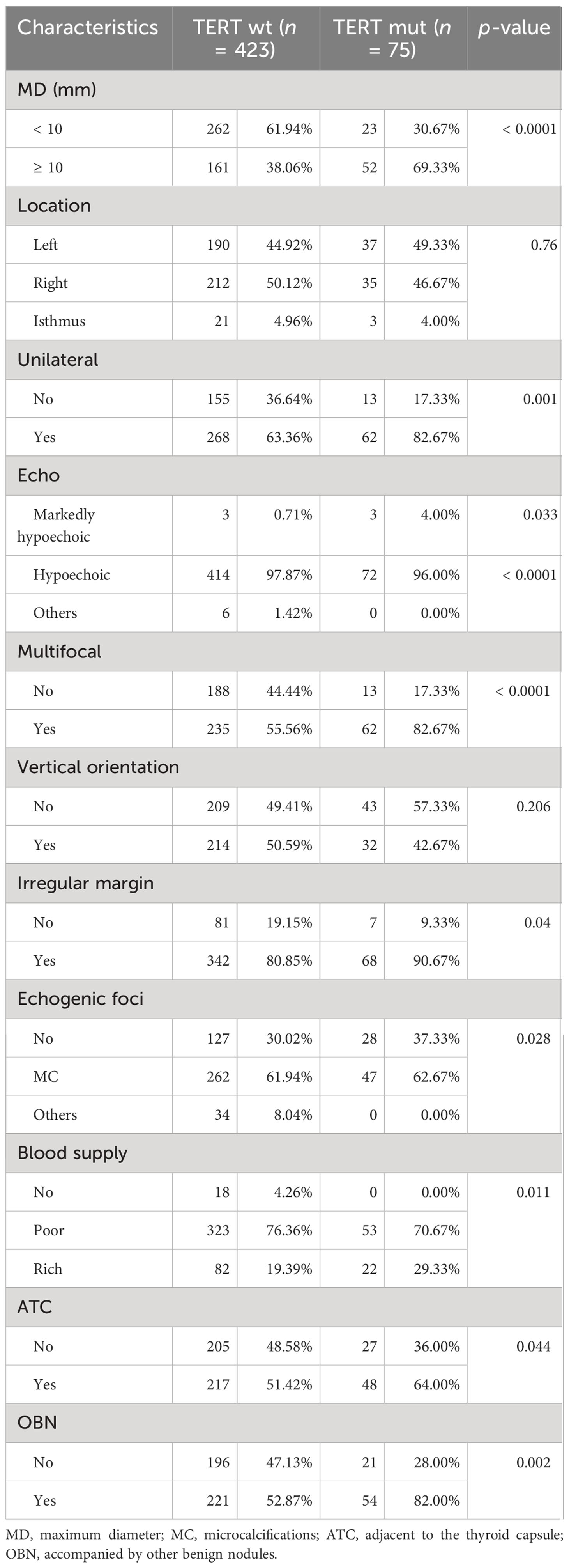
Table 2 US characteristics of thyroid nodules.
Lasso regression and multivariate analysis
Univariate analysis was used to compare the clinical features and US characteristics of the patients, as well as the US characteristics of the nodules, between the two groups. Preliminary results showed that risk factors associated with the development of TERT promoter mutations in PTC were older age, male, low TSH, normal Tgab and Tpoab, low Tg, cervical lymph node metastasis in the lateral neck region, number of malignant thyroid nodules, maximum diameter of ≥ 10 mm, unilateral, markedly hypoechoic, irregular margin, nonmicrocalcification, rich blood supply, adjacent to the thyroid capsule, and accompanied by other benign nodules. To exclude the effect of multicollinearity, we used lasso 10-fold cross-validation to screen for true risk factors from the candidate variables identified by univariate analysis ( Figure 1 ). After correction, risk factors analyzed by univariate analysis were male, older age, number of malignant thyroid nodules, cervical lymph node metastasis in the lateral neck region, maximal diameter of ≥ 10 mm, unilateral, multifocal, markedly hypoechoic, nonmicrocalcification, adjacent to the thyroid capsule, and accompanied by other benign nodules. Further multivariate analysis revealed that the independent risk factors for TERT promoter mutation were older age (OR = 1.07; p = 0.002), maximum diameter of ≥ 10 mm (OR = 3.94; p < 0.0001), unilateral (OR = 4.15; p < 0.0001), multifocal (OR = 7.69; p < 0.0001), adjacent to the thyroid capsule (OR = 1.94; p = 0.044), and accompanied by other benign nodules (OR = 1.94; p = 0.039) ( Tables 3 , 4 ).
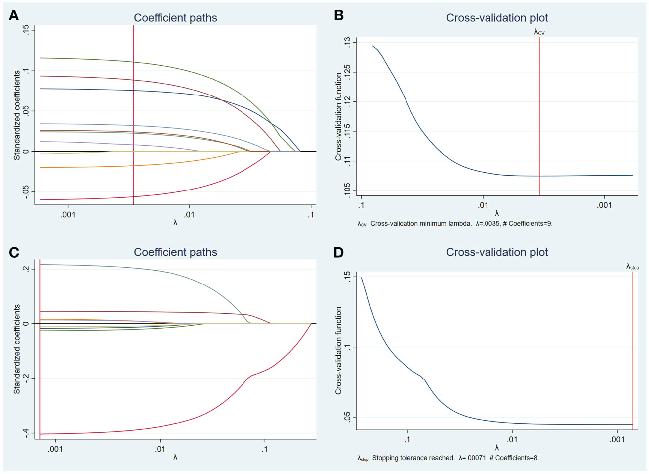
Figure 1 Lasso regression for risk factors from univariate analysis. Lasso regression screening was performed on nodule features that showed differences in the results of the preliminary univariate analysis, and the colored lines from right (blue) to left (purple) in order represent the following factors: maximum diameter of ≥ 10 mm, multifocal, unilateral, microcalcification, accompanied by other benign nodules, adjacent to the thyroid capsule, markedly hypoechoic, irregular margin, rich blood supply (A) . Selection of the tuning parameter lambda (B) . Lasso regression was performed to screen for patient characteristics that showed differences, and the first four colored lines from right (red) to left (green) represent, in order, the following indicators: age, gender, cervical lymph node metastasis in the lateral neck region, and number of malignant thyroid nodules, while others represent TSH, Tgab, Tpoab, and Tg (C) . Selection of the tuning parameter lambda (D) .
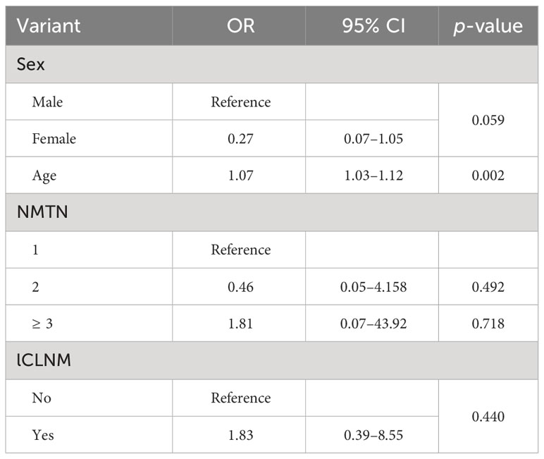
Table 3 Clinical and US feature analyses of patients.
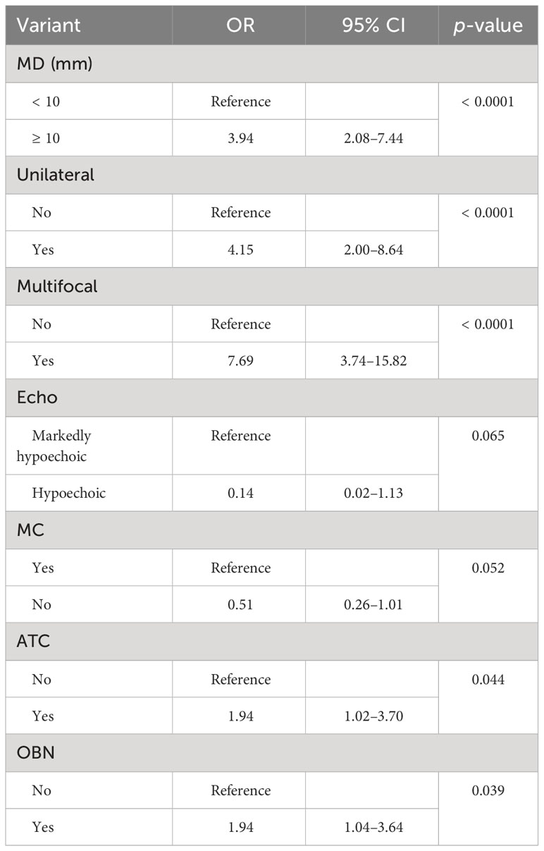
Table 4 US feature screening based on multifactor logistic regression.
Predictive model
Six clinical and US features were identified as independent risk factors for predicting the occurrence of TERT promoter mutations in PTC. These include older age, maximum diameter of ≥ 10 mm, unilateral, multifocal, adjacent to the thyroid capsule, and accompanied by other benign nodules. Based on these factors, a predictive model was established with an area under the curve (AUC) of the receiver operating characteristic of 0.839 ( Figure 2 ).
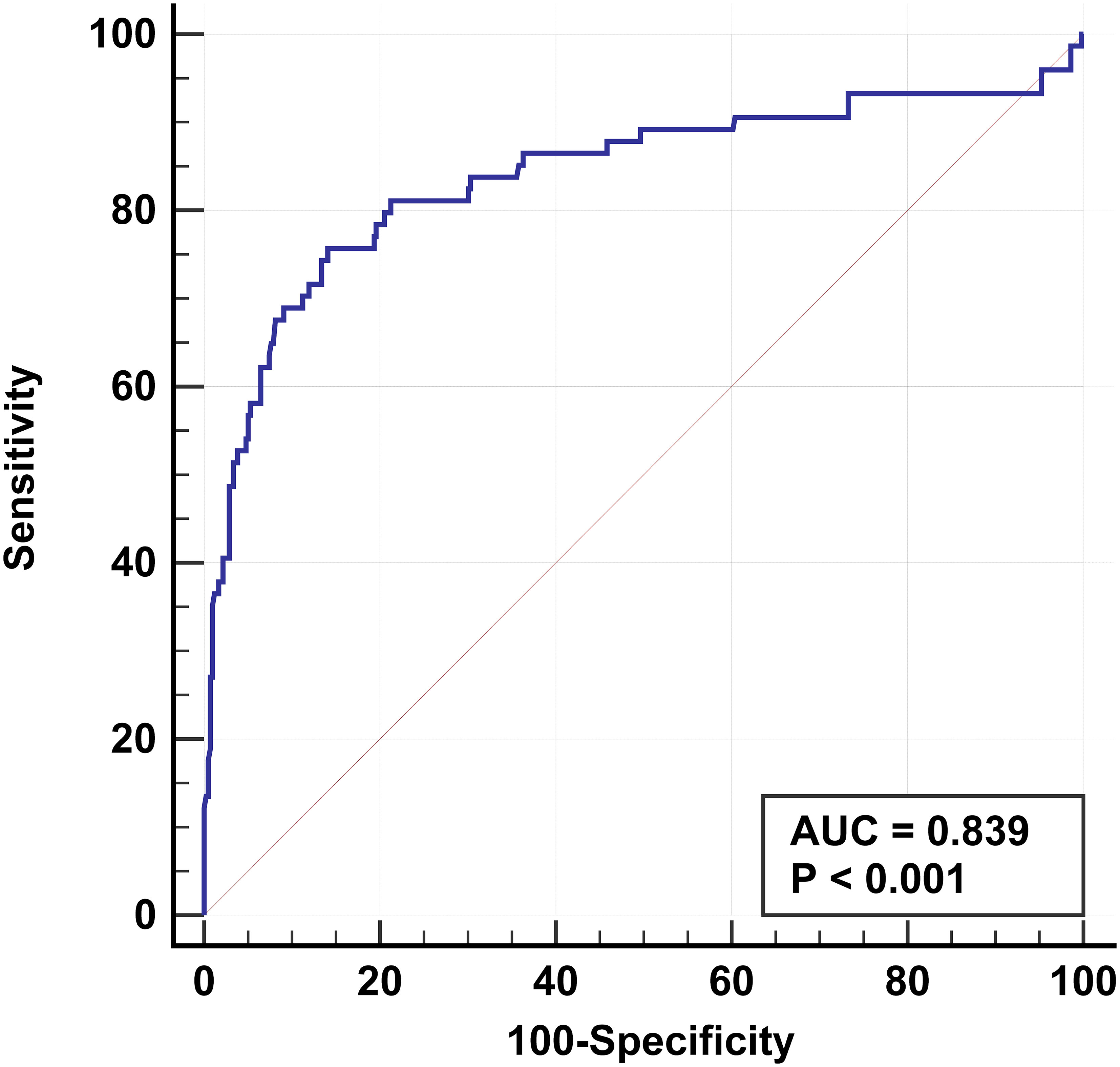
Figure 2 Receiver operating characteristic based on results of multifactorial analysis. The receiver operating characteristics are established for older age, multifocal, maximum diameter of ≥ 10 mm, unilateral, adjacent to the thyroid capsule, and accompanied by other benign nodules. The AUC is 0.839.
We examined the TERT promoter status in 365 PTC patients confirmed by pathology. The results of the univariate analysis suggest that the TERT promoter mutation may be associated with several factors, including older age, male, low TSH, normal Tgab, normal Tpoab, low Tg, cervical lymph node metastasis in the lateral neck region, number of malignant thyroid nodules, maximum diameter of ≥ 10 mm, unilateral, markedly hypoechoic, irregular margin, nonmicrocalcification, rich blood supply, adjacent to the thyroid capsule, and accompanied by other benign nodules. However, after correction using machine learning and further multivariate analysis, we discovered that older age, maximum diameter of ≥ 10 mm, unilateral, multifocality, adjacent to the thyroid capsule, and accompanied by other benign nodules were independent risk factors for predicting TERT promoter mutation. Finally, we established a prediction model for TERT promoter mutations based on the above independent risk factors. The model achieved an AUC of 0.839, indicating good predictive performance, and may become a promising predictive tool for clinical practice.
Our study showed that for older patients with PTC, the risk of TERT promoter mutation was higher (OR = 1.07; p < 0.05), and the risk of TERT mutation increased with each 1-year increase in age, that is, by 7%. Additionally, maximum diameter of ≥ 10 mm, unilateral, multifocal, and adjacent to the thyroid capsule were identified as independent risk factors for TERT promoter mutation in PTC patients. Furthermore, our study also counted and compared the presence of other benign nodules in patients with PTC and found that differences emerged between the two groups. This confirmed that the presence of other benign nodules could serve as an independent risk factor for predicting the occurrence of TERT promoter mutations in PTCs.
It is worth noting that when counting the variables, we recorded the number of malignant thyroid nodules in the PTC patients and compared the multifocality in the ultrasound features of the nodules between the two groups. Although both variables referred to the same thing and showed differences in the initial univariate analysis, the number of malignant thyroid nodules was excluded after eliminating the effect of multicollinearity. The multifocality ultimately remained an independent risk factor for predicting the TERT promoter mutations.
Previous studies have demonstrated that age, maximum diameter of the nodules, multifocality, and adjacent to the thyroid capsule are independent risk factors for TERT promoter mutations, which are consistent with our findings ( 21 – 23 ). Especially age, which has been widely reported in many studies, was consistently validated ( 24 – 26 ). In addition, other studies have suggested that indicators such as males, irregular margins, and vertical orientation can predict TERT promoter mutations ( 27 , 28 ). In our study, the results of univariate analysis showed a possible relationship between TERT promoter mutations and male and irregular margins. However, these two variables were excluded for multiple covariance. In contrast, vertical orientation did not show significant differences in our study. While unilateral and accompanied by other benign nodules have rarely been analyzed before, even fewer studies have suggested an association with the presence of mutations in the TERT promoter. This study found that these two are independent risk factors for TERT promoter mutations.
A meta-analysis that included 51 studies indicated that cervical lymph node metastasis was associated with TERT promoter mutations, which was additionally confirmed by a meta-analysis that included a total of 2,035 patients from eight studies ( 29 , 30 ). There is also a study suggesting that cervical lymph node metastasis is not associated with TERT promoter mutations ( 31 ). Therefore, the correlation between cervical lymph node metastasis and TERT promoter mutations remains controversial. Our results suggest that cervical lymph node metastasis detected by US was not an independent risk factor for the TERT promoter mutation.
In this study, a predictive model based on relevant clinical and US features can serve as an auxiliary tool for the precise management of PTC patients. The combination of clinical and US features can help predict TERT promoter mutations in a noninvasive way, replacing FNA to reduce patient pain and medical costs. Based on previous research reports and the results of this study, the mutation of the TERT promoter was found to be associated with the adjacent thyroid capsule and adverse prognostic features such as high recurrence rate and mortality rate. Therefore, detection of TERT promoter mutations can assist in rationally selecting appropriate treatment strategies for those high-risk populations, such as establishing more frequent follow-up plans. Conversely, individuals at relatively low risk can opt for more conservative treatment to reduce overtreatment.
There are some limitations to this study. First, the retrospective study design might limit the analysis of additional potential variables. Secondly, the predictive model lacks external validation. Additionally, although the sample size has notably increased compared to prior studies, a larger prospective cohort could provide more robust and persuasive findings. Therefore, future research will prioritize rigorous prospective, multicenter studies that focus on validating and optimizing this predictive model.
In conclusion, our study demonstrated that older age, maximum diameter of ≥ 10 mm, unilateral, multifocal, adjacent to the thyroid capsule, and accompanied by other benign nodules were independent risk factors for TERT promoter mutations in PTC. The prediction model based on these characteristics was of high predictive value. The correct acquisition and interpretation of the patient’s clinical characteristics as well as the US characteristics of the nodule are important and may aid in predicting the TERT promoter status, providing a valuable reference for subsequent patient management and treatment options.
Data availability statement
The original contributions presented in the study are included in the article/supplementary material. Further inquiries can be directed to the corresponding authors.
Ethics statement
The studies involving humans were approved by the ethics review committee of Ruijin Hospital, Shanghai Jiaotong University School of Medicine. The studies were conducted in accordance with the local legislation and institutional requirements. The requirement for obtaining informed consent from patients was waived because of its retrospective nature.
Author contributions
YH: Writing – review & editing, Writing – original draft, Visualization, Validation, Supervision, Software, Resources, Project administration, Methodology, Investigation, Formal analysis, Data curation, Conceptualization. SX: Software, Conceptualization, Writing – review & editing, Visualization, Validation, Supervision, Resources, Project administration, Methodology, Investigation. LD: Methodology, Formal analysis, Writing – review & editing. ZP: Investigation, Writing – review & editing, Resources, Formal analysis, Data curation. LZ: Methodology, Validation, Resources, Writing – review & editing. WZ: Visualization, Validation, Supervision, Project administration, Methodology, Funding acquisition, Writing – review & editing, Resources, Investigation.
The author(s) declare that financial support was received for the research, authorship, and/or publication of this article. This work was supported by the National Natural Science Foundation of China (82071923).
Acknowledgments
We thank our colleagues for their support in this work.
Conflict of interest
The authors declare that the research was conducted in the absence of any commercial or financial relationships that could be construed as a potential conflict of interest.
Publisher’s note
All claims expressed in this article are solely those of the authors and do not necessarily represent those of their affiliated organizations, or those of the publisher, the editors and the reviewers. Any product that may be evaluated in this article, or claim that may be made by its manufacturer, is not guaranteed or endorsed by the publisher.
1. Sung H, Ferlay J, Siegel RL, Laversanne M, Soerjomataram I, Jemal A, et al. Global cancer statistics 2020: GLOBOCAN estimates of incidence and mortality worldwide for 36 cancers in 185 countries. CA Cancer J Clin . (2021) 71:209–49. doi: 10.3322/caac.21660
PubMed Abstract | CrossRef Full Text | Google Scholar
2. Pizzato M, Li M, Vignat J, Laversanne M, Singh D, La Vecchia C, et al. The epidemiological landscape of thyroid cancer worldwide: GLOBOCAN estimates for incidence and mortality rates in 2020. Lancet Diabetes Endocrinol . (2022) 10:264–72. doi: 10.1016/S2213-8587(22)00035-3
3. Aschebrook-Kilfoy B, Ward MH, Sabra MM, Devesa SS. Thyroid cancer incidence patterns in the United States by histologic type, 1992-2006. Thyroid . (2011) 21:125–34. doi: 10.1089/thy.2010.0021
4. Network CGAR. Integrated genomic characterization of papillary thyroid carcinoma. Cell . (2014) 159:676–90. doi: 10.1016/j.cell.2014.09.050
5. Yu P, Qu N, Zhu R, Hu J, Han P, Wu J, et al. TERT accelerates BRAF mutant-induced thyroid cancer dedifferentiation and progression by regulating ribosome biogenesis. Sci Adv . (2023) 9:eadg7125. doi: 10.1126/sciadv.adg7125
6. Pu W, Shi X, Yu P, Zhang M, Liu Z, Tan L, et al. Single-cell transcriptomic analysis of the tumor ecosystems underlying initiation and progression of papillary thyroid carcinoma. Nat Commun . (2021) 12:6058. doi: 10.1038/s41467-021-26343-3
7. Landa I, Ibrahimpasic T, Boucai L, Sinha R, Knauf JA, Shah RH, et al. Genomic and transcriptomic hallmarks of poorly differentiated and anaplastic thyroid cancers. J Clin Invest . (2016) 126:1052–66. doi: 10.1172/JCI85271
8. Cabanillas ME, Ryder M, Jimenez C. Targeted therapy for advanced thyroid cancer: kinase inhibitors and beyond. Endocr Rev . (2019) 40:1573–604. doi: 10.1210/er.2019-00007
9. Yuan X, Dai M, Xu D. TERT promoter mutations and GABP transcription factors in carcinogenesis: More foes than friends. Cancer Lett . (2020) 493:1–9. doi: 10.1016/j.canlet.2020.07.003
10. Huang FW, Hodis E, Xu MJ, Kryukov GV, Chin L, Garraway LA. Highly recurrent TERT promoter mutations in human melanoma. Science . (2013) 339:957–9. doi: 10.1126/science.1229259
11. Horn S, Figl A, Rachakonda PS, Fischer C, Sucker A, Gast A, et al. TERT promoter mutations in familial and sporadic melanoma. Science . (2013) 339:959–61. doi: 10.1126/science.1230062
12. Melo M, da Rocha AG, Vinagre J, Batista R, Peixoto J, Tavares C, et al. TERT promoter mutations are a major indicator of poor outcome in differentiated thyroid carcinomas. J Clin Endocrinol Metab . (2014) 99:E754–65. doi: 10.1210/jc.2013-3734
13. Bullock M, Ren Y, O’Neill C, Gill A, Aniss A, Sywak M, et al. TERT promoter mutations are a major indicator of recurrence and death due to papillary thyroid carcinomas. Clin Endocrinol (Oxf) . (2016) 85:283–90. doi: 10.1111/cen.12999
14. Xing M, Liu R, Liu X, Murugan AK, Zhu G, Zeiger MA, et al. BRAF V600E and TERT promoter mutations cooperatively identify the most aggressive papillary thyroid cancer with highest recurrence. J Clin Oncol . (2014) 32:2718–26. doi: 10.1200/JCO.2014.55.5094
15. Lee SE, Hwang TS, Choi YL, Han HS, Kim WS, Jang MH, et al. Prognostic significance of TERT promoter mutations in papillary thyroid carcinomas in a BRAF(V600E) mutation-prevalent population. Thyroid . (2016) 26:901–10. doi: 10.1089/thy.2015.0488
16. Song YS, Lim JA, Choi H, Won JK, Moon JH, Cho SW, et al. Prognostic effects of TERT promoter mutations are enhanced by coexistence with BRAF or RAS mutations and strengthen the risk prediction by the ATA or TNM staging system in differentiated thyroid cancer patients. Cancer . (2016) 122:1370–9. doi: 10.1002/cncr.29934
17. Liu R, Bishop J, Zhu G, Zhang T, Ladenson PW, Xing M. Mortality risk stratification by combining BRAF V600E and TERT promoter mutations in papillary thyroid cancer: genetic duet of BRAF and TERT promoter mutations in thyroid cancer mortality. JAMA Oncol . (2017) 3:202–8. doi: 10.1001/jamaoncol.2016.3288
18. Patel KN, Yip L, Lubitz CC, Grubbs EG, Miller BS, Shen W, et al. The american association of endocrine surgeons guidelines for the definitive surgical management of thyroid disease in adults. Ann Surg . (2020) 271:e21–93. doi: 10.1097/SLA.0000000000003580
19. Han D, Min D, Xie R, Wang Z, Xiao G, Wang X, et al. Molecular testing raises thyroid nodule fine needle aspiration diagnostic value. Endocr Connect . (2023) 12:e230135. doi: 10.1530/EC-23-0135
20. Li X, Li E, Du J, Wang J, Zheng B. BRAF mutation analysis by ARMS-PCR refines thyroid nodule management. Clin Endocrinol (Oxf) . (2019) 91:834–41. doi: 10.1111/cen.14079
21. Kim MJ, Kim JK, Kim GJ, Kang SW, Lee J, Jeong JJ, et al. TERT promoter and BRAF V600E mutations in papillary thyroid cancer: A single-institution experience in Korea. Cancers (Basel) . (2022) 14:4928. doi: 10.3390/cancers14194928
22. Nakao T, Matsuse M, Saenko V, Rogounovitch T, Tanaka A, Suzuki K, et al. Preoperative detection of the TERT promoter mutations in papillary thyroid carcinomas. Clin Endocrinol (Oxf) . (2021) 95:790–9. doi: 10.1111/cen.14567
23. Choi YS, Choi SW, Yi JW. Prospective analysis of TERT promoter mutations in papillary thyroid carcinoma at a single institution. J Clin Med . (2021) 10:2179. doi: 10.3390/jcm10102179
24. Sun J, Zhang J, Lu J, Gao J, Ren X, Teng L, et al. BRAF V600E and TERT promoter mutations in papillary thyroid carcinoma in chinese patients. PloS One . (2016) 11:e0153319. doi: 10.1371/journal.pone.0153319
25. Na HY, Yu HW, Kim W, Moon JH, Ahn CH, Choi SI, et al. Clinicopathological indicators for TERT promoter mutation in papillary thyroid carcinoma. Clin Endocrinol (Oxf) . (2022) 97:106–15. doi: 10.1111/cen.14728
26. Kim TH, Kim YE, Ahn S, Kim JY, Ki CS, Oh YL, et al. TERT promoter mutations and long-term survival in patients with thyroid cancer. Endocr Relat Cancer . (2016) 23:813–23. doi: 10.1530/ERC-16-0219
27. Shi H, Guo LH, Zhang YF, Fu HJ, Zheng JY, Wang HX, et al. Suspicious ultrasound and clinicopathological features of papillary thyroid carcinoma predict the status of TERT promoter. Endocrine . (2020) 68:349–57. doi: 10.1007/s12020-020-02214-7
28. Kim TH, Ki CS, Hahn SY, Oh YL, Jang HW, Kim SW, et al. Ultrasonographic prediction of highly aggressive telomerase reverse transcriptase (TERT) promoter-mutated papillary thyroid cancer. Endocrine . (2017) 57:234–40. doi: 10.1007/s12020-017-1340-3
29. Yang J, Gong Y, Yan S, Chen H, Qin S, Gong R. Association between TERT promoter mutations and clinical behaviors in differentiated thyroid carcinoma: a systematic review and meta-analysis. Endocrine . (2020) 67:44–57. doi: 10.1007/s12020-019-02117-2
30. Yin DT, Yu K, Lu RQ, Li X, Xu J, Lei M, et al. Clinicopathological significance of TERT promoter mutation in papillary thyroid carcinomas: a systematic review and meta-analysis. Clin Endocrinol (Oxf) . (2016) 85:299–305. doi: 10.1111/cen.13017
31. Du Y, Zhang S, Zhang G, Hu J, Zhao L, Xiong Y, et al. Mutational profiling of Chinese patients with thyroid cancer. Front Endocrinol (Lausanne) . (2023) 14:1156999. doi: 10.3389/fendo.2023.1156999
Keywords: papillary thyroid cancer, thyroid nodule, TERT, ultrasound, molecular marker
Citation: Hu Y, Xu S, Dong L, Pan Z, Zhang L and Zhan W (2024) Clinical features combined with ultrasound characteristics to predict TERT promoter mutations in papillary thyroid carcinoma: a single-center study over the past 5 years. Front. Endocrinol. 15:1322731. doi: 10.3389/fendo.2024.1322731
Received: 16 October 2023; Accepted: 26 February 2024; Published: 18 March 2024.
Reviewed by:
Copyright © 2024 Hu, Xu, Dong, Pan, Zhang and Zhan. This is an open-access article distributed under the terms of the Creative Commons Attribution License (CC BY) . The use, distribution or reproduction in other forums is permitted, provided the original author(s) and the copyright owner(s) are credited and that the original publication in this journal is cited, in accordance with accepted academic practice. No use, distribution or reproduction is permitted which does not comply with these terms.
*Correspondence: Weiwei Zhan, [email protected] ; Lu Zhang, [email protected]
Disclaimer: All claims expressed in this article are solely those of the authors and do not necessarily represent those of their affiliated organizations, or those of the publisher, the editors and the reviewers. Any product that may be evaluated in this article or claim that may be made by its manufacturer is not guaranteed or endorsed by the publisher.
BME Alum Guides Iliac Vein Fluid Dynamics Research He Began as an Undergraduate
To build and validate patient-specific computational fluid dynamics models of the iliac veins, the U-M team used computed tomography and ultrasound data.
Michele Santillan
A team of researchers working with C. Alberto Figueroa, the Edward B. Diethrich Professor of Vascular Surgery, Professor, Biomedical Engineering, discovered that patients with iliac vein compression syndrome (IVCS) have elevated shear rates, which may explain why they have a higher risk of experiencing venous blood clots. The U-M research findings were published in recent issues of Frontiers in Bioengineering and Biotechnology and the Journal of Vascular Surgery Venous and Lymphatics Disorders .
More than 20 percent of the population has IVCS, which is associated with left leg pain, swelling and clotting. Dr. Figueroa noted that IVCS originates as an anatomical abnormality, when one of the veins, specifically the left common iliac, is more compressed than normal. This extra compression combines with additional risk factors to increase the likelihood that a patient may experience recurring episodes of blood clots.
To build and validate patient-specific computational fluid dynamics models of the iliac veins, the U-M team used computed tomography and ultrasound data. Based on their results, the researchers propose that non-invasive measurement of shear rate may help with risk stratification of patients with moderate compression in which current treatment is highly variable. More investigation is needed to determine the prognostic value of shear rate as a clinical metric and to understand the mechanisms of clot formation in IVCS patients.
The first author of this report is BME alum Ismael Assi (BSE, ‘22), who has researched this topic for more than three years, beginning as an undergraduate. Assi and Dr. Figueroa recently discussed the backstory of how the study came together.
“The very origin of this project came from the PhD work of BME student Sabrina Lynch, who developed methods to simulate flow on the venous system, where properties of blood are different from those that we see on the arterial side,” Dr. Figueroa said. “At the time, Ismael was a sophomore. He started closely working with Sabrina and eventually took over this project, looking specifically at patients with iliac vein compression syndrome, his own.”
Dr. Figueroa continued: “Ismael made it into a very clinically oriented study and demonstrated tremendous initiative. He started working with vascular surgeons, interventional radiologists, and also with nurses and other staff in the diagnostic vascular unit (DVU). He basically developed a prospective study entirely by himself–including securing an IRB (Institutional Review Board) approval.” Dr. Figueroa noted that Assi worked in tandem with clinicians and was engaged in all aspects of the study, including patient recruitment.
“It was very much a team effort,” Assi said. We collaborated with nurses, sonographers, interventional radiologists, surgeons and engineers. You had everyone from mechanical engineering to clinical researchers involved, and I think that’s what really helped to bring the project together.”
Assi noted that his work with a diverse group of professionals taught him how to organize his thoughts, improve his writing skills and interact with journal reviewers. He was especially thankful for all the mentorship he received from Drs. Figueroa, Lynch, Obi, Wakefield, Williams, and all the sonographers at the diagnostic vascular unit.
“You learn so much by doing a research project,” Assi said. “Especially when it’s a longitudinal one over several years. I look back at the mistakes I made, and they’re very valuable because I was learning how to communicate with people from different backgrounds and skill sets, how to acquire data and how to organize meetings. These are all valuable skills that I learned in research that I wouldn’t have learned in other avenues. I would definitely encourage students to seek out research to explore their interests. There’s nothing like it.”
Assi added that he was inspired by hearing the stories of patients and what they went through when they had blood clots. “Hopefully, I will be able to make a difference in the treatment of these patients,” he said. “Many people have this compression, so the medical community is working to gain a better understanding of when it is indicated to treat patients. Do you only treat symptomatic patients with anticoagulation? When do you stent patients? When do you not treat patients? There are a lot of questions that are unanswered.”
This study consisted of a group of four patients with venous compression and four control patients with no compression. “The goal was to look for a metric that could stratify patients,” Assi said. “How can you measure the severity of a patient’s disease? That question requires further clinical validation. Right now, we found that shear rate, which is a surrogate index for the activation of platelets, can be used to differentiate healthy patients versus patients who have compressed iliac veins.”
“The results are quite statistically strong,” added Dr. Figueroa, “in the sense that these parameters or these biomarkers of the shear rate showed a very strong significance between patients with normal iliac vein anatomy and patients who have compression syndrome. Even for the iliac vein compression syndrome examples, there was a very substantial difference between the contralateral vein, which is the vein on the other side. There’s an ongoing study where measurements are being taken for both diameters and ultrasound-derived velocities to look at a much larger group of patients. One of the things that we are trying to do is integrate something into the clinical workflow that you can almost get a quick answer for without having to go through all the sequence of steps in the computational analysis that we do, which is quite time consuming.”
BME alum Assi knew about the breadth of professional options available to biomedical engineering majors, but his research experience ultimately inspired him to attend medical school. “I think one of the big reasons I chose medical school was a patient who was a former nurse who had a blood clot in her leg,” Assi said. “Because she was a nurse, she knew exactly what was going on, so she went right to the emergency room. She told me her whole story, but I think another aspect that was very important was that her daughter had had a blood clot in her carotid artery, which is in the neck. The patients’ dad had also passed away from a blood clot. When I heard about how her entire family was unfortunately afflicted by blood clotting, it was very motivating. My interest in the project just increased that much more because I really wanted to make a difference in the lives of patients like her.”
Related Topics

IMAGES
VIDEO
COMMENTS
Ultrasound articles from across Nature Portfolio. Atom. RSS Feed. Ultrasound is a non-invasive imaging technique that uses the differential reflectance of acoustic waves at ultrasonic frequencies ...
The objective of this study was to determine the learning curve of tracheal−esophageal ultrasound by prehospital medical and paramedical staff. Paul-Georges Reuter, Chris Ballouz, Thomas Loeb, Tomislav Petrovic and Frédéric Lapostolle. The Ultrasound Journal 2023 15 :20. Original article Published on: 1 May 2023.
Medical ultrasound represented by ultrasound imaging is generally believed to be well-known area in research, because ultrasound imaging systems has been widely distributed and actively used in the world since the 1960s. ... Hence, this special issue is designed to bring in various ultrasound-related topics in medicine. We also tried to invite ...
Introduction. Making waves at the frontiers of medicine, the rise of clinical ultrasound in today's 21 st century highlights its many robust applications in medicine and guiding patient-centered management for patients at the bedside. Illuminated through photography and long exposure light technique, "Riding the waves" captures the serenity of the sea at sunrise (Figure 1); the waves and ...
Jan. 31, 2023 — New research found that using focused-ultrasound-mediated liquid biopsy in a mouse model released more tau proteins and another biomarker into the blood than without the ...
Since Ultrasonography was launched as an international peer-reviewed academic journal dedicated to medical ultrasound (US) in January 2014, it has been an honor and privilege for me to serve as the journal's Honorary Editor [].The purpose of this editorial update is to provide readers with some recent information and to share my views on the journal and its future directions.
Ultrasound Imaging - Current Topics presents complex and current topics in ultrasound imaging in a simplified format. It is easy to read and exemplifies the range of experiences of each contributing author. ... Open Access is an initiative that aims to make scientific research freely available to all. To date our community has made over 100 ...
After years of research, an NIH-funded team has developed a wearable cardiac ultrasound imager that can non-invasively capture real-time images of the human heart. The prototype patch, which is about the size of a postage stamp, can be worn during exercise, providing valuable cardiac information when the heart is under stress.
One of the most common uses of ultrasound is during pregnancy, to monitor the growth and development of the fetus, but there are many other uses, including imaging the heart, blood vessels, eyes, thyroid, brain, breast, abdominal organs, skin, and muscles. Ultrasound images are displayed in either 2D, 3D, or 4D (which is 3D in motion).
Focused ultrasound (FUS) is a noninvasive therapeutic technology with multiple pediatric clinical applications. The ability of focused ultrasound to target tissues deep in the body without exposing children to the morbidities associated with conventional surgery, interventional procedures, or radiation offers significant advantages. In 2021, there are 10 clinical pediatric focused ultrasound ...
Computed Tomography (CT) is the standard reference in the emergency for evalua... Lucio Cagini, Sabrina Gravante, Corrado Maria Malaspina, Elviro Cesarano, Melchiorre Giganti, Alberto Rebonato, Paolo Fonio and Michele Scialpi. Critical Ultrasound Journal 2013 5 (Suppl 1) :S9. Research Published on: 15 July 2013.
Keywords: medical ultrasound imaging, therapeutic ultrasound, non-destructive evaluation, nonlinear ultrasonics, physics of ultrasound, underwater ultrasound, ultrasonic transducers . Important Note: All contributions to this Research Topic must be within the scope of the section and journal to which they are submitted, as defined in their mission statements.
Advanced ultrasound techniques constitute a suite of new technologies that employ microbubbles to provide contrast and enhance flow visualization, elastography to measure tissue stiffness, ultrafast Doppler to deliver high spatiotemporal resolution of flow, three- and four-dimensional technique to generate accurate spatiotemporal representation ...
Transparent ultrasound chip improves cell stimulation and imaging. Ultrasound scans, best known for monitoring pregnancies or imaging organs, can also be used to stimulate cells and direct cell function. A team of Penn State researchers has developed an easier, more effective way to harness the technology for biomedical applications.
Ultrasound and Color Doppler-Guided Surgery. The purpose of the study is to examine the opinions of the trainees attending a training course concerning the use of technology. We will write. a custom essay specifically for you by our professional experts. 809 writers online.
Keywords: Ultrasound device, Transducer, acoustic material, neuromodulation, brain therapy, neuroimaging . Important Note: All contributions to this Research Topic must be within the scope of the section and journal to which they are submitted, as defined in their mission statements.Frontiers reserves the right to guide an out-of-scope manuscript to a more suitable section or journal at any ...
The therapeutic use of ultrasound emerged early as an area of interest through the development of High-Intensity Focused Ultrasound (HIFU). The development of ultrasound as a therapeutic tool is the subject of much varied research in the cardiovascular field. The tissue modifications that ultrasound can induce are dependent on its frequency, energy, and focalization or not.
List of topics. Topics for thesis and projects are given below. Most of the topics can be adjusted to the students qualifications and wishes. Don't hesitate to take contact with the corresponding supervisor - we're looking forward to a discussion with you!
A thesis or dissertation, as some people would like to call it, is an integral part of the Radiology curriculum, be it MD, DNB, or DMRD. We have tried to aggregate radiology thesis topics from various sources for reference. Not everyone is interested in research, and writing a Radiology thesis can be daunting.
Abstract. Ultrasound (US) is acoustic energy that interacts with human tissues, thus, producing bioeffects that may be hazardous, especially in sensitive organs (i.e., brain, eye, heart, lung, and digestive tract) and embryos/fetuses. Two basic mechanisms of US interaction with biological systems have been identified: thermal and non-thermal.
About this Research Topic. Submission closed. Musculoskeletal ultrasound is becoming an important and effective tool for the diagnosis of various soft tissue and joint disorders. It is accurate, safe, affordable, efficient, and dynamic. Musculoskeletal ultrasound is also a useful tool for guided therapy. The advances of musculoskeletal imaging ...
The goals of research in ultrasound usage in space environments are: (1) Determine accuracy of ultrasound in novel clinical conditions. (2) Determine optimal training methodologies, (3) Determine microgravity associated changes and (4) Develop intuitive ultrasound catalog to enhance autonomous medical care.
The state-of-the-art ultrasound machine that's pushed around on a cart, with cables and multiple probes dangling, is being wheeled aside permanently in favor of handheld probes that send images ...
With the use of a modified Delphi and Hanlon process of prioritization methodology, a ranked list of priority pediatric emergency medicine point‐of‐care ultrasound research topics was generated and could be used to inform future international multicenter studies and trials.
This article is part of the Research Topic Advances in precision medicine in the management of thyroid nodules and thyroid cancer View all 7 articles. Clinical features combined with ultrasound characteristics to predict TERT promoter mutations in papillary thyroid carcinoma: a single-center study over the past 5 years ...
The U-M research findings were published in recent issues of Frontiers in Bioengineering and Biotechnology and the Journal of Vascular Surgery Venous ... the U-M team used computed tomography and ultrasound data. Based on their results, the researchers propose that non-invasive measurement of shear rate may help with risk stratification of ...