Appointments at Mayo Clinic
- Pregnancy week by week
- Fetal presentation before birth
The way a baby is positioned in the uterus just before birth can have a big effect on labor and delivery. This positioning is called fetal presentation.
Babies twist, stretch and tumble quite a bit during pregnancy. Before labor starts, however, they usually come to rest in a way that allows them to be delivered through the birth canal headfirst. This position is called cephalic presentation. But there are other ways a baby may settle just before labor begins.
Following are some of the possible ways a baby may be positioned at the end of pregnancy.

Head down, face down
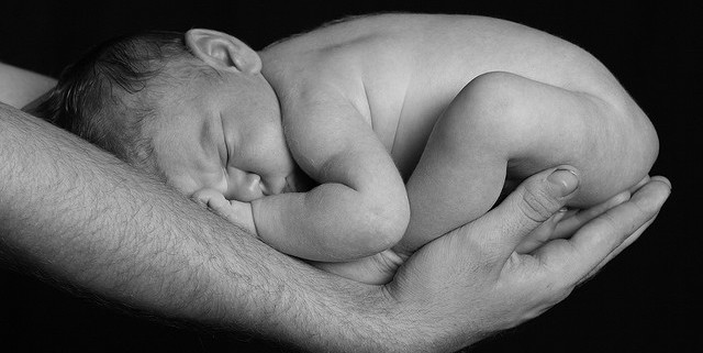
When a baby is head down, face down, the medical term for it is the cephalic occiput anterior position. This the most common position for a baby to be born in. With the face down and turned slightly to the side, the smallest part of the baby's head leads the way through the birth canal. It is the easiest way for a baby to be born.

Head down, face up
When a baby is head down, face up, the medical term for it is the cephalic occiput posterior position. In this position, it might be harder for a baby's head to go under the pubic bone during delivery. That can make labor take longer.
Most babies who begin labor in this position eventually turn to be face down. If that doesn't happen, and the second stage of labor is taking a long time, a member of the health care team may reach through the vagina to help the baby turn. This is called manual rotation.
In some cases, a baby can be born in the head-down, face-up position. Use of forceps or a vacuum device to help with delivery is more common when a baby is in this position than in the head-down, face-down position. In some cases, a C-section delivery may be needed.

Frank breech
When a baby's feet or buttocks are in place to come out first during birth, it's called a breech presentation. This happens in about 3% to 4% of babies close to the time of birth. The baby shown below is in a frank breech presentation. That's when the knees aren't bent, and the feet are close to the baby's head. This is the most common type of breech presentation.
If you are more than 36 weeks into your pregnancy and your baby is in a frank breech presentation, your health care professional may try to move the baby into a head-down position. This is done using a procedure called external cephalic version. It involves one or two members of the health care team putting pressure on your belly with their hands to get the baby to roll into a head-down position.
If the procedure isn't successful, or if the baby moves back into a breech position, talk with a member of your health care team about the choices you have for delivery. Most babies in a frank breech position are born by planned C-section.

Complete and incomplete breech
A complete breech presentation, as shown below, is when the baby has both knees bent and both legs pulled close to the body. In an incomplete breech, one or both of the legs are not pulled close to the body, and one or both of the feet or knees are below the baby's buttocks. If a baby is in either of these positions, you might feel kicking in the lower part of your belly.
If you are more than 36 weeks into your pregnancy and your baby is in a complete or incomplete breech presentation, your health care professional may try to move the baby into a head-down position. This is done using a procedure called external cephalic version. It involves one or two members of the health care team putting pressure on your belly with their hands to get the baby to roll into a head-down position.
If the procedure isn't successful, or if the baby moves back into a breech position, talk with a member of your health care team about the choices you have for delivery. Many babies in a complete or incomplete breech position are born by planned C-section.

When a baby is sideways — lying horizontal across the uterus, rather than vertical — it's called a transverse lie. In this position, the baby's back might be:
- Down, with the back facing the birth canal.
- Sideways, with one shoulder pointing toward the birth canal.
- Up, with the hands and feet facing the birth canal.
Although many babies are sideways early in pregnancy, few stay this way when labor begins.
If your baby is in a transverse lie during week 37 of your pregnancy, your health care professional may try to move the baby into a head-down position. This is done using a procedure called external cephalic version. External cephalic version involves one or two members of your health care team putting pressure on your belly with their hands to get the baby to roll into a head-down position.
If the procedure isn't successful, or if the baby moves back into a transverse lie, talk with a member of your health care team about the choices you have for delivery. Many babies who are in a transverse lie are born by C-section.

If you're pregnant with twins and only the twin that's lower in the uterus is head down, as shown below, your health care provider may first deliver that baby vaginally.
Then, in some cases, your health care team may suggest delivering the second twin in the breech position. Or they may try to move the second twin into a head-down position. This is done using a procedure called external cephalic version. External cephalic version involves one or two members of the health care team putting pressure on your belly with their hands to get the baby to roll into a head-down position.
Your health care team may suggest delivery by C-section for the second twin if:
- An attempt to deliver the baby in the breech position is not successful.
- You do not want to try to have the baby delivered vaginally in the breech position.
- An attempt to move the baby into a head-down position is not successful.
- You do not want to try to move the baby to a head-down position.
In some cases, your health care team may advise that you have both twins delivered by C-section. That might happen if the lower twin is not head down, the second twin has low or high birth weight as compared to the first twin, or if preterm labor starts.

- Landon MB, et al., eds. Normal labor and delivery. In: Gabbe's Obstetrics: Normal and Problem Pregnancies. 8th ed. Elsevier; 2021. https://www.clinicalkey.com. Accessed May 19, 2023.
- Holcroft Argani C, et al. Occiput posterior position. https://www.updtodate.com/contents/search. Accessed May 19, 2023.
- Frequently asked questions: If your baby is breech. American College of Obstetricians and Gynecologists https://www.acog.org/womens-health/faqs/if-your-baby-is-breech. Accessed May 22, 2023.
- Hofmeyr GJ. Overview of breech presentation. https://www.updtodate.com/contents/search. Accessed May 22, 2023.
- Strauss RA, et al. Transverse fetal lie. https://www.updtodate.com/contents/search. Accessed May 22, 2023.
- Chasen ST, et al. Twin pregnancy: Labor and delivery. https://www.updtodate.com/contents/search. Accessed May 22, 2023.
- Cohen R, et al. Is vaginal delivery of a breech second twin safe? A comparison between delivery of vertex and non-vertex second twins. The Journal of Maternal-Fetal & Neonatal Medicine. 2021; doi:10.1080/14767058.2021.2005569.
- Marnach ML (expert opinion). Mayo Clinic. May 31, 2023.
Products and Services
- A Book: Obstetricks
- A Book: Mayo Clinic Guide to a Healthy Pregnancy
- 3rd trimester pregnancy
- Fetal development: The 3rd trimester
- Overdue pregnancy
- Pregnancy due date calculator
- Prenatal care: 3rd trimester
Mayo Clinic does not endorse companies or products. Advertising revenue supports our not-for-profit mission.
- Opportunities
Mayo Clinic Press
Check out these best-sellers and special offers on books and newsletters from Mayo Clinic Press .
- Mayo Clinic on Incontinence - Mayo Clinic Press Mayo Clinic on Incontinence
- The Essential Diabetes Book - Mayo Clinic Press The Essential Diabetes Book
- Mayo Clinic on Hearing and Balance - Mayo Clinic Press Mayo Clinic on Hearing and Balance
- FREE Mayo Clinic Diet Assessment - Mayo Clinic Press FREE Mayo Clinic Diet Assessment
- Mayo Clinic Health Letter - FREE book - Mayo Clinic Press Mayo Clinic Health Letter - FREE book
- Healthy Lifestyle
Your gift holds great power – donate today!
Make your tax-deductible gift and be a part of the cutting-edge research and care that's changing medicine.
- Getting Pregnant
- Registry Builder
- Baby Products
- Birth Clubs
- See all in Community
- Ovulation Calculator
- How To Get Pregnant
- How To Get Pregnant Fast
- Ovulation Discharge
- Implantation Bleeding
- Ovulation Symptoms
- Pregnancy Symptoms
- Am I Pregnant?
- Pregnancy Tests
- See all in Getting Pregnant
- Due Date Calculator
- Pregnancy Week by Week
- Pregnant Sex
- Weight Gain Tracker
- Signs of Labor
- Morning Sickness
- COVID Vaccine and Pregnancy
- Fetal Weight Chart
- Fetal Development
- Pregnancy Discharge
- Find Out Baby Gender
- Chinese Gender Predictor
- See all in Pregnancy
- Baby Name Generator
- Top Baby Names 2023
- Top Baby Names 2024
- How to Pick a Baby Name
- Most Popular Baby Names
- Baby Names by Letter
- Gender Neutral Names
- Unique Boy Names
- Unique Girl Names
- Top baby names by year
- See all in Baby Names
- Baby Development
- Baby Feeding Guide
- Newborn Sleep
- When Babies Roll Over
- First-Year Baby Costs Calculator
- Postpartum Health
- Baby Poop Chart
- See all in Baby
- Average Weight & Height
- Autism Signs
- Child Growth Chart
- Night Terrors
- Moving from Crib to Bed
- Toddler Feeding Guide
- Potty Training
- Bathing and Grooming
- See all in Toddler
- Height Predictor
- Potty Training: Boys
- Potty training: Girls
- How Much Sleep? (Ages 3+)
- Ready for Preschool?
- Thumb-Sucking
- Gross Motor Skills
- Napping (Ages 2 to 3)
- See all in Child
- Photos: Rashes & Skin Conditions
- Symptom Checker
- Vaccine Scheduler
- Reducing a Fever
- Acetaminophen Dosage Chart
- Constipation in Babies
- Ear Infection Symptoms
- Head Lice 101
- See all in Health
- Second Pregnancy
- Daycare Costs
- Family Finance
- Stay-At-Home Parents
- Breastfeeding Positions
- See all in Family
- Baby Sleep Training
- Preparing For Baby
- My Custom Checklist
- My Registries
- Take the Quiz
- Best Baby Products
- Best Breast Pump
- Best Convertible Car Seat
- Best Infant Car Seat
- Best Baby Bottle
- Best Baby Monitor
- Best Stroller
- Best Diapers
- Best Baby Carrier
- Best Diaper Bag
- Best Highchair
- See all in Baby Products
- Why Pregnant Belly Feels Tight
- Early Signs of Twins
- Teas During Pregnancy
- Baby Head Circumference Chart
- How Many Months Pregnant Am I
- What is a Rainbow Baby
- Braxton Hicks Contractions
- HCG Levels By Week
- When to Take a Pregnancy Test
- Am I Pregnant
- Why is Poop Green
- Can Pregnant Women Eat Shrimp
- Insemination
- UTI During Pregnancy
- Vitamin D Drops
- Best Baby Forumla
- Postpartum Depression
- Low Progesterone During Pregnancy
- Baby Shower
- Baby Shower Games
Breech, posterior, transverse lie: What position is my baby in?

Fetal presentation, or how your baby is situated in your womb at birth, is determined by the body part that's positioned to come out first, and it can affect the way you deliver. At the time of delivery, 97 percent of babies are head-down (cephalic presentation). But there are several other possibilities, including feet or bottom first (breech) as well as sideways (transverse lie) and diagonal (oblique lie).
Fetal presentation and position
During the last trimester of your pregnancy, your provider will check your baby's presentation by feeling your belly to locate the head, bottom, and back. If it's unclear, your provider may do an ultrasound or an internal exam to feel what part of the baby is in your pelvis.
Fetal position refers to whether the baby is facing your spine (anterior position) or facing your belly (posterior position). Fetal position can change often: Your baby may be face up at the beginning of labor and face down at delivery.
Here are the many possibilities for fetal presentation and position in the womb.
Medical illustrations by Jonathan Dimes
Head down, facing down (anterior position)
A baby who is head down and facing your spine is in the anterior position. This is the most common fetal presentation and the easiest position for a vaginal delivery.
This position is also known as "occiput anterior" because the back of your baby's skull (occipital bone) is in the front (anterior) of your pelvis.
Head down, facing up (posterior position)
In the posterior position , your baby is head down and facing your belly. You may also hear it called "sunny-side up" because babies who stay in this position are born facing up. But many babies who are facing up during labor rotate to the easier face down (anterior) position before birth.
Posterior position is formally known as "occiput posterior" because the back of your baby's skull (occipital bone) is in the back (posterior) of your pelvis.
Frank breech
In the frank breech presentation, both the baby's legs are extended so that the feet are up near the face. This is the most common type of breech presentation. Breech babies are difficult to deliver vaginally, so most arrive by c-section .
Some providers will attempt to turn your baby manually to the head down position by applying pressure to your belly. This is called an external cephalic version , and it has a 58 percent success rate for turning breech babies. For more information, see our article on breech birth .
Complete breech
A complete breech is when your baby is bottom down with hips and knees bent in a tuck or cross-legged position. If your baby is in a complete breech, you may feel kicking in your lower abdomen.
Incomplete breech
In an incomplete breech, one of the baby's knees is bent so that the foot is tucked next to the bottom with the other leg extended, positioning that foot closer to the face.
Single footling breech
In the single footling breech presentation, one of the baby's feet is pointed toward your cervix.
Double footling breech
In the double footling breech presentation, both of the baby's feet are pointed toward your cervix.
Transverse lie
In a transverse lie, the baby is lying horizontally in your uterus and may be facing up toward your head or down toward your feet. Babies settle this way less than 1 percent of the time, but it happens more commonly if you're carrying multiples or deliver before your due date.
If your baby stays in a transverse lie until the end of your pregnancy, it can be dangerous for delivery. Your provider will likely schedule a c-section or attempt an external cephalic version , which is highly successful for turning babies in this position.
Oblique lie
In rare cases, your baby may lie diagonally in your uterus, with his rump facing the side of your body at an angle.
Like the transverse lie, this position is more common earlier in pregnancy, and it's likely your provider will intervene if your baby is still in the oblique lie at the end of your third trimester.
Was this article helpful?
What to know if your baby is breech
What's a sunny-side up baby?

How your twins’ fetal positions affect labor and delivery

What happens to your baby right after birth

BabyCenter's editorial team is committed to providing the most helpful and trustworthy pregnancy and parenting information in the world. When creating and updating content, we rely on credible sources: respected health organizations, professional groups of doctors and other experts, and published studies in peer-reviewed journals. We believe you should always know the source of the information you're seeing. Learn more about our editorial and medical review policies .
Ahmad A et al. 2014. Association of fetal position at onset of labor and mode of delivery: A prospective cohort study. Ultrasound in obstetrics & gynecology 43(2):176-182. https://www.ncbi.nlm.nih.gov/pubmed/23929533 Opens a new window [Accessed September 2021]
Gray CJ and Shanahan MM. 2019. Breech presentation. StatPearls. https://www.ncbi.nlm.nih.gov/books/NBK448063/ Opens a new window [Accessed September 2021]
Hankins GD. 1990. Transverse lie. American Journal of Perinatology 7(1):66-70. https://www.ncbi.nlm.nih.gov/pubmed/2131781 Opens a new window [Accessed September 2021]
Medline Plus. 2020. Your baby in the birth canal. U.S. National Library of Medicine. https://medlineplus.gov/ency/article/002060.htm Opens a new window [Accessed September 2021]

Where to go next

Enter search terms to find related medical topics, multimedia and more.
Advanced Search:
- Use “ “ for exact phrases.
- For example: “pediatric abdominal pain”
- Use – to remove results with certain keywords.
- For example: abdominal pain -pediatric
- Use OR to account for alternate keywords.
- For example: teenager OR adolescent
Fetal Presentation, Position, and Lie (Including Breech Presentation)
, MD, Children's Hospital of Philadelphia
Variations in Fetal Position and Presentation
- 3D Models (0)
- Calculators (0)
- Lab Test (0)

Presentation refers to the part of the fetus’s body that leads the way out through the birth canal (called the presenting part). Usually, the head leads the way, but sometimes the buttocks (breech presentation), shoulder, or face leads the way.
Position refers to whether the fetus is facing backward (occiput anterior) or forward (occiput posterior). The occiput is a bone at the back of the baby's head. Therefore, facing backward is called occiput anterior (facing the mother’s back and facing down when the mother lies on her back). Facing forward is called occiput posterior (facing toward the mother's pubic bone and facing up when the mother lies on her back).
Lie refers to the angle of the fetus in relation to the mother and the uterus. Up-and-down (with the baby's spine parallel to mother's spine, called longitudinal) is normal, but sometimes the lie is sideways (transverse) or at an angle (oblique).
For these aspects of fetal positioning, the combination that is the most common, safest, and easiest for the mother to deliver is the following:
Head first (called vertex or cephalic presentation)
Facing backward (occiput anterior position)
Spine parallel to mother's spine (longitudinal lie)
Neck bent forward with chin tucked
Arms folded across the chest
If the fetus is in a different position, lie, or presentation, labor may be more difficult, and a normal vaginal delivery may not be possible.
Variations in fetal presentation, position, or lie may occur when
The fetus is too large for the mother's pelvis (fetopelvic disproportion).

The fetus has a birth defect Overview of Birth Defects Birth defects, also called congenital anomalies, are physical abnormalities that occur before a baby is born. They are usually obvious within the first year of life. The cause of many birth... read more .
There is more than one fetus (multiple gestation).

Position and Presentation of the Fetus
Some variations in position and presentation that make delivery difficult occur frequently.
Occiput posterior position
In occiput posterior position (sometimes called sunny-side up), the fetus is head first (vertex presentation) but is facing forward (toward the mother's pubic bone—that is, facing up when the mother lies on her back). This is a very common position that is not abnormal, but it makes delivery more difficult than when the fetus is in the occiput anterior position (facing toward the mother's spine—that is facing down when the mother lies on her back).
Breech presentation
In breech presentation, the baby's buttocks or sometimes the feet are positioned to deliver first (before the head).
When delivered vaginally, babies that present buttocks first are more at risk of injury or even death than those that present head first.
The reason for the risks to babies in breech presentation is that the baby's hips and buttocks are not as wide as the head. Therefore, when the hips and buttocks pass through the cervix first, the passageway may not be wide enough for the head to pass through. In addition, when the head follows the buttocks, the neck may be bent slightly backwards. The neck being bent backward increases the width required for delivery as compared to when the head is angled forward with the chin tucked, which is the position that is easiest for delivery. Thus, the baby’s body may be delivered and then the head may get caught and not be able to pass through the birth canal. When the baby’s head is caught, this puts pressure on the umbilical cord in the birth canal, so that very little oxygen can reach the baby. Brain damage due to lack of oxygen is more common among breech babies than among those presenting head first.
Breech presentation is more likely to occur in the following circumstances:
Labor starts too soon (preterm labor).
Sometimes the doctor can turn the fetus to be head first before labor begins by doing a procedure that involves pressing on the pregnant woman’s abdomen and trying to turn the baby around. Trying to turn the baby is called an external cephalic version and is usually done at 37 or 38 weeks of pregnancy. Sometimes women are given a medication (such as terbutaline ) during the procedure to prevent contractions.
Other presentations
In face presentation, the baby's neck arches back so that the face presents first rather than the top of the head.
In brow presentation, the neck is moderately arched so that the brow presents first.
Usually, fetuses do not stay in a face or brow presentation. These presentations often change to a vertex (top of the head) presentation before or during labor. If they do not, a cesarean delivery is usually recommended.
In transverse lie, the fetus lies horizontally across the birth canal and presents shoulder first. A cesarean delivery is done, unless the fetus is the second in a set of twins. In such a case, the fetus may be turned to be delivered through the vagina.
Was This Page Helpful?

Test your knowledge
Brought to you by Merck & Co, Inc., Rahway, NJ, USA (known as MSD outside the US and Canada)—dedicated to using leading-edge science to save and improve lives around the world. Learn more about the MSD Manuals and our commitment to Global Medical Knowledge .
- Permissions
- Cookie Settings
- Terms of use
- Veterinary Edition

- IN THIS TOPIC
Enter search terms to find related medical topics, multimedia and more.
Advanced Search:
- Use “ “ for exact phrases.
- For example: “pediatric abdominal pain”
- Use – to remove results with certain keywords.
- For example: abdominal pain -pediatric
- Use OR to account for alternate keywords.
- For example: teenager OR adolescent
Fetal Presentation, Position, and Lie (Including Breech Presentation)
, MD, Children's Hospital of Philadelphia
- 3D Models (0)
- Calculators (0)

Abnormal fetal lie or presentation may occur due to fetal size, fetal anomalies, uterine structural abnormalities, multiple gestation, or other factors. Diagnosis is by examination or ultrasonography. Management is with physical maneuvers to reposition the fetus, operative vaginal delivery Operative Vaginal Delivery Operative vaginal delivery involves application of forceps or a vacuum extractor to the fetal head to assist during the second stage of labor and facilitate delivery. Indications for forceps... read more , or cesarean delivery Cesarean Delivery Cesarean delivery is surgical delivery by incision into the uterus. The rate of cesarean delivery was 32% in the United States in 2021 (see March of Dimes: Delivery Method). The rate has fluctuated... read more .
Terms that describe the fetus in relation to the uterus, cervix, and maternal pelvis are
Fetal presentation: Fetal part that overlies the maternal pelvic inlet; vertex (cephalic), face, brow, breech, shoulder, funic (umbilical cord), or compound (more than one part, eg, shoulder and hand)
Fetal position: Relation of the presenting part to an anatomic axis; for transverse presentation, occiput anterior, occiput posterior, occiput transverse
Fetal lie: Relation of the fetus to the long axis of the uterus; longitudinal, oblique, or transverse
Normal fetal lie is longitudinal, normal presentation is vertex, and occiput anterior is the most common position.
Abnormal fetal lie, presentation, or position may occur with
Fetopelvic disproportion (fetus too large for the pelvic inlet)
Fetal congenital anomalies
Uterine structural abnormalities (eg, fibroids, synechiae)
Multiple gestation
Several common types of abnormal lie or presentation are discussed here.

Transverse lie
Fetal position is transverse, with the fetal long axis oblique or perpendicular rather than parallel to the maternal long axis. Transverse lie is often accompanied by shoulder presentation, which requires cesarean delivery.
Breech presentation
There are several types of breech presentation.
Frank breech: The fetal hips are flexed, and the knees extended (pike position).
Complete breech: The fetus seems to be sitting with hips and knees flexed.
Single or double footling presentation: One or both legs are completely extended and present before the buttocks.
Types of breech presentations
Breech presentation makes delivery difficult ,primarily because the presenting part is a poor dilating wedge. Having a poor dilating wedge can lead to incomplete cervical dilation, because the presenting part is narrower than the head that follows. The head, which is the part with the largest diameter, can then be trapped during delivery.
Additionally, the trapped fetal head can compress the umbilical cord if the fetal umbilicus is visible at the introitus, particularly in primiparas whose pelvic tissues have not been dilated by previous deliveries. Umbilical cord compression may cause fetal hypoxemia.

Predisposing factors for breech presentation include
Preterm labor Preterm Labor Labor (regular uterine contractions resulting in cervical change) that begins before 37 weeks gestation is considered preterm. Risk factors include prelabor rupture of membranes, uterine abnormalities... read more
Multiple gestation Multifetal Pregnancy Multifetal pregnancy is presence of > 1 fetus in the uterus. Multifetal (multiple) pregnancy occurs in up to 1 of 30 deliveries. Risk factors for multiple pregnancy include Ovarian stimulation... read more
Uterine abnormalities
Fetal anomalies
If delivery is vaginal, breech presentation may increase risk of
Umbilical cord prolapse

Perinatal death
It is best to detect abnormal fetal lie or presentation before delivery. During routine prenatal care, clinicians assess fetal lie and presentation with physical examination in the late third trimester. Ultrasonography can also be done. If breech presentation is detected, external cephalic version can sometimes move the fetus to vertex presentation before labor, usually at 37 or 38 weeks. This technique involves gently pressing on the maternal abdomen to reposition the fetus. A dose of a short-acting tocolytic ( terbutaline 0.25 mg subcutaneously) may help. The success rate is about 50 to 75%. For persistent abnormal lie or presentation, cesarean delivery is usually done at 39 weeks or when the woman presents in labor.

Face or brow presentation
In face presentation, the head is hyperextended, and position is designated by the position of the chin (mentum). When the chin is posterior, the head is less likely to rotate and less likely to deliver vaginally, necessitating cesarean delivery.
Brow presentation usually converts spontaneously to vertex or face presentation.
Occiput posterior position
The most common abnormal position is occiput posterior.
The fetal neck is usually somewhat deflexed; thus, a larger diameter of the head must pass through the pelvis.
Progress may arrest in the second phase of labor. Operative vaginal delivery Operative Vaginal Delivery Operative vaginal delivery involves application of forceps or a vacuum extractor to the fetal head to assist during the second stage of labor and facilitate delivery. Indications for forceps... read more or cesarean delivery Cesarean Delivery Cesarean delivery is surgical delivery by incision into the uterus. The rate of cesarean delivery was 32% in the United States in 2021 (see March of Dimes: Delivery Method). The rate has fluctuated... read more is often required.
Position and Presentation of the Fetus
If a fetus is in the occiput posterior position, operative vaginal delivery or cesarean delivery is often required.
In breech presentation, the presenting part is a poor dilating wedge, which can cause the head to be trapped during delivery, often compressing the umbilical cord.
For breech presentation, usually do cesarean delivery at 39 weeks or during labor, but external cephalic version is sometimes successful before labor, usually at 37 or 38 weeks.
Drugs Mentioned In This Article
Was This Page Helpful?

Test your knowledge
Brought to you by Merck & Co, Inc., Rahway, NJ, USA (known as MSD outside the US and Canada) — dedicated to using leading-edge science to save and improve lives around the world. Learn more about the Merck Manuals and our commitment to Global Medical Knowledge.
- Permissions
- Cookie Settings
- Terms of use
- Veterinary Manual

- IN THIS TOPIC
Fetal Presentation: Baby’s First Pose
Share this post

Occiput Anterior
Occiput posterior, transverse position, complete breech, frank breech, changing fetal presentation, baby positions.
The position in which your baby develops is called the “fetal presentation.” During most of your pregnancy, the baby will be curled up in a ball – that’s why we call it the “fetal position.” The baby might flip around over the course of development, which is why you can sometimes feel a foot poking into your side or an elbow prodding your bellybutton. As you get closer to delivery, the baby will change positions and move lower in your uterus in preparation. Over the last part of your pregnancy, your doctor or medical care provider will monitor the baby’s position to keep an eye out for any potential problems.
In the occiput anterior position, the baby is pointed headfirst toward the birth canal and is facing down – toward your back. This is the easiest possible position for delivery because it allows the crown of the baby’s head to pass through first, followed by the shoulders and the rest of the body. The crown of the head is the narrowest part, so it can lead the way for the rest of the head.
The baby’s head will move slowly downward as you get closer to delivery until it “engages” with your pelvis. At that point, the baby’s head will fit snugly and won’t be able to wobble around. That’s exactly where you want to be just before labor. The occiput anterior position causes the least stress on your little one and the easiest labor for you.
In the occiput posterior position, the baby is pointed headfirst toward the birth canal but is facing upward, toward your stomach. This can trap the baby’s head under your pubic bone, making it harder to get out through the birth canal. In most cases, a baby in the occiput posterior position will either turn around naturally during the course of labor or your doctor or midwife may help it along manually or with forceps.
In a transverse position, the baby is sideways across the birth canal rather than head- or feet-first. It’s rare for a baby to stay in this position all the way up to delivery, but your doctor may attempt to gently push on your abdomen until the baby is in a more favorable fetal presentation. If you go into labor while the baby is in a transverse position, your medical care provider will likely recommend a c-section to avoid stressing or injuring the baby.
Breech Presentation
If the baby’s legs or buttocks are leading the way instead of the head, it’s called a breech presentation. It’s much harder to deliver in this position – the baby’s limbs are unlikely to line up all in the right direction and the birth canal likely won’t be stretched enough to allow the head to pass. Breech presentation used to be extremely dangerous for mothers and children both, and it’s still not easy, but medical intervention can help.
Sometimes, the baby will turn around and you’ll be able to deliver vaginally. Most healthcare providers, however, recommend a cesarean section for all breech babies because of the risks of serious injury to both mother and child in a breech vaginal delivery.
A complete breech position refers to the baby being upside down for delivery – feet first and head up. The baby’s legs are folded up and the feet are near the buttocks.
In a frank breech position, the baby’s legs are extended and the baby’s buttocks are closest to the birth canal. This is the most common breech presentation .
By late in your pregnancy, your baby can already move around – you’re probably feeling those kicks! Unfortunately, your little one doesn’t necessarily know how to aim for the birth canal. If the baby isn’t in the occiput anterior position by about 32 weeks, your doctor or midwife will typically recommend trying adjust the fetal presentation. They’ll use monitors to keep an eye on the baby and watch for signs of stress as they push and lift on your belly to coax your little one into the right spot. Your doctor may also advise you to try certain exercises at home to encourage the baby to move into the proper position. For example, getting on your hands and knees for a few minutes every day can help bring the baby around. You can also put cushions on your chairs to make sure your hips are always elevated, which can help move things into the right place. It’s important to start working on the proper fetal position early, as it becomes much harder to adjust after about 37 weeks when there’s less room to move around.
In many cases, the baby will eventually line up properly before delivery. Sometimes, however, the baby is still in the wrong spot by the time you go into labor. Your doctor or midwife may be able to move the baby during labor using forceps or ventouse . If that’s not possible, it’s generally safer for you and the baby if you deliver by c-section.
Image Credit and License
Leave a Reply
Your email address will not be published. Required fields are marked *
Save my name, email, and website in this browser for the next time I comment.
- Stages of Pregnancy
- Foods to Avoid
- Medicines to Avoid
- Pregnancy Road Map
Birth Injuries
- Cerebral Palsy
- Brachial Plexus Injuries & Erb’s Palsy
- Brain Damage
- Meconium Aspiration
- Bone Fractures
- Nerve Damage
Newborn Care
- Baby Development
Legal Issues
- Birth Injury vs. Birth Defect
- Birth Injury Lawsuits
- Proving Your Case
- Elements Of A Case

- Email Address *
- Phone Number *
- Email This field is for validation purposes and should be left unchanged.
Variation in fetal presentation
- Report problem with article
- View revision history
Citation, DOI, disclosures and article data
At the time the article was created The Radswiki had no recorded disclosures.
At the time the article was last revised Yuranga Weerakkody had no financial relationships to ineligible companies to disclose.
- Delivery presentations
- Variation in delivary presentation
- Abnormal fetal presentations
There can be many variations in the fetal presentation which is determined by which part of the fetus is projecting towards the internal cervical os . This includes:
cephalic presentation : fetal head presenting towards the internal cervical os, considered normal and occurs in the vast majority of births (~97%); this can have many variations which include
left occipito-anterior (LOA)
left occipito-posterior (LOP)
left occipito-transverse (LOT)
right occipito-anterior (ROA)
right occipito-posterior (ROP)
right occipito-transverse (ROT)
straight occipito-anterior
straight occipito-posterior
breech presentation : fetal rump presenting towards the internal cervical os, this has three main types
frank breech presentation (50-70% of all breech presentation): hips flexed, knees extended (pike position)
complete breech presentation (5-10%): hips flexed, knees flexed (cannonball position)
footling presentation or incomplete (10-30%): one or both hips extended, foot presenting
other, e.g one leg flexed and one leg extended
shoulder presentation
cord presentation : umbilical cord presenting towards the internal cervical os
- 1. Fox AJ, Chapman MG. Longitudinal ultrasound assessment of fetal presentation: a review of 1010 consecutive cases. Aust N Z J Obstet Gynaecol. 2006;46 (4): 341-4. doi:10.1111/j.1479-828X.2006.00603.x - Pubmed citation
- 2. Merz E, Bahlmann F. Ultrasound in obstetrics and gynecology. Thieme Medical Publishers. (2005) ISBN:1588901475. Read it at Google Books - Find it at Amazon
Incoming Links
- Obstetric curriculum
- Cord presentation
- Polyhydramnios
- Footling presentation
- Normal obstetrics scan (third trimester singleton)
Promoted articles (advertising)
ADVERTISEMENT: Supporters see fewer/no ads
By Section:
- Artificial Intelligence
- Classifications
- Imaging Technology
- Interventional Radiology
- Radiography
- Central Nervous System
- Gastrointestinal
- Gynaecology
- Haematology
- Head & Neck
- Hepatobiliary
- Interventional
- Musculoskeletal
- Paediatrics
- Not Applicable
Radiopaedia.org
- Feature Sponsor
- Expert advisers

- Pregnancy Classes

Breech Births
In the last weeks of pregnancy, a baby usually moves so his or her head is positioned to come out of the vagina first during birth. This is called a vertex presentation. A breech presentation occurs when the baby’s buttocks, feet, or both are positioned to come out first during birth. This happens in 3–4% of full-term births.
What are the different types of breech birth presentations?
- Complete breech: Here, the buttocks are pointing downward with the legs folded at the knees and feet near the buttocks.
- Frank breech: In this position, the baby’s buttocks are aimed at the birth canal with its legs sticking straight up in front of his or her body and the feet near the head.
- Footling breech: In this position, one or both of the baby’s feet point downward and will deliver before the rest of the body.
What causes a breech presentation?
The causes of breech presentations are not fully understood. However, the data show that breech birth is more common when:
- You have been pregnant before
- In pregnancies of multiples
- When there is a history of premature delivery
- When the uterus has too much or too little amniotic fluid
- When there is an abnormally shaped uterus or a uterus with abnormal growths, such as fibroids
- The placenta covers all or part of the opening of the uterus placenta previa
How is a breech presentation diagnosed?
A few weeks prior to the due date, the health care provider will place her hands on the mother’s lower abdomen to locate the baby’s head, back, and buttocks. If it appears that the baby might be in a breech position, they can use ultrasound or pelvic exam to confirm the position. Special x-rays can also be used to determine the baby’s position and the size of the pelvis to determine if a vaginal delivery of a breech baby can be safely attempted.
Can a breech presentation mean something is wrong?
Even though most breech babies are born healthy, there is a slightly elevated risk for certain problems. Birth defects are slightly more common in breech babies and the defect might be the reason that the baby failed to move into the right position prior to delivery.
Can a breech presentation be changed?
It is preferable to try to turn a breech baby between the 32nd and 37th weeks of pregnancy . The methods of turning a baby will vary and the success rate for each method can also vary. It is best to discuss the options with the health care provider to see which method she recommends.
Medical Techniques
External Cephalic Version (EVC) is a non-surgical technique to move the baby in the uterus. In this procedure, a medication is given to help relax the uterus. There might also be the use of an ultrasound to determine the position of the baby, the location of the placenta and the amount of amniotic fluid in the uterus.
Gentle pushing on the lower abdomen can turn the baby into the head-down position. Throughout the external version the baby’s heartbeat will be closely monitored so that if a problem develops, the health care provider will immediately stop the procedure. ECV usually is done near a delivery room so if a problem occurs, a cesarean delivery can be performed quickly. The external version has a high success rate and can be considered if you have had a previous cesarean delivery.
ECV will not be tried if:
- You are carrying more than one fetus
- There are concerns about the health of the fetus
- You have certain abnormalities of the reproductive system
- The placenta is in the wrong place
- The placenta has come away from the wall of the uterus ( placental abruption )
Complications of EVC include:
- Prelabor rupture of membranes
- Changes in the fetus’s heart rate
- Placental abruption
- Preterm labor
Vaginal delivery versus cesarean for breech birth?
Most health care providers do not believe in attempting a vaginal delivery for a breech position. However, some will delay making a final decision until the woman is in labor. The following conditions are considered necessary in order to attempt a vaginal birth:
- The baby is full-term and in the frank breech presentation
- The baby does not show signs of distress while its heart rate is closely monitored.
- The process of labor is smooth and steady with the cervix widening as the baby descends.
- The health care provider estimates that the baby is not too big or the mother’s pelvis too narrow for the baby to pass safely through the birth canal.
- Anesthesia is available and a cesarean delivery possible on short notice
What are the risks and complications of a vaginal delivery?
In a breech birth, the baby’s head is the last part of its body to emerge making it more difficult to ease it through the birth canal. Sometimes forceps are used to guide the baby’s head out of the birth canal. Another potential problem is cord prolapse . In this situation the umbilical cord is squeezed as the baby moves toward the birth canal, thus slowing the baby’s supply of oxygen and blood. In a vaginal breech delivery, electronic fetal monitoring will be used to monitor the baby’s heartbeat throughout the course of labor. Cesarean delivery may be an option if signs develop that the baby may be in distress.
When is a cesarean delivery used with a breech presentation?
Most health care providers recommend a cesarean delivery for all babies in a breech position, especially babies that are premature. Since premature babies are small and more fragile, and because the head of a premature baby is relatively larger in proportion to its body, the baby is unlikely to stretch the cervix as much as a full-term baby. This means that there might be less room for the head to emerge.
Want to Know More?
- Creating Your Birth Plan
- Labor & Birth Terms to Know
- Cesarean Birth After Care
Compiled using information from the following sources:
- ACOG: If Your Baby is Breech
- William’s Obstetrics Twenty-Second Ed. Cunningham, F. Gary, et al, Ch. 24.
- Danforth’s Obstetrics and Gynecology Ninth Ed. Scott, James R., et al, Ch. 21.
BLOG CATEGORIES
- Can I get pregnant if… ? 3
- Child Adoption 19
- Fertility 54
- Pregnancy Loss 11
- Breastfeeding 29
- Changes In Your Body 5
- Cord Blood 4
- Genetic Disorders & Birth Defects 17
- Health & Nutrition 2
- Is it Safe While Pregnant 54
- Labor and Birth 65
- Multiple Births 10
- Planning and Preparing 24
- Pregnancy Complications 68
- Pregnancy Concerns 62
- Pregnancy Health and Wellness 149
- Pregnancy Products & Tests 8
- Pregnancy Supplements & Medications 14
- The First Year 41
- Week by Week Newsletter 40
- Your Developing Baby 16
- Options for Unplanned Pregnancy 18
- Paternity Tests 2
- Pregnancy Symptoms 5
- Prenatal Testing 16
- The Bumpy Truth Blog 7
- Uncategorized 4
- Abstinence 3
- Birth Control Pills, Patches & Devices 21
- Women's Health 34
- Thank You for Your Donation
- Unplanned Pregnancy
- Getting Pregnant
- Healthy Pregnancy
- Privacy Policy
Share this post:
Similar post.

Episiotomy: Advantages & Complications

Retained Placenta

What is Dilation in Pregnancy?
Track your baby’s development, subscribe to our week-by-week pregnancy newsletter.
- The Bumpy Truth Blog
- Fertility Products Resource Guide
Pregnancy Tools
- Ovulation Calendar
- Baby Names Directory
- Pregnancy Due Date Calculator
- Pregnancy Quiz
Pregnancy Journeys
- Partner With Us
- Corporate Sponsors
An official website of the United States government
Here’s how you know
Official websites use .gov A .gov website belongs to an official government organization in the United States.
Secure .gov websites use HTTPS A lock ( Lock Locked padlock icon ) or https:// means you’ve safely connected to the .gov website. Share sensitive information only on official, secure websites.

- Health Topics
- Drugs & Supplements
- Medical Tests
- Medical Encyclopedia
- About MedlinePlus
- Customer Support
Breech - series—Types of breech presentation
- Go to slide 1 out of 7
- Go to slide 2 out of 7
- Go to slide 3 out of 7
- Go to slide 4 out of 7
- Go to slide 5 out of 7
- Go to slide 6 out of 7
- Go to slide 7 out of 7

There are three types of breech presentation: complete, incomplete, and frank.
Complete breech is when both of the baby's knees are bent and his feet and bottom are closest to the birth canal.
Incomplete breech is when one of the baby's knees is bent and his foot and bottom are closest to the birth canal.
Frank breech is when the baby's legs are folded flat up against his head and his bottom is closest to the birth canal.
There is also footling breech where one or both feet are presenting.
Review Date 11/21/2022
Updated by: LaQuita Martinez, MD, Department of Obstetrics and Gynecology, Emory Johns Creek Hospital, Alpharetta, GA. Also reviewed by David C. Dugdale, MD, Medical Director, Brenda Conaway, Editorial Director, and the A.D.A.M. Editorial team.
Related MedlinePlus Health Topics
- Childbirth Problems

Obstetric and Newborn Care I
10.02 key terms related to fetal positions.
a. “Lie” of an Infant.
Lie refers to the position of the spinal column of the fetus in relation to the spinal column of the mother. There are two types of lie, longitudinal and transverse. Longitudinal indicates that the baby is lying lengthwise in the uterus, with its head or buttocks down. Transverse indicates that the baby is lying crosswise in the uterus.
b. Presentation/Presenting Part.
Presentation refers to that part of the fetus that is coming through (or attempting to come through) the pelvis first.
(1) Types of presentations (see figure 10-1). The vertex or cephalic (head), breech, and shoulder are the three types of presentations. In vertex or cephalic, the head comes down first. In breech, the feet or buttocks comes down first, and last–in shoulder, the arm or shoulder comes down first. This is usually referred to as a transverse lie.

(2) Percentages of presentations.
(a) Head first is the most common-96 percent.
(b) Breech is the next most common-3.5 percent.
(c) Shoulder or arm is the least common-5 percent.
(3) Specific presentation may be evaluated by several ways.
(a) Abdominal palpation-this is not always accurate.
(b) Vaginal exam–this may give a good indication but not infallible.
(c) Ultrasound–this confirms assumptions made by previous methods.
(d) X-ray–this confirms the presentation, but is used only as a last resort due to possible harm to the fetus as a result of exposure to radiation.
c. Attitude.
This is the degree of flexion of the fetus body parts (body, head, and extremities) to each other. Flexion is resistance to the descent of the fetus down the birth canal, which causes the head to flex or bend so that the chin approaches the chest.
(1) Types of attitude (see figure 10-2).

(a) Complete flexion. This is normal attitude in cephalic presentation. With cephalic, there is complete flexion at the head when the fetus “chin is on his chest.” This allows the smallest cephalic diameter to enter the pelvis, which gives the fewest mechanical problems with descent and delivery.
(b) Moderate flexion or military attitude. In cephalic presentation, the fetus head is only partially flexed or not flexed. It gives the appearance of a military person at attention. A larger diameter of the head would be coming through the passageway.
(c) Poor flexion or marked extension. In reference to the fetus head, it is extended or bent backwards. This would be called a brow presentation. It is difficult to deliver because the widest diameter of the head enters the pelvis first. This type of cephalic presentation may require a C/Section if the attitude cannot be changed.
(d) Hyperextended. In reference to the cephalic position, the fetus head is extended all the way back. This allows a face or chin to present first in the pelvis. If there is adequate room in the pelvis, the fetus may be delivered vaginally.
(2) Areas to look at for flexion.
(a) Head-discussed in previous paragraph, 10-2c(1).
(b) Thighs-flexed on the abdomen.
(c) Knees-flexed at the knee joints.
(d) Arches of the feet-rested on the anterior surface of the legs.
(e) Arms-crossed over the thorax.
(3) Attitude of general flexion. This is when all of the above areas are flexed appropriately as described.

d. Station.
This refers to the depth that the presenting part has descended into the pelvis in relation to the ischial spines of the mother’s pelvis. Measurement of the station is as follows:
(1) The degree of advancement of the presenting part through the pelvis is measured in centimeters.
(2) The ischial spines is the dividing line between plus and minus stations.
(3) Above the ischial spines is referred to as -1 to -5, the numbers going higher as the presenting part gets higher in the pelvis (see figure10-3).
(4) The ischial spines is zero (0) station.
(5) Below the ischial spines is referred to +1 to +5, indicating the lower the presenting part advances.
e. Engagement.
This refers to the entrance of the presenting part of the fetus into the true pelvis or the largest diameter of the presenting part into the true pelvis. In relation to the head, the fetus is said to be engaged when it reaches the midpelvis or at a zero (0) station. Once the fetus is engaged, it (fetus) does not go back up. Prior to engagement occurring, the fetus is said to be “floating” or ballottable.
f. Position.
This is the relationship between a predetermined point of reference or direction on the presenting part of the fetus to the pelvis of the mother.
(1) The maternal pelvis is divided into quadrants.
(a) Right and left side, viewed as the mother would.
(b) Anterior and posterior. This is a line cutting the pelvis in the middle from side to side. The top half is anterior and the bottom half is posterior.
(c) The quadrants never change, but sometimes it is confusing because the student or physician’s viewpoint changes.
NOTE: Remember that when you are describing the quadrants, view them as the mother would.
(2) Specific points on the fetus.
(a) Cephalic or head presentation.
1 Occiput (O). This refers to the Y sutures on the top of the head.
2 Brow or fronto (F). This refers to the diamond sutures or anterior fontanel on the head.
3 Face or chin presentation (M). This refers to the mentum or chin.
(b) Breech or butt presentation.
1 Sacrum or coccyx (S). This is the point of reference.
2 Breech birth is associated with a higher perinatal mortality.
(c) Shoulder presentation.
1 This would be seen with a transverse lie.
2. Scapula (Sc) or its upper tip, the acromion (A) would be used for the point of reference.
(3) Coding of positions.
(a) Coding simplifies explaining the various positions.
1 The first letter of the code tells which side of the pelvis the fetus reference point is on (R for right, L for left).
2 The second letter tells what reference point on the fetus is being used (Occiput-O, Fronto-F, Mentum-M, Breech-S, Shoulder-Sc or A).
3 The last letter tells which half of the pelvis the reference point is in (anterior-A, posterior-P, transverse or in the middle-T).

(b) Each presenting part has the possibility of six positions. They are normally recognized for each position–using “occiput” as the reference point.
1 Left occiput anterior (LOA).
2 Left occiput posterior (LOP).
3 Left occiput transverse (LOT).
4 Right occiput anterior (ROA).
5. Right occiput posterior (ROP).
6 Right occiput transverse (ROT).
(c) A transverse position does not use a first letter and is not the same as a transverse lie or presentation.
1 Occiput at sacrum (O.S.) or occiput at posterior (O.P.).
2 Occiput at pubis (O.P.) or occiput at anterior (O.A.).
(4) Types of breech presentations (see figure10-4).
(a) Complete or full breech. This involves flexion of the fetus legs. It looks like the fetus is sitting in a tailor fashion. The buttocks and feet appear at the vaginal opening almost simultaneously.
A–Complete. B–Frank. C–Incomplete.

(b) Frank and single breech. The fetus thighs are flexed on his abdomen. His legs are against his trunk and feet are in his face (foot-in-mouth posture). This is the most common and easiest breech presentation to deliver.
(c) Incomplete breech. The fetus feet or knees will appear first. His feet are labeled single or double footing, depending on whether 1 or 2 feet appear first.
(5) Observations about positions (see figure 10-5).
(a) LOA and ROA positions are the most common and permit relatively easy delivery.
(b) LOP and ROP positions usually indicate labor may be longer and harder, and the mother will experience severe backache.

(c) Knowing positions will help you to identify where to look for FHT’s.
1 Breech. This will be upper R or L quad, above the umbilicus.
2 Vertex. This will be lower R or L quad, below the umbilicus.
(d) An occiput in the posterior quadrant means that you will feel lumpy fetal parts, arms and legs (see figure 10-5 A). If delivered in that position, the infant will come out looking up.
(e) An occiput in the anterior quadrant means that you will feel a more smooth back (see figure 10-5 B). If delivered in that position, the infant will come out looking down at the floor.
Distance Learning for Medical and Nursing Professionals

Contemporary Obstetrics and Gynecology for Developing Countries pp 193–201 Cite as
Breech Presentation and Delivery
- Uche A. Menakaya 5 , 6
- First Online: 06 August 2021
1228 Accesses
Breech presentation refers to the presence of the fetal buttocks, knees or feet at the lower pole of the gravid uterus during pregnancy. At term, up to 4% of pregnancies are breech. The term breech foetus faces peculiar challenges in resource restricted countries with its lack of consensus on management and limited investments in health care systems and training of health care providers. This chapter describes the different types of breech presentation, the risk factors for term breech presentation and the antenatal management options including external cephalic version available to women presenting with a term breech foetus. The chapter also describes the techniques for performing external cephalic version and the maneuvers critical for a successful vaginal breech delivery and highlights the limitations of the evidence for and against vaginal breech delivery in the sub-Saharan continent.
- Term breech
- Caesarean section
- External cephalic version
- Vaginal breech delivery
This is a preview of subscription content, log in via an institution .
Buying options
- Available as PDF
- Read on any device
- Instant download
- Own it forever
- Available as EPUB and PDF
- Compact, lightweight edition
- Dispatched in 3 to 5 business days
- Free shipping worldwide - see info
- Durable hardcover edition
Tax calculation will be finalised at checkout
Purchases are for personal use only
Hofmeyr GJ, Lockwood CJ, Barss VA. Overview of issues related to breech presentation: Uptodate Topic 6776 Version 24.0.
Google Scholar
Scheer K, Nubar J. Variation of fetal presentation with gestational age. Am J Obstet Gynecol. 1976;125(2):269–70.
Article CAS PubMed Google Scholar
Hickok DE, Gordon DC, Milberg JA, Williams MA, Daling JR. The frequency of breech presentation by gestational age at birth: a large population-based study. Am J Obstet Gynecol. 1992;166(3):851–2.
Albrechtsen S, Rasmussen S, Dalaker K, Irgens LM. Reproductive career after breech presentation: subsequent pregnancy rates, inter pregnancy interval, and recurrence. Obstet Gynecol. 1998;92(3):345.
CAS PubMed Google Scholar
Ford JB, Roberts CL, Nassar N, Giles W, Morris JM. Recurrence of breech presentation in consecutive pregnancies. BJOG. 2010;117(7):830.
Article CAS PubMed PubMed Central Google Scholar
Nordtveit TI, Melve KK, Albrechtsen S, Skjaerven R. Maternal and paternal contribution to intergenerational recurrence of breech delivery: population based cohort study. BMJ. 2008;336(7649):872.
Article PubMed PubMed Central Google Scholar
Hofmeyr GJ. Abnormal fetal presentation and position. In: Chapter 34, Turnbull's obstetrics; 2000.
Thorp JM Jr, Jenkins T, Watson W. Utility of Leopold maneuvers in screening for malpresentation. Obstet Gynecol. 1991;78(3 Pt 1):394.
PubMed Google Scholar
Grootscholten K, Kok M, Oei SG, Mol BW, van der Post JA. External cephalic version-related risks: a meta-analysis. Obstet Gynecol. 2008;112(5):1143.
Article PubMed Google Scholar
Hofmeyr GJ, Kulier R, West HM. External cephalic version for breech presentation at term. Cochrane Database Syst Rev. 2015 Apr;4:CD000083.
De Hundt M, Velzel J, de Groot CJ, Mol BW, Kok M. Mode of delivery after successful external cephalic version: a systematic review and meta-analysis. Obstet Gynecol. 2014;123(6):1327.
Article PubMed CAS Google Scholar
Tan JM, Macario A, Carvalho B, Druzin ML, El-Sayed YY. Cost-effectiveness of external cephalic version for term breech presentation. BMC Pregnancy Childbirth. 2010;10:3.
Gifford DS, Keeler E, Kahn KL. Reductions in cost and caesarean rate by routine use of external cephalic version: a decision analysis. Obstet Gynecol. 1995;85(6):930–6.
Ben-Meir A, Erez Y, Sela HY, Shveiky D, Tsafrir A, Ezra Y. Prognostic parameters for successful external cephalic version. J Matern Fetal Neonatal Med. 2008;21(9):660.
Kok M, Cnossen J, Gravendeel L, van der Post J, Opmeer B, Mol BW. Clinical factors to predict the outcome of external cephalic version: a meta-analysis. Am J Obstet Gynecol. 2008;199(6):630.e1.
Article Google Scholar
Ebner F, Friedl TW, Leinert E, Schramm A, Reister F, Lato K, Janni W, De Gregorio N. Predictors for a successful external cephalic version: a single centre experience. Arch Gynecol Obstet. 2016;293(4):749.
Buhimschi CS, Buhimschi IA, Wehrum MJ, Molaskey-Jones S, Sfakianaki AK, Pettker CM, Thung S, Campbell KH, Dulay AT, Funai EF, Bahtiyar MO. Ultrasonographic evaluation of myometrial thickness and prediction of a successful external cephalic version. Obstet Gynecol. 2011;118(4):913–20.
Fortunato SJ, Mercer LJ, Guzick DS. External cephalic version with tocolysis: factors associated with success. Obstet Gynecol. 1988;72(1):59.
Boucher M, Bujold E, Marquette GP, Vezina Y. The relationship between amniotic fluid index and successful external cephalic version: a 14-year experience. Am J Obstet Gynecol. 2003;189(3):751.
Hofmeyr GJ, Sadan O, Myer IG, Galal KC, Simko G. External cephalic version and spontaneous version rates: ethnic and other determinants. Br J Obstet Gynaecol. 1986;93(1):13.
Hofmeyr GJ. Effect of external cephalic version in late pregnancy on breech presentation and caesarean section rate: a controlled trial. Br J Obstet Gynaecol. 1983;90(5):392.
Collins S, Ellaway P, Harrington D, Pandit M, Impey LW. The complications of external cephalic version: results from 805 consecutive attempts. BJOG. 2007;114(5):636.
Holmes WR, Hofmeyr GJ. Management of breech presentation in areas with high prevalence of HIV infection. Int J Gynaecol Obstet. 2004;87(3):272.
American College of Obstetricians and Gynaecologists. External cephalic version. Practice Bulletin No. 161. Obstet Gynecol. 2016;127:e54.
Royal College of Obstetrics and Gynaecology. External Cephalic Version (ECV) and Reducing the Incidence of Breech Presentation (Green-top Guideline No. 20a). https://www.rcog.org.uk/en/guidelines-researchservices/guidelines/gtg20a/ . Accessed on 12 May 2016.
Ben-Meir A, Elram T, Tsafrir A, Elchalal U, Ezra Y. The incidence of spontaneous version after failed external cephalic version. Am J Obstet Gynecol. 2007;196(2):157.e1.
Hutton EK, Hannah ME, Ross SJ, Delisle MF, Carson GD, Windrim R, Ohlsson A, Willan AR, Gafni A, Sylvestre G, Natale R, Barrett Y, Pollard JK, Dunn MS, Turtle P. Early ECV2 Trial Collaborative Group. The Early External Cephalic Version (ECV) 2 Trial: an international multicentre randomised controlled trial of timing of ECV for breech pregnancies. BJOG. 2011;118(5):564.
Hutton EK, Hofmeyr GJ, Dowswell T. External cephalic version for breech presentation before term. Cochrane Database Syst Rev. 2015;7:CD000084.
Hutton EK, Kaufman K, Hodnett E, Amankwah K, Hewson SA, McKay D, Szalai JP, Hannah ME. External cephalic version beginning at 34 weeks' gestation versus 37 weeks' gestation: a randomized multicenter trial. Am J Obstet Gynecol. 2003;189(1):245.
Nassar N, Roberts CL, Raynes-Greenow CH, Barratt A, Peat B. Decision aid for breech presentation trial collaborators evaluation of a decision aid for women with breech presentation at term: a randomised controlled trial [ISRCTN14570598]. BJOG. 2007;114(3):325.
Cluver C, Gyte GM, Sinclair M, Dowswell T, Hofmeyr GJ. Interventions for helping to turn term breech babies to head first presentation when using external cephalic version. Cochrane Database Syst Rev. 2015 Feb;2:CD000184.
Rosman AN, Vlemmix F, Fleuren MA, Rijnders ME, Beuckens A, Opmeer BC, Mol BW, van Zwieten MC, Kok M. Patients' and professionals' barriers and facilitators to external cephalic version for breech presentation at term, a qualitative analysis in the Netherlands. Midwifery. 2014;30(3):324.
Hofmeyr GJ. Sonnendecker EW Cardiotocographic changes after external cephalic version. Br J Obstet Gynaecol. 1983;90(10):914.
Menakaya UA, Trivedi A. Qualitative assessment of women experiences with ECV. Women Birth. 2013;26(1):41–4.
ACOG Committee on Obstetric Practice ACOG Committee Opinion No. 340. Mode of term singleton breech delivery. Obstet Gynecol. 2006;108 (1):235.
Royal College of Obstetricians and Gynaecologist (RCOG). The management of breech presentation. London: RCOG; 2006.
Westgren M, Edvall H, Nordström L, Svalenius E, Ranstam J. Spontaneous cephalic version of breech presentation in the last trimester. Br J Obstet Gynaecol. 1985;92(1):19.
Coyle ME, Smith CA, Peat B. Cephalic version by moxibustion for breech presentation. Cochrane Database Syst Rev. 2012;5:CD003928.
Hofmeyr GJ, Kulier R. Cephalic version by postural management for breech presentation. Cochrane Database Syst Rev. 2012;10:CD000051.
Glezerman M. Five years to the term breech trial: the rise and fall of a randomized controlled trial. Am J Obstet Gynecol. 2006;194(1):20.
Menticoglou SM. Why vaginal breech delivery should still be offered. J Obstet Gynaecol Can. 2006;28(5):380.
Hannah ME, Hannah WJ, Hewson SA, Hodnett ED, Saigal S, Willan AR. Planned caesarean section versus planned vaginal birth for breech presentation at term: a randomised multicentre trial. Term Breech Trial Collaborative Group. Lancet. 2000;356(9239):1375.
Vlemmix F, Bergenhenegouwen L, Schaaf JM, Ensing S, Rosman AN, Ravelli AC, Van Der Post JA, Verhoeven A, Visser GH, Mol BW, Kok M. Term breech deliveries in the Netherlands: did the increased caesarean rate affect neonatal outcome? A population-based cohort study. Acta Obstet Gynecol Scand. 2014;93(9):888.
Whyte H, Hannah ME, Saigal S, Hannah WJ, Hewson S, Amankwah K, Cheng M, Gafni A, Guselle P, Helewa M, Hodnett ED, Hutton E, Kung R, McKay D, Ross S, Willan A, Term Breech Trial Collaborative Group. Outcomes of children at 2 years after planned cesarean birth versus planned vaginal birth for breech presentation at term: The International Randomized Term Breech Trial. Am J Obstet Gynecol. 2004;191(3):864–71.
Adegbola O, Akindele OM. Outcome of term singleton breech deliveries at a University Teaching Hospital in Lagos. Nigeria Niger Postgrad Med J. 2009;16(2):154–7.
Hartnack Tharin JE, Rasmussen S, Krebs L. Consequences of the term breech trial in Denmark. Acta Obstet Gynecol Scand. 2011;90(7):767.
Vistad I, Klungsøyr K, Albrechtsen S, Skjeldestad FE. Neonatal outcome of singleton term breech deliveries in Norway from 1991 to 2011. Acta Obstet Gynecol Scand. 2015;94(9):997.
Lyons J, Pressey T, Bartholomew S, Liu S, Liston RM, Joseph KS. Canadian Perinatal Surveillance System (Public Health Agency of Canada) Delivery of breech presentation at term gestation in Canada, 2003-2011. Obstet Gynecol. 2015;125(5):1153.
Goffinet F, Carayol M, Foidart JM, Alexander S, Uzan S, Subtil D, Bréart G, PREMODA Study Group. Is planned vaginal delivery for breech presentation at term still an option? Results of an observational prospective survey in France and Belgium. Am J Obstet Gynecol. 2006;194(4):1002.
Kotaska A, Menticoglou S, Gagnon R, Farine D, Basso M, Bos H, Delisle MF, Grabowska K, Hudon L, Mundle W, Murphy-Kaulbeck L, Ouellet A, Pressey T, Roggensack A. Maternal Fetal Medicine Committee, Society of Obstetricians and Gynaecologists of Canada. Vaginal delivery of breech presentation. J Obstet Gynaecol Can. 2009;31 (6):557.
Goffinet F, Blondel B, Breart G. Breech presentation: questions raised by the controlled trial by Hannah et al. on systematic use of caesarean section for breech presentations. J Gynecol Obstet Biol Reprod. 2001;30:187–90.
CAS Google Scholar
Glezerman M. Five years to the term breech trial: the risk and fall of a randomised controlled trial. Am J Obstet Gynecol. 2006;194:20–5.
Hauth JC, Cunningham FG. Vaginal breech delivery is still justified. Obstet Gynecol. 2002;99:1115–6.
Hull AD, Moore TR. Multiple repeat caesareans and the threat of placenta accreta: incidence, diagnosis, management. Clin Perinatol. 2011;38(2):285.
Moore WT, Steptoe PP. The experience of the John Hopkins hospital with breech presentation: an analysis of 1444 cases. South Med J. 1943;36:295.
Piper EB, Bachman C. The prevention of fetal injuries in breech delivery. J Am Med Assoc. 1929;92:217–21.
DeLee JB. Year book of obstetrics and gynaecology. Chicago: Year Book Medical Publishers; 1939.
Azria E, Le Meaux JP, Khoshnood B, Alexander S, Subtil D, Goffinet F. PREMODA Study Group Factors associated with adverse perinatal outcomes for term breech fetuses with planned vaginal delivery. Am J Obstet Gynecol. 2012;207(4):285.e1–9. Epub 2012 Aug 17
Alarab M, Regan C, O'Connell MP, Keane DP, O'Herlihy C, Foley ME. Singleton vaginal breech delivery at term: still a safe option. Obstet Gynecol. 2004;103(3):407.
Hofmeyr GJ, Lockwood CJ, Barss VA. Delivery of the fetus in breech presentation. Uptodate.com 2016 Topic 5384 version 18.
De Hundt M, Vlemmix F, Bais JM, Hutton EK, de Groot CJ, Mol BW, Kok M. Risk factors for developmental dysplasia of the hip: a meta-analysis. Eur J Obstet Gynecol Reprod Biol. 2012;165(1):8.
Kenfack B, Ateudjieu J, Ymele FF, et al. Does the advice to Assumethe knee-chest position at the 36th to 37th weeks of gestation reduce the incidence of breech presentation at delivery? Clin Mother Child Health. 2012;9:1–5.
Dobbit JS, Foumane P, Tochie JN, Mamoudou F, Mazou N, Temgoua MN, Tankeu R, Aletum V, Mboudou E. Maternal and neonatal outcomes of vaginal breech delivery for singleton term pregnancies in a carefully selected Cameroonian population: a cohort study. BMJ Open. 2017;7(11):e017198.
Download references
Author information
Authors and affiliations.
JUNIC Specialist Imaging and Women’s Centre, Coombs, ACT, Australia
Uche A. Menakaya
Calvary Public Hospital, Bruce, ACT, Australia
You can also search for this author in PubMed Google Scholar
Corresponding author
Correspondence to Uche A. Menakaya .
Editor information
Editors and affiliations.
Centre of Excellence in Reproductive Health Innovation, Department of Obstetrics and Gynaecology, University of Benin, Benin City, Nigeria
Friday Okonofua
College of Health Sciences, Chicago State University, Chicago, IL, USA
Joseph A. Balogun
Roswell Park Cancer Institute, Buffalo, NY, USA
Kunle Odunsi
Clinical Obstetrics and Gynaecology, Weil Cornell Medicine, Doha, Qatar
Victor N. Chilaka
Rights and permissions
Reprints and permissions
Copyright information
© 2021 The Author(s), under exclusive license to Springer Nature Switzerland AG
About this chapter
Cite this chapter.
Menakaya, U.A. (2021). Breech Presentation and Delivery. In: Okonofua, F., Balogun, J.A., Odunsi, K., Chilaka, V.N. (eds) Contemporary Obstetrics and Gynecology for Developing Countries . Springer, Cham. https://doi.org/10.1007/978-3-030-75385-6_17
Download citation
DOI : https://doi.org/10.1007/978-3-030-75385-6_17
Published : 06 August 2021
Publisher Name : Springer, Cham
Print ISBN : 978-3-030-75384-9
Online ISBN : 978-3-030-75385-6
eBook Packages : Medicine Medicine (R0)
Share this chapter
Anyone you share the following link with will be able to read this content:
Sorry, a shareable link is not currently available for this article.
Provided by the Springer Nature SharedIt content-sharing initiative
- Publish with us
Policies and ethics
- Find a journal
- Track your research
Fastest Nurse Insight Engine
- MEDICAL ASSISSTANT
- Abdominal Key
- Anesthesia Key
- Basicmedical Key
- Otolaryngology & Ophthalmology
- Musculoskeletal Key
- Obstetric, Gynecology and Pediatric
- Oncology & Hematology
- Plastic Surgery & Dermatology
- Clinical Dentistry
- Radiology Key
- Thoracic Key
- Veterinary Medicine
- Gold Membership
Labor and Birth
Labor and Birth Just the facts In this chapter, you’ll learn: types of fetal presentations and positions ways in which labor can be stimulated signs and symptoms of labor stages and cardinal movements of labor nursing responsibilities during labor and birth, including ways to provide comfort and support. A look at labor and birth Labor and birth is physically and emotionally straining for a woman. As the patient’s body undergoes physical changes to help the fetus pass through the cervix, she may also feel discomfort, pain, panic, irritability, and loss of control. To ensure the safest outcome for the mother and child, you must fully understand the stages of labor as well as the factors affecting its length and difficulty. With an understanding of the labor and birth process, you’ll be better able to provide supportive measures that promote relaxation and help increase the patient’s sense of control. Fetal presentation Fetal presentation is the relationship of the fetus to the cervix. It can be assessed through vaginal examination, abdominal inspection and palpation, sonography, or auscultation of fetal heart tones. By knowing the fetal presentation, you can anticipate which part of the fetus will first pass through the cervix during delivery. How long and how hard Fetal presentation can affect the length and difficulty of labor as well as how the fetus is delivered. For example, if the fetus is in a breech presentation (the fetus’s soft buttocks are presenting first), the force exerted against the cervix by uterine contractions is less than it would be if the fetus’s firm head presented first. The decreased force against the cervix decreases the effectiveness of the uterine contractions that help open the cervix and push the fetus through the birth canal. Presenting difficulties Sometimes, the fetus’s presenting part is too large to pass through the mother’s pelvis or the fetus is in a position that’s undeliverable. In such cases, cesarean birth may be necessary. In addition to the usual risks associated with surgery, an abnormal fetal presentation increases the risk of complications for the mother and fetus. Factors determining fetal presentation The primary factors that determine fetal presentation during birth are fetal attitude, lie, and position. Fetal attitude Fetal attitude (degree of flexion) is the relationship of the fetal body parts to one another. It indicates whether the presenting parts of the fetus are in flexion (complete or moderate) or extension (partial or full). What’s in an attitude? Complete flexion Moderate flexion Partial extension Full extension In complete flexion, the head of the fetus is tucked down onto the chest, with the chin touching the sternum. Moderate flexion (aka military position or sinciput), the head of the fetus is slightly flexed but held straighter than in complete flexion. The chin doesn’t touch the chest. In partial extension, the head of the fetus is extended, with the head pushed slightly backward so that the brow becomes the first part of the fetus to pass through the pelvis during birth. In complete extension, the head and neck of the fetus are hyperextended and the occiput touches the fetus’s upper back. The back is usually arched, which increases the degree of hyperextension. This position is commonly called the fetal position. Many fetuses assume this attitude early in labor but convert to complete flexion as labor progresses. This is an uncommon fetal position and a vaginal birth is unlikely. Commonly, this skull diameter is too large to pass through the pelvis. The occiput is the presenting part. The top of the head is the presenting part. The brow or forehead is the presenting part. The mentum (chin) is the presenting part. Fetal lie The relationship of the fetal spine to the maternal spine is referred to as fetal lie. Fetal lie can be described as longitudinal, transverse, or oblique. Fetal position Fetal position is the relationship of the presenting part of the fetus to a specific quadrant of the mother’s pelvis. It’s important to define fetal position because it influences the progression of labor and whether surgical intervention is needed. Spelling it out Fetal position is defined using three letters. The first letter designates whether the presenting part is facing the woman’s right (R) or left (L) side. The second letter or letters refer to the presenting part of the fetus: the occiput (O), mentum (M), sacrum (Sa), or scapula or acromion process (A). The third letter designates whether the presenting part is pointing to the anterior (A), posterior (P), or transverse (T) section of the mother’s pelvis. The most common fetal positions are left occiput anterior (LOA) and right occiput anterior (ROA). (See Fetal position abbreviations , page 298 .) Duration determinant Commonly, the duration of labor and birth is shortest when the fetus is in the LOA or ROA position. When the fetal position is posterior, such as left occiput posterior (LOP), labor tends to be longer and more painful for the woman because the fetal head puts pressure on her sacral nerves. (See Determining fetal position .) Which way do I Lie? Longitudinal Transverse Oblique The fetal spine is parallel to the maternal spine. The fetal spine is at a 90-degree angle to the maternal spine. The fetal spine is at a 45-degree angle to the maternal spine. Approximately 99% of all fetuses are in this position. The presenting part can be either vertex or breech. Occurs in less than 1% of all deliveries and is considered abnormal. The presenting part can be a shoulder, an iliac crest, a hand, or an elbow. Also considered abnormal and is rare. The presenting part can also be a shoulder, an iliac crest, a hand, or an elbow. Fetal position abbreviations Here’s a list of presentations that are used when documenting vertex presentations. Although it is possible to apply the same abbreviation system to breech (sacrum), face (mentum) and shoulder (acromion process) presentation, it is rarely done due to those presentations precipitating a cesarean section delivery. Vertex presentations (occiput) LOA, left occipitoanterior ROA, right occipitoanterior LOP, left occipitoposterior ROP, right occipitoposterior LOT, left occipitotransverse ROT, right occipitotransverse Types of fetal presentation Fetal presentation refers to the part of the fetus that presents into the birth canal first. It’s determined by fetal attitude, lie, and position. Fetal presentation should be determined in the early stages of labor in case an abnormal presentation endangers the mother and the fetus. (See Classifying fetal presentation , pages 300 and 301.) The four main types of fetal presentation are: cephalic breech shoulder compound. Cephalic presentation When the fetus is in cephalic presentation, the head is the first part to contact the cervix and expel from the uterus during delivery. About 95% of all fetuses are in cephalic presentation at birth. The four types of cephalic presentation are vertex, brow, face, and mentum (chin). Determining fetal position Fetal position is determined by the relationship of a specific presenting part (occiput, sacrum, mentum [chin], or sinciput [deflected vertex]) to the four quadrants (anterior, posterior, right, or left) of the maternal pelvis. For example, a fetus whose occiput (O) is the presenting part and who’s located in the right (R) and anterior (A) quadrant of the maternal pelvis is identified as ROA. These illustrations show the possible positions of a fetus in vertex presentation. Vertex In the vertex cephalic presentation, the most common presentation overall, the fetus is in a longitudinal lie with an attitude of complete flexion. The parietal bones (between the two fontanels) are the presenting part of the fetus. This presentation is considered optimal for fetal descent through the pelvis. Classifying fetal presentation Fetal presentation may be broadly classified as cephalic, shoulder, compound, or breech. Almost all births are cephalic presentations. Breech births are the second most common type. Cephalic In the cephalic, or head-down, presentation, the position of the fetus may be further classified by the presenting skull landmark, such as vertex, brow, sinciput, or mentum (chin). Shoulder Although a fetus may adopt one of several shoulder presentations, examination can’t differentiate among them; thus, all transverse lies are considered shoulder presentations. Compound In compound presentation, an extremity prolapses alongside the major presenting part so that two presenting parts appear in the pelvis at the same time. Breech In the breech, or head-up, presentation, the position of the fetus may be further classified as frank, where the hips are flexed and knees remain straight; complete, where the knees and hips are flexed; kneeling, where the knees are flexed and the hips remain extended; and incomplete, where one or both hips remain extended and one or both feet or knees lie below the breech; or footling, where one or both feet extend below the breech. Brow In brow presentation, the fetus’s brow or forehead is the presenting part. The fetus is in a longitudinal lie and exhibits an attitude of partial flexion. Although this isn’t the optimal presentation for a fetus, few suffer serious complications from the delivery. In fact, many brow presentations convert to vertex presentations during descent through the pelvis. Face The face type of cephalic presentation is unfavorable for the mother and the fetus. In this presentation, the fetus is in a longitudinal lie and exhibits an attitude of complete extension. Because the face is the presenting part of the fetal head, severe edema and facial distortion may occur from the pressure of uterine contractions during labor. Faced with potential complications If labor is allowed to progress, careful monitoring of both the fetus and the mother is necessary to reduce the risk of compromise. Labor may be prolonged and ineffective in some instances, and vaginal birth may not be possible because the presenting part has a larger diameter than the pelvic outlet. Attempts to manually convert the face presentation to a more favorable position are rarely successful and are associated with high perinatal mortality and maternal morbidity. Mentum The mentum, or chin, type of cephalic presentation is also unfavorable for the mother and the fetus. In this presentation, the fetus is in a longitudinal lie with an attitude of complete extension. The presenting part of the fetus is the chin, which may lead to severe edema and facial distortion from the pressure of the uterine contractions during labor. The widest diameter of the fetal head is presenting through the pelvis because of the extreme extension of the head. If labor is allowed to progress, careful monitoring of both the fetus and the mother is necessary to reduce the risk of compromise. Labor is usually prolonged and ineffective. Vaginal delivery is usually impossible because the fetus can’t pass through the ischial spines. Breech presentation Although 25% of all fetuses are in breech presentation at week 30 of gestation, most turn spontaneously at 32 to 34 weeks’ gestation. However, breech presentation occurs at term in about 3% of births. Labor is usually prolonged with breech presentation because of ineffective cervical dilation caused by decreased pressure on the cervix and delayed descent of the fetus. It gets complicated In addition to prolonging labor, the breech presentation increases the risk of complications. In the fetus, cord prolapse; anoxia; intracranial hemorrhage caused by rapid molding of the head; neck trauma; and shoulder, arm, hip, and leg dislocations or fractures may occur. Complications that may occur in the mother include perineal tears and cervical lacerations during delivery and infection from premature rupture of the membranes. How will I know? A breech presentation can be identified by abdominal and cervical examination. The signs of breech presentation include: fetal head is felt at the uterine fundus during an abdominal examination fetal heart tones are heard above the umbilicus soft buttocks or feet are palpated during a cervical examination. Once, twice, three types more The three types of breech presentation are complete, frank, and incomplete. Complete breech In a complete breech presentation, the fetus’s buttocks and the feet are the presenting parts. The fetus is in a longitudinal lie and is in complete flexion. The fetus is sitting crossed-legged and both legs are drawn up (hips flexed) with the anterior of the thighs pressed tightly against the abdomen; the lower legs are crossed with the calves pressed against the posterior of the thighs; and the feet are tightly flexed against the outer aspect of the posterior thighs. Although considered an abnormal fetal presentation, complete breech is the least difficult of the breech presentations. Frank breech In a frank breech presentation, the fetus’s buttocks are the presenting part. The fetus is in a longitudinal lie and is in moderate flexion. Both legs are drawn up (hips flexed) with the anterior of the thighs pressed against the body; the knees are fully extended and resting on the upper body with the lower legs stretched upward; the arms may be flexed over or under the legs; and the feet are resting against the head. The attitude is moderate. Incomplete breech In an incomplete breech presentation, also called a footling breech, one or both of the knees or legs are the presenting parts. If one leg is extended, it’s called a single-footling breech (the other leg may be flexed in the normal attitude); if both legs are extended, it’s called a double-footling breech. The fetus is in a longitudinal lie. At least one of the thighs and one of the lower legs are extended with little or no hip flexion. Perhaps expect prolapse A footling breech is the most difficult of the breech deliveries. Cord prolapse is common in a footling breech because of the space created by the extended leg. A cesarean birth may be necessary to reduce the risk of fetal or maternal mortality. Shoulder presentation Although common in multiple pregnancies, the shoulder presentation of the fetus is an abnormal presentation that occurs in less than 1% of deliveries. In this presentation, the shoulder, iliac crest, hand, or elbow is the presenting part. The fetus is in a transverse lie, and the attitude may range from complete flexion to complete extension. Lacking space and support In the multiparous woman, shoulder presentation may be caused by the relaxation of the abdominal walls. If the abdominal walls are relaxed, the unsupported uterus falls forward, causing the fetus to turn horizontally. Other causes of shoulder presentation may include pelvic contraction (the vertical space in the pelvis is smaller than the horizontal space) or placenta previa (the low-lying placenta decreases the vertical space in the uterus). Early identification and intervention are critical when the fetus is in a shoulder presentation. Abdominal and cervical examination and sonography are used to confirm whether the mother’s abdomen has an abnormal or distorted shape. Attempts to turn the fetus may be unsuccessful unless the fetus is small or preterm. A cesarean delivery may be necessary to reduce the risk of fetal or maternal death. Compound presentation In a compound presentation, an extremity presents with another major presenting part, usually the head. In this type of presentation, the extremity prolapses alongside the major presenting part so that they present simultaneously. Engagement Engagement occurs when the presenting part of the fetus passes into the pelvis to the point where, in cephalic presentation, the biparietal diameter of the fetal head is at the level of the midpelvis (or at the level of the ischial spines). Vaginal and cervical examinations are used to assess the degree of engagement before and during labor. A good sign Because the ischial spines are usually the narrowest area of the female pelvis, an engagement indicates that the pelvic inlet is large enough for the fetus to pass through (because the widest part of the fetus has already passed through the narrowest part of the pelvis). Floating away In the primipara, nonengagement of the presenting part at the onset of labor may indicate a complication, such as cephalopelvic disproportion, abnormal presentation or position, or an abnormality of the fetal head. The nonengaged presenting part is described as floating. In the multipara, nonengagement is common at the onset of labor; however, the presenting part quickly becomes engaged as labor progresses. Station Station is the relationship of the presenting part of the fetus to the mother’s ischial spines. If the fetus is at station 0, the fetus is considered to be at the level of the ischial spines. The fetus is considered engaged when it reaches station 0. Grand central stations Fetal station is measured in centimeters. The measurement is called minus when it’s above the level of the ischial spines and plus when it’s below that level. Station measurements range from — 1 to — 3 cm (minus station) and + 1 to + 4 cm (plus station). A crowning achievement When the station is measured at + 4 cm, the presenting part of the fetus is at the perineum—commonly known as crowning. (See Assessing fetal engagement and station , page 306 .) Advice from the experts Assessing fetal engagement and station During a cervical examination, you’ll assess the extent of the fetal presenting part into the pelvis. This is referred to as fetal engagement. After you have determined fetal engagement, palpate the presenting part and grade the fetal station (where the presenting part lies in relation to the ischial spines of the maternal pelvis). If the presenting part isn’t fully engaged into the pelvis, you won’t be able to assess station. Station grades range from —3 (3 cm above the maternal ischial spines) to +4 (4 cm below the maternal ischial spines, causing the perineum to bulge). A zero grade indicates that the presenting part lies level with the ischial spines. A look at labor stimulation For some patients, it’s necessary to stimulate labor. The stimulation of labor may involve induction (artificially starting labor) or augmentation (assisting a labor that started spontaneously). Although induction and augmentation involve the same methods and risks, they’re performed for different reasons. Many high-risk pregnancies must be induced because the safety of the mother or fetus is in jeopardy. Medical problems that justify induction of labor include preeclampsia, eclampsia, severe hypertension, diabetes, Rh sensitization, prolonged rupture of the membranes (over 24 hours), and a postmature fetus (a fetus that’s 42 weeks’ gestation or older). Augmentation of labor may be necessary if the contractions are too weak or infrequent to be effective. Conditions for labor stimulation Before stimulating labor, the fetus must be: in longitudinal lie (the long axis of the fetus is parallel to the long axis of the mother) at least 39 weeks’ gestation or have fetal lung maturity established The ripe type In addition to the above fetal criteria, the mother must have a ripe cervix before labor is induced. A ripe cervix is soft and supple to the touch rather than firm. Softening of the cervix allows for cervical effacement, dilation, and effective coordination of contractions. Using Bishop score, you can determine whether a cervix is ripe enough for induction. (See Bishop score , page 308 .) When it isn’t so great to stimulate Stimulation of labor should be done with caution in women age 35 and older and in those with grand parity or uterine scars. Labor should not be stimulated if, but not limited to: transverse fetal position umbilical cord prolapse active genital herpes infections women who have had previous myomectomy (fibroid removal) from the inside of the uterus stimulation of the uterus increases the risk of such complications as placenta previa, abruptio placentae, uterine rupture, and decreased fetal blood supply caused by the increased intensity or duration of contractions. Methods of labor stimulation If labor is to be induced or augmented, one method or a combination of methods may be used. Methods of labor stimulation include breast stimulation, amniotomy, oxytocin administration, and ripening agent application. Breast stimulation In breast stimulation, the nipples are massaged to induce labor. Stimulation results in the release of oxytocin, which causes contractions that sometimes result in labor. The patient or her partner can help with breast stimulation by: applying a water-soluble lubricant to the nipple area (to prevent irritation) gently rolling the nipple through the patient’s clothing. Too much, too soon? One drawback of breast stimulation is that the amount of oxytocin being released by the woman’s body can’t be controlled. In some cases (rarely), too much oxytocin leads to excessive uterine stimulation (tachysystole or tetanic contractions), which impairs fetal or placental blood flow, causing fetal distress. Bishop score Bishop score is a tool that you can use to assess whether a woman is ready for labor. A score ranging from 0 to 3 is given for each of five factors: cervical dilation, length (effacement), consistency, position, and station. If the woman’s score exceeds 8, the cervix is considered suitable for induction. Factor Score Cervical dilation • Cervix dilated <1 cm 0 • Cervix dilated 1 to 2 cm 1 • Cervix dilated 2 to 4 cm 2 • Cervix dilated >4 cm 3 Cervical length (effacement) • Cervical length >4 cm (0% effaced) 0 • Cervical length 2 to 4 cm (0% to 50% effaced) 1 • Cervical length 1 to 2 cm (50% to 75% effaced) 2 • Cervical length <1 cm (>75% effaced) 3 Cervical consistency • Firm cervical consistency 0 • Average cervical consistency 1 • Soft cervical consistency 2 Cervical position • Posterior cervical position 0 • Middle or anterior cervical position 1 Zero station notation (presenting part level) • Presenting part at ischial spines —3 cm 0 • Presenting part at ischial spines —1 cm 1 • Presenting part at ischial spines +1 cm 2 • Presenting part at ischial spines +2 cm 3 Modifiers Add 1 point to score for: Preeclampsia Each prior vaginal delivery Subtract 1 point from score for: Postdates pregnancy Nulliparity Premature or prolonged rupture of membranes Adapted with permission from Bishop, E. H. (1964). Pelvic scoring for elective induction. Obstetrics and Gynecology, 24, 266-268. Amniotomy Amniotomy (artificial rupturing of the membranes) is performed to augment or induce labor when the membranes haven’t ruptured spontaneously. This procedure allows the fetal head to contact the cervix more directly, thus increasing the efficiency of contractions. Amniotomy is virtually painless for both the mother and the fetus because the membranes don’t have nerve endings. System requirements To perform amniotomy, the fetus must be in the vertex presentation with the cervix dilated to at least 2 cm; additionally, the head should be well applied to the cervix to help prevent umbilical cord prolapse. Nurse need to be aware of the potential for umbilical cord prolapse during an amniotomy if the head is not fully engaged into the pelvis at zero station. Let it flow, let it flow, let it flow During amniotomy, the woman is placed in a dorsal recumbent position. An amniohook (a long, thin instrument similar to a crochet hook) is inserted into the vagina to puncture the membranes. If puncture is properly performed, amniotic fluid gushes out. Advice from the experts Complications of amniotomy Umbilical cord prolapse—a life-threatening complication of amniotomy—is an emergency that requires immediate cesarean birth to prevent fetal death. It occurs when amniotic fluid, gushing from the ruptured sac, sweeps the cord down through the cervix. Prolapse risk is higher if the fetal head isn’t engaged in the pelvis before rupture occurs. Cord prolapse can lead to cord compression as the fetal presenting part presses the cord against the pelvic brim. Immediate action must be taken to relieve the pressure and prevent fetal anoxia and fetal distress. Here are some options: Insert a gloved hand into the vagina and gently push the fetal presenting part away from the cord. Place the woman in Trendelenburg position to tilt the presenting part backward into the pelvis and relieve pressure on the cord. Administer oxygen to the mother by face mask to improve oxygen flow to the fetus. If the cord has prolapsed to the point that it’s visible outside the vagina, don’t attempt to push the cord back in. This can add to the compression and may cause kinking. Cover the exposed portion with a compress soaked with sterile saline solution to prevent drying, which could result in atrophy of the umbilical vessels. Persevere if it isn’t clear Normal amniotic fluid is clear. Bloody or meconium-stained amniotic fluid is considered abnormal and requires careful, continuous monitoring of the mother and fetus. Bloody amniotic fluid may indicate a bleeding problem. Meconium-stained amniotic fluid may indicate fetal distress. If the fluid is meconium-stained, note whether the staining is thin, moderate, thick, or particulate. Take a whiff Amniotic fluid has a scent described as either a sweet smell or odorless. A foul smell indicated the presence of an infection and the patient needs further evaluation. Prolapse potential Amniotomy increases the risk to the fetus because there’s a possibility that a portion of the umbilical cord will prolapse with the amniotic fluid. Fetal heart rate (FHR) should be monitored during and after the procedure to make sure that umbilical cord prolapse didn’t occur. (See Complications of amniotomy , page 309 .) Oxytocin administration Synthetic oxytocin (Pitocin) is used to induce or augment labor. It may be used in patients with gestational hypertension, prolonged gestation, maternal diabetes, Rh sensitization, premature or prolonged rupture of membranes, and incomplete or inevitable abortion. Oxytocin is also used to evaluate for fetal distress after 31 weeks’ gestation and to control bleeding and enhance uterine contractions after the placenta is delivered. Oxytocin is always administered I.V. with an infusion pump. Throughout administration, FHR and uterine contractions should be assessed, monitored, and documented according to National Institutes of Child Health and Human Development (NICHD) criteria. First things first Prior to the start of an infusion you should have at least a 15-minute strip of both FHR and uterine activity to establish a reassuring FHR. There also should be a Bishop score documented as a measure of ensuring the cervix is ripe for labor. Additionally, a set of maternal vital signs should also be obtained. Nursing interventions Here’s how to administer oxytocin: Start a primary I.V. line. Insert the tubing of the administration set through the infusion pump, and set the drip rate to administer the oxytocin at a starting infusion rate of 0.5 to 2 mU/minute. The maximum dosage of oxytocin is 20 mU/minute. Typically, oxytocin is diluted 10 units in 500 ml or 20 units in 1,000 ml of an isotonic solution; lactated Ringer is the most common. This dilution results in a dosage of 2 mU/minute for every 3 ml/hour of I.V. fluid infused. An alternative dosing is 30 units diluted in 500 ml and the dosage becomes 1 mU/minute for every 1 ml/hour of I.V. fluid infused. Piggyback ride The oxytocin solution is then piggybacked to the primary I.V. line, through the lowest possible access point on the I.V. tubing. If a problem occurs, such as a nonreassuring FHR pattern or uterine tachysystole, stop the piggyback infusion immediately and resume the primary line. Immediate action Because oxytocin begins acting immediately, be prepared to start monitoring uterine contractions. Increase the oxytocin dosage as ordered—but never increase the dose more than 1 to 2 mU/minute every 15 to 60 minutes. Typically, the dosage continues at a rate that maintains a regular pattern (uterine contractions occur every 2 to 3 minutes lasting less than 2 minutes duration). If more is in store Before each increase, be sure to assess contractions, maternal vital signs, fetal heart rhythm, and FHR. If you’re using an external fetal monitor, the uterine activity strip or grid should show contractions occurring every 2 to 3 minutes. The contractions should last for about 60 seconds and be followed by uterine relaxation. If you’re using an internal uterine pressure catheter (IUPC), look for an optimal baseline value ranging from 5 to 15 mm Hg. Your goal is to verify uterine relaxation between contractions. Assist with comfort measures, such as repositioning the patient on her other side, as needed. Following through Continue assessing maternal and fetal responses to the oxytocin. Maternal assessment should include blood pressure, pulse, and a pain assessment Review the infusion rate to prevent uterine tachysystole. To manage tachysystole, discontinue the infusion and administer oxygen. (See Complications of oxytocin administration , page 312 .) To reduce uterine irritability, try to increase uterine blood flow. Do this by changing the patient’s position and increasing the infusion rate of the primary I.V. line. After tachysystole resolves, resume the oxytocin infusion per your facility’s policy. Advice from the experts Complications of oxytocin administration Oxytocin can cause uterine tachysystole. This, in turn, may progress to tetanic contractions, which last longer than 2 minutes. Signs of tachysystole include contractions that are less than 2 minutes apart and last 90 seconds or longer, uterine pressure that doesn’t return to baseline between contractions, and intrauterine pressure that rises over 75 mm Hg. What else to watch for Other potential complications include fetal distress, abruptio placentae, uterine rupture, and water intoxication. Water intoxication, which can cause maternal seizures or coma, can result because the antidiuretic effect of oxytocin causes decreased urine flow. Stop signs Watch for the following signs of oxytocin administration complications. If any indication of any potential complications exists, stop the oxytocin administration, administer oxygen via face mask, and notify the doctor immediately. Fetal distress Signs of fetal distress include: late decelerations bradycardia. Abruptio placentae Signs of abruptio placentae include: sharp, stabbing uterine pain pain over and above the uterine contraction pain heavy bleeding hard, boardlike uterus. Also watch for signs of shock, including rapid, weak pulse; falling blood pressure; cold and clammy skin; and dilation of the nostrils. Uterine rupture Signs of uterine rupture include: sudden, severe pain during a uterine contractions tearing sensation absent fetal heart sounds. Also watch for signs of shock, including rapid, weak pulse; falling blood pressure; cold and clammy skin; and dilation of the nostrils. Water intoxication Signs and symptoms of water intoxication include: headache and vomiting (usually seen first) hypertension peripheral edema shallow or labored breathing dyspnea tachypnea lethargy confusion change in level of consciousness. Ripening agent application If a woman’s cervix isn’t soft and supple, a ripening agent may be applied to it to stimulate labor. Drugs containing prostaglandin E 2 —such as dinoprostone (Cervidil, Prepidil, Prostin E2)—are commonly used to ripen the cervix. These drugs initiate the breakdown of the collagen that keeps the cervix tightly closed. The ripening agent can be: applied to the interior surface of the cervix with a catheter or suppository applied to a diaphragm that’s then placed against the cervix inserted vaginally. Additional doses may be applied every 3 to 6 hours; however, two or three doses are usually enough to cause ripening. The woman should remain flat after application to prevent leakage of the medication. Success half the time The success of this labor stimulation method varies with the agent used. After just a single application of a ripening agent, about 50% of women go into labor spontaneously and deliver within 24 hours. Those women who don’t go into labor require a different method of labor stimulation. Prostaglandin should be removed before amniotomy. Use this drug with caution in women with asthma, glaucoma, and renal or cardiac disease. Not to be ignored Prior to application of the ripening agent, a 15-minute strip of FHR and uterine activity should be completed as a baseline. Although the ripening agent is applied, carefully monitor the patient’s uterine activity. If uterine tachysystole occurs or if labor begins, the prostaglandin agent should be removed. The patient should also be monitored for adverse effects of prostaglandin application, including headache, vomiting, fever, diarrhea, and hypertension. FHR and uterine activity should be monitored continuously between 30 minutes and 2 hours after vaginal insertion, dependent upon the agent used. Onset of labor True labor begins when the woman has bloody show, her membranes rupture, and she has painful contractions of the uterus that cause effacement and dilation of the cervix. The actual mechanism that triggers this process is unknown. Before the onset of true labor, preliminary signs appear that indicate the beginning of the birthing process. Although not considered to be a true stage of labor, these signs signify that true labor isn’t far away. Preliminary signs and symptoms of labor Preliminary signs and symptoms of labor include lightening, increased level of activity, Braxton Hicks contractions, and ripening of the cervix. Subjective signs, such as restlessness, anxiety, and sleeplessness, may also occur. (See Labor: True or false?) Lightening Lightening is the descent of the fetal head into the pelvis. The uterus lowers and moves into a more anterior position, and the contour of the abdomen changes. In primiparas, these changes commonly occur about 2 weeks before birth. In multiparas, these changes can occur on the day labor begins or after labor starts. More pressure here, less pressure there Lightening increases pressure on the bladder, which may cause urinary frequency. In addition, leg pain may occur if the shifting of the fetus and uterus increases pressure on the sciatic nerve. The mother may also notice an increase in vaginal discharge because of the pressure of the fetus on the cervix. However, breathing becomes easier for the woman after lightening because pressure on the diaphragm is decreased. Advice from the experts Labor: True or false? Use this chart to help differentiate between the signs and symptoms of true labor and those of false labor. Signs and symptoms True labor False labor Cervical changes Cervix softens and dilates No cervical dilation or effacement Level of discomfort Intense Mild Location of contractions Start in the back and spread to the abdomen Abdomen or groin Uterine consistency when palpated Hard as a board; can’t be indented Easily indented with a finger Regularity of contractions Regular with increasing frequency and duration Irregular; no discernible pattern; tends to decrease in intensity and frequency with activity Frequency and duration of contractions affected by position or activity No Yes Ruptured membranes Possible No Increased level of activity After having endured increased fatigue for most of the third trimester, it’s common for a woman to experience a sudden increase in energy before true labor starts. This phenomenon is sometimes referred to as “nesting” because, in many cases, the woman directs this energy toward last-minute activities, such as organizing the baby’s room, cleaning and straightening her home, and preparing other children in the household for the new arrival. A built-in energy source The woman’s increase in activity may be caused by a decrease in placental progesterone production (which may also be partly responsible for the onset of labor) that results in an increase in the release of epinephrine. This epinephrine increase gives the woman extra energy for labor. Braxton Hicks contractions Braxton Hicks contractions are mild contractions of the uterus that occur throughout pregnancy. They may become extremely strong a few days to a month before labor begins, which may cause some women, especially a primipara, to misinterpret them as true labor. Several characteristics, however, distinguish Braxton Hicks contractions from labor contractions. Patternless Braxton Hicks contractions are irregular. There’s no pattern to the length of time between them and they vary widely in their strength. They gradually increase in frequency and intensity throughout the pregnancy, but they maintain an irregular pattern. In addition, Braxton Hicks contractions can be diminished by increasing activity or by eating, drinking, or changing position. Labor contractions can’t be diminished by these activities. Painless Braxton Hicks contractions are commonly painless—especially early in pregnancy. Many women feel only a tightening of the abdomen in the first or second trimester. If the woman does feel pain from these contractions, it’s felt only in the abdomen and the groin—usually not in the back. This is a major difference from the contractions of labor. No softening or stretching Probably, the most important differentiation between Braxton Hicks contractions and true labor contractions is that Braxton Hicks contractions don’t cause progressive effacement or dilation of the cervix. The uterus can still be indented with a finger during a contraction, which indicates that the contractions aren’t efficient enough for effacement or dilation to occur. Ripening of the cervix Ripening of the cervix refers to the process in which the cervix softens to prepare for dilation and effacement. It’s thought to be the result of hormone-mediated biochemical events that initiate breakdown of the collagen in the cervix, thus causing it to soften and become flexible. As the cervix ripens, it also changes position by tipping forward in the vagina. Ripening of the cervix doesn’t produce outwardly observable signs or symptoms. The ripeness of the cervix is determined during a pelvic examination, usually in the last weeks of the third trimester. Signs of true labor Signs of true labor include uterine contractions, bloody show, and spontaneous rupture of membranes. Uterine contractions The involuntary uterine contractions of true labor help effacement and dilation of the uterus and push the fetus through the birth canal. Although uterine contractions are irregular when they begin, as labor progresses they become regular with a predictable pattern. Early contractions occur anywhere from 5 to 30 minutes apart and last about 30 to 45 seconds. The interval between the contractions allows blood flow to resume to the placenta, which supplies oxygen to the fetus and removes waste products. As labor progresses, the contractions increase in frequency, duration, and intensity. During the transition phase of the first stage of labor—when contractions reach their maximum intensity, frequency, and duration— they each last 60 to 90 seconds and recur every 2 to 3 minutes.
Share this:
- Click to share on Twitter (Opens in new window)
- Click to share on Facebook (Opens in new window)
Related posts:
- Complications of the Postpartum Period
- High-Risk Neonatal Conditions
- Prenatal Care
- Complications of Labor and Birth
Stay updated, free articles. Join our Telegram channel
Comments are closed for this page.

Full access? Get Clinical Tree


An official website of the United States government
The .gov means it's official. Federal government websites often end in .gov or .mil. Before sharing sensitive information, make sure you're on a federal government site.
The site is secure. The https:// ensures that you are connecting to the official website and that any information you provide is encrypted and transmitted securely.
- Publications
- Account settings
- Browse Titles
NCBI Bookshelf. A service of the National Library of Medicine, National Institutes of Health.
StatPearls [Internet]. Treasure Island (FL): StatPearls Publishing; 2024 Jan-.

StatPearls [Internet].
External cephalic version.
Meaghan M. Shanahan ; Daniel J. Martingano ; Caron J. Gray .
Affiliations
Last Update: December 13, 2023 .
- Continuing Education Activity
In carefully selected patients, an external cephalic version (ECV) may be an alternative to cesarean delivery for fetal malpresentation at term. ECV is a noninvasive procedure that manipulates fetal position through the abdominal wall of the gravida. With the global cesarean section rate reaching 34%, fetal malpresentation ranks as the third most common indication for cesarean delivery, accounting for nearly 17% of cases. Studies suggest a 60% mean success rate for ECV, emphasizing its cost-effectiveness and potential to decrease cesarean delivery rates significantly. While particularly crucial in resource-limited settings where access to medical services during labor is constrained or cesarean delivery is unavailable or unsafe, ECV presents a viable option to improve rates of vaginal delivery in singleton gestations in all settings.
This activity reviews the indications, contraindications, necessary equipment, preferred personnel, procedural technique, risks, and benefits of ECV and highlights the role of the interprofessional team in caring for patients who may benefit from this procedure.
- Select suitable candidates for an external cephalic version based on their clinical history and presentation.
- Screen patients effectively regarding the risks and benefits of an external cephalic version.
- Apply best practices when performing an external cephalic version.
- Develop and implement effective interpersonal team strategies to improve outcomes for patients undergoing external cephalic version.
- Introduction
The global cesarean section rate has increased from approximately 23% to 34% in the past decade. Fetal malpresentation is now the third-most common indication for cesarean delivery, encompassing nearly 17% of cases. Almost one-fourth of all fetuses are in a breech presentation at 28 weeks gestational age; this number decreases to between 3% and 4% at term. In current clinical practice, most pregnancies with a breech fetus are delivered by cesarean section.
Individual and institutional efforts are increasing to reduce the overall cesarean delivery rate, particularly for nulliparous patients with term, singleton, and vertex gestations. [1] [2] An alternative to cesarean delivery for fetal malpresentation at term is an external cephalic version (ECV), a procedure to correct fetal malpresentation. ECV may be indicated when the fetus is breech or in an oblique or transverse lie after 37 0/7 weeks gestation. [3] The overall success rate for ECV approaches 60%, is cost-effective, and can lead to decreased cesarean delivery rates. [4] ECV is of particular importance in resource-poor environments, where patients may have limited access to medical services during labor and delivery or where cesarean delivery is unavailable or unsafe.
- Anatomy and Physiology
ECV can be attempted when managing breech presentations or fetuses with a transverse or oblique lie. Three types of breech presentation are established concerning fetal attitude: complete, frank, and incomplete, which is sometimes referred to as footling breech. In complete breech, the fetal pelvis engages with the maternal pelvic inlet, and the fetal hips and knees are flexed. In frank breech, the fetal pelvis engages with the maternal pelvic inlet, the fetal hips are flexed, the knees are extended, and the feet are near the head. In incomplete or footling breech, one (single footling) or both (double footling) feet are extended below the level of the fetal pelvis.
A fetus with a transverse lie is positioned with their long axis, defined as the spine, at a right angle to the long axis of the gravida. The fetal head may be to the right or left side of the maternal spine. The fetus may be facing up or down. The long axis of the fetus characterizes an oblique lie at any angle to the maternal long axis that is not 90°. An oblique fetus is usually positioned with their head in the right or left lower quadrants, although this is not universal.
- Indications
ECV may be indicated in carefully selected patients. The fetus must be at or beyond 36 0/7 weeks of gestation with malpresentation, and there must be no absolute contraindications to vaginal delivery, such as placenta previa, vasa previa, or a history of classical cesarean delivery. Fetal status must be reassuring, and preprocedural nonstress testing is recommended. While ECV may be performed as early as 36 0/7 weeks gestation, many practitioners will delay ECV until 37 0/7 weeks gestation to ensure delivery of a term fetus.
ECV is more successful in multigravidas, those with a complete breech or transverse or oblique presentation, an unengaged presenting part, adequate amniotic fluid, and a posterior placenta. Nulliparous patients and those with an anterior, lateral, or cornual placenta have lower success rates. Patients with advanced cervical dilatation, obesity, oligohydramnios, or ruptured membranes also have lower success rates. Additionally, if the fetus weighs less than 2500 g, is at a low station with an engaged presenting part, is frank breech, or the spine is posterior, the success of ECV is decreased. [5]
Evidence supports the use of parenteral tocolysis, most often with the beta-2-agonist medication terbutaline, to improve the success of ECV; most studies evaluating the various aspects of ECV aspects include using a tocolytic agent. [6] [7] [8] [9] Data regarding the improved success of ECV incorporating regional anesthesia is inconsistent.
- Contraindications
Any contraindication to vaginal delivery would also be a contraindication to ECV. These contraindications include but are not limited to placenta previa, vasa previa, active genital herpes outbreak, or a history of classical cesarean delivery. A history of low transverse cesarean delivery is not an absolute contraindication to ECV. [10] The overall success rate of ECV in patients with a previous cesarean birth ranges from 50% to 84%; no cases of uterine rupture during ECV were reported in the four trials evaluating this outcome in patients with a prior cesarean delivery. [11] [12] [13] [14]
Antepartum ECV is contraindicated in multiple gestations, although it can be utilized for twin gestations that would otherwise be suitable candidates for breech extraction. [15] [16]
Patients with severe oligohydramnios, nonreassuring fetal monitoring, a hyperextended fetal head, significant fetal or uterine anomaly, fetal growth restriction, and maternal hypertension carry a low likelihood of successful ECV and a significantly increased risk of poor fetal outcomes; ECV in such situations requires careful consideration.
If a gravida who is otherwise a suitable candidate for ECV presents in early labor with fetal malpresentation, ECV may be a reasonable option if the presenting part is unengaged, the amniotic fluid index is within the normal range, and there are no contraindications to ECV or vaginal delivery. Data from the Nationwide Inpatient Sample from 1998 to 2011 noted a success rate of 65% for ECV performed in carefully selected patients during the admission for delivery. [17] ECV performed in this circumstance resulted in a significantly lower cesarean birth rate and hospital stay of greater than 7 days compared to patients with a persistent breech presentation at the time of delivery. [17]
External cephalic versions should be attempted only in settings where cesarean delivery services are readily available. Therefore, the required equipment for ECV includes all such requirements for cesarean delivery, including anesthesia services. Access to tocolytic agents, bedside ultrasonography, and external fetal heart rate monitoring equipment is also required. Following ECV, fetal status must be assessed; nonstress testing is preferred. If nonstress testing is unavailable, Doppler indices of the umbilical artery, middle cerebral artery, and ductus venosus may be performed. [18]
The personnel typically required to perform an ECV include:
- Obstetrician
- Labor and delivery nurse.
ECV may only be performed in a setting where cesarean delivery services are readily available. Personnel typically required for cesarean delivery include:
- Surgical first assistant
- Anesthesia personnel
- Surgical technician or operating room nurse
- Circulating or operating room nurse
- Pediatric personnel
- Note: for cesarean delivery, labor and delivery nurses may serve as surgical technicians, circulating, or operating room roles.
- Preparation
Before attempting ECV, informed consent must be obtained; this should include tocolysis and neuraxial analgesia if those procedures will be performed. Some clinicians will obtain consent from the patient for potential emergency cesarean delivery at this time, although this practice is not universal. Additionally, an ultrasound examination should be performed to verify fetal presentation, exclude fetal and uterine anomalies, locate the placental position, and evaluate the amniotic fluid index. Many clinicians will evaluate preprocedural fetal status with a nonstress test.
The current evidence supports the administration of terbutaline 0.25mg subcutaneously 15 to 30 minutes before the ECV but does not support using calcium channel blockers or nitroglycerin for preprocedural tocolysis. [19] While multiple studies report the increased success of ECV in patients who are administered epidural or spinal neuraxial anesthesia, overall data is insufficient to warrant a universal recommendation; neuraxial anesthesia may improve success rates for ECV in situations where tocolysis alone was unsuccessful. [20]
- Technique or Treatment
The gravida should be supine with a leftward tilt using a wedge support to relieve pressure on the great vessels. ECV is best performed using a 2-handed approach.
If the fetal presentation is breech, lift the breech out of the pelvis with one hand and apply downward pressure to the posterior fetal head to attempt a forward roll. If a forward roll is unsuccessful, a backward roll can be attempted. If the fetus is in either a transverse or oblique presentation, similar manipulation of the fetus is used to try to move the fetal head to the pelvis. [21]
Fetal well-being should be evaluated intermittently with Doppler or real-time ultrasonography during ECV. ECV should be abandoned if there is significant fetal bradycardia, patient discomfort, or if a version is not achieved easily. After a successful or unsuccessful ECV, external fetal heart rate monitoring should be performed for 30 to 60 minutes. If the gravida is Rh negative, anti-D immune globulin should be administered.
Immediate induction of labor to minimize reversion is not recommended. If the initial attempt at ECV is unsuccessful, additional attempts can be made during the same admission or at a later date.
- Complications
Complications of ECV are rare and occur in only 1% to 2% of attempts. The most common complication associated with ECV is fetal heart rate abnormalities, particularly bradycardia, occurring at a rate of 4.7% to 20%; these abnormalities usually are transient and improve upon completion or abandonment of the procedure.
More severe complications of ECV occur at a rate of less than 1% and include premature rupture of membranes, cord prolapse, vaginal bleeding, placental abruption, fetomaternal hemorrhage, emergent cesarean delivery, and stillbirth. Many of these rare complications require emergent cesarean delivery; some clinicians choose to perform ECV in the operating room, although this is neither necessary nor universal. [22]
ECV is associated with changes in Doppler indices that may reflect decreased placental perfusion. It appears these changes are short-lived and have no detrimental effects on the outcomes of uncomplicated pregnancies. A recent prospective study investigating the effects of ECV on fetal circulation in the antepartum period noted no differences in the Doppler evaluation of the middle cerebral artery or ductus venosus; all studied patients remained stable and were discharged home after the procedure. [18]
- Clinical Significance
Some data indicate that only 20% to 30% of eligible candidates are offered ECV. [23] Patients who undergo a successful ECV procedure have a lower cesarean delivery rate than patients who do not but are still at a higher risk of cesarean delivery than patients with cephalic fetuses who do not require ECV. ECV is cost-effective if the probability of a successful ECV exceeds 32%. Overall, ECV is successful in 58% of attempts, reduces the risk for CS by two-thirds, and enables 80% of these patients to deliver vaginally. [24]
- Enhancing Healthcare Team Outcomes
ECV is not a benign procedure and is most successful when performed under the care of an interprofessional team. Labor and delivery nurses play an integral role in the success of ECV as they frequently assist in the procedure, prepare the patient for ECV, and implement external fetal monitoring before, during, and after the procedure. Additionally, the support of emergent operating room staff promotes the safe delivery of a healthy fetus should complications arise during the ECV procedure. Clear and concise anticipatory interprofessional communication improves safety and outcomes for the gravida and the fetus should complications occur.
- Review Questions
- Access free multiple choice questions on this topic.
- Comment on this article.
Disclosure: Meaghan Shanahan declares no relevant financial relationships with ineligible companies.
Disclosure: Daniel Martingano declares no relevant financial relationships with ineligible companies.
Disclosure: Caron Gray declares no relevant financial relationships with ineligible companies.
This book is distributed under the terms of the Creative Commons Attribution-NonCommercial-NoDerivatives 4.0 International (CC BY-NC-ND 4.0) ( http://creativecommons.org/licenses/by-nc-nd/4.0/ ), which permits others to distribute the work, provided that the article is not altered or used commercially. You are not required to obtain permission to distribute this article, provided that you credit the author and journal.
- Cite this Page Shanahan MM, Martingano DJ, Gray CJ. External Cephalic Version. [Updated 2023 Dec 13]. In: StatPearls [Internet]. Treasure Island (FL): StatPearls Publishing; 2024 Jan-.
In this Page
Bulk download.
- Bulk download StatPearls data from FTP
Related information
- PMC PubMed Central citations
- PubMed Links to PubMed
Similar articles in PubMed
- Value of routine ultrasound examination at 35-37 weeks' gestation in diagnosis of non-cephalic presentation. [Ultrasound Obstet Gynecol. 2020] Value of routine ultrasound examination at 35-37 weeks' gestation in diagnosis of non-cephalic presentation. De Castro H, Ciobanu A, Formuso C, Akolekar R, Nicolaides KH. Ultrasound Obstet Gynecol. 2020 Feb; 55(2):248-256.
- External cephalic version at 38 weeks' gestation at a specialized German single center. [PLoS One. 2021] External cephalic version at 38 weeks' gestation at a specialized German single center. Zielbauer AS, Louwen F, Jennewein L. PLoS One. 2021; 16(8):e0252702. Epub 2021 Aug 30.
- External cephalic version in singleton pregnancies at term: a retrospective analysis. [Gynecol Obstet Invest. 2008] External cephalic version in singleton pregnancies at term: a retrospective analysis. Zeck W, Walcher W, Lang U. Gynecol Obstet Invest. 2008; 66(1):18-21. Epub 2008 Jan 30.
- Review [Breech Presentation: CNGOF Guidelines for Clinical Practice - External Cephalic Version and other Interventions to turn Breech Babies to Cephalic Presentation]. [Gynecol Obstet Fertil Senol. 2...] Review [Breech Presentation: CNGOF Guidelines for Clinical Practice - External Cephalic Version and other Interventions to turn Breech Babies to Cephalic Presentation]. Ducarme G. Gynecol Obstet Fertil Senol. 2020 Jan; 48(1):81-94. Epub 2019 Oct 31.
- Review Association between hospitals' cesarean delivery rates for breech presentation and their success rates for external cephalic version. [Eur J Obstet Gynecol Reprod Bi...] Review Association between hospitals' cesarean delivery rates for breech presentation and their success rates for external cephalic version. Athiel Y, Girault A, Le Ray C, Goffinet F. Eur J Obstet Gynecol Reprod Biol. 2022 Mar; 270:156-163. Epub 2022 Jan 13.
Recent Activity
- External Cephalic Version - StatPearls External Cephalic Version - StatPearls
Your browsing activity is empty.
Activity recording is turned off.
Turn recording back on
Connect with NLM
National Library of Medicine 8600 Rockville Pike Bethesda, MD 20894
Web Policies FOIA HHS Vulnerability Disclosure
Help Accessibility Careers
Labour and Delivery Care Module: 8. Abnormal Presentations and Multiple Pregnancies
Study session 8 abnormal presentations and multiple pregnancies, introduction.
In previous study sessions of this module, you have been introduced to the definitions, signs, symptoms and stages of normal labour, and about the ‘normal’ vertex presentation of the fetus during delivery. In this study session, you will learn about the most common abnormal presentations (breech, shoulder, face or brow), their diagnostic criteria and the required actions you need to take to prevent complications developing during labour. Taking prompt action may save the life of the mother and her baby if the delivery becomes obstructed because the baby is in an abnormal presentation. We will also tell you about twin births and the complications that may result if the two babies become ‘locked’ together, preventing either of them from being born.
Learning Outcomes for Study Session 8
After studying this session, you should be able to:
8.1 Define and use correctly all of the key words printed in bold . (SAQs 8.1 and 8.2)
8.2 Describe how you would identify a fetus in the vertex presentation and distinguish this from common malpresentations and malpositions. (SAQs 8.1 and 8.2)
8.3 Describe the causes and complications for the fetus and the mother of fetal malpresentation during full term labour. (SAQ 8.3)
8.4 Describe how you would identify a multiple pregnancy and the complications that may arise. (SAQ 8.4)
8.5 Explain when and how you would refer a woman in labour due to abnormal fetal presentation or multiple pregnancy. (SAQ 8.4)
8.1 Normal and abnormal presentations
8.1.1 vertex presentation.
In about 95% of deliveries, the part of the fetus which arrives first at the mother’s pelvic brim is the highest part of the fetal head, which is called the vertex (Figure 8.1). This presentation is called the vertex presentation . Notice that the baby’s chin is tucked down towards its chest, so that the vertex is the leading part entering the mother’s pelvis. The baby’s head is said to be ‘well-flexed’ in this position.

During early pregnancy, the baby is the other way up — with its bottom pointing down towards the mother’s cervix — which is called the breech presentation . This is because during its early development, the head of the fetus is bigger than its buttocks; so in the majority of cases, the head occupies the widest cavity, i.e. the fundus (rounded top) of the uterus. As the fetus grows larger, the buttocks become bigger than the head and the baby spontaneously reverses its position, so its buttocks occupy the fundus. In short, in early pregnancy, the majority of fetuses are in the breech presentation and later in pregnancy most of them make a spontaneous transition to the vertex presentation.
8.1.2 Malpresentations
You will learn about obstructed labour in Study Session 9.
When the baby presents itself in the mother’s pelvis in any position other than the vertex presentation, this is termed an abnormal presentation, or m alpresentation . The reason for referring to this as ‘abnormal’ is because it is associated with a much higher risk of obstruction and other birth complications than the vertex presentation. The most common types of malpresentation are termed breech, shoulder, face or brow. We will discuss each of these in turn later. Notice that the baby can be ‘head-down’ but in an abnormal presentation, as in face or brow presentations, when the baby’s face or forehead (brow) is the presenting part.
8.1.3 Malposition
Although it may not be so easy for you to identify this, the baby can also be in an abnormal position even when it is in the vertex presentation. In a normal delivery, when the baby’s head has engaged in the mother’s pelvis, the back of the baby’s skull (the occiput ) points towards the front of the mother’s pelvis (the pubic symphysis ), where the two pubic bones are fused together. This orientation of the fetal skull is called the occipito-anterior position (Figure 8.2a). If the occiput (back) of the fetal skull is towards the mother’s back, this occipito-posterior position (Figure 8.2b) is a vertex malposition , because it is more difficult for the baby to be born in this orientation. The good thing is that more than 90% of babies in vertex malpositions undergo rotation to the occipito-anterior position and are delivered normally.
You learned the directional positions: anterior/in front of and posterior/behind or in the back of, in the Antenatal Care Module, Part 1, Study Session 3.
Note that the fetal skull can also be tilted to the left or to the right in either the occipito-anterior or occipito-posterior positions.

8.2 Causes and consequences of malpresentations and malpositions
In the majority of individual cases it may not be possible to identify what caused the baby to be in an abnormal presentation or position during delivery. However, the general conditions that are thought to increase the risk of malpresentation or malposition are listed below:
Multiple pregnancy is the subject of Section 8.7 of this study session. You learned about placenta previa in the Antenatal Care Module, Study Session 21.
- Abnormally increased or decreased amount of amniotic fluid
- A tumour (abnormal tissue growth) in the uterus preventing the spontaneous inversion of the fetus from breech to vertex presentation during late pregnancy
- Abnormal shape of the pelvis
- Laxity (slackness) of muscular layer in the walls of the uterus
- Multiple pregnancy (more than one baby in the uterus)
- Placenta previa (placenta partly or completely covering the cervical opening).
If the baby presents at the dilating cervix in an abnormal presentation or malposition, it will more difficult (and may be impossible) for it to complete the seven cardinal movements that you learned about in Study Sessions 3 and 5. As a result, birth is more difficult and there is an increased risk of complications, including:
You learned about PROM in Study Session 17 of the Antenatal Care Module, Part 2.
- Premature rupture of the fetal membranes (PROM)
- Premature labour
- Slow, erratic, short-lived contractions
- Uncoordinated and extremely painful contractions, with slow or no progress of labour
- Prolonged and obstructed labour, leading to a ruptured uterus (see Study Sessions 9 and 10 of this Module)
- Postpartum haemorrhage (see Study Session 11)
- Fetal and maternal distress, which may lead to the death of the baby and/or the mother.
With these complications in mind, we now turn your attention to the commonest types of malpresentation and how to recognise them.
8.3 Breech presentation
In a b reech presentation , the fetus lies with its buttocks in the lower part of the uterus, and its buttocks and/or the feet are the presenting parts during delivery. Breech presentation occurs on average in 3–4% of deliveries after 34 weeks of pregnancy.
When is the breech position the normal position for the fetus?
During early pregnancy the baby’s bottom points down towards the mother’s cervix, and its head (the largest part of the fetus at this stage of development) occupies the fundus (rounded top) of the uterus, which is the widest part of the uterine cavity.
8.3.1 Causes of breech presentation
You can see a transverse lie in Figure 8.7 later in this study session.
In the majority of cases there is no obvious reason why the fetus should present by the breech at full term. In practice, what is commonly observed is the association of breech presentation at delivery with a transverse lie earlier in the pregnancy, i.e. the fetus lies sideways across the mother’s abdomen, facing a sideways implanted placenta. It is thought that when the placenta is in front of the baby’s face, it may obstruct the normal process of inversion, when the baby turns head-down as it gets bigger during the pregnancy. As a result, the fetus turns in the other direction and ends in the breech presentation. Some other circumstances that are thought to favour a breech presentation during labour include:
- Premature labour, beginning before the baby undergoes spontanous inversion from breech to vertex presentation
- Multiple pregnancy, preventing the normal inversion of one or both babies
- Polyhydramnios: excessive amount of amniotic fluid, which makes it more difficult for the fetal head to ‘engage’ with the mother’s cervix (polyhydramnios is pronounced ‘poll-ee-hy-dram-nee-oss’. Hydrocephaly is pronounced ‘hy-droh-keff-all-ee’)
- Hydrocephaly (‘water on the brain’) i.e. an abnormally large fetal head due to excessive accumulation of fluid around the brain
- Placenta praevia
- Breech delivery in the previous pregnancy
- Abnormal formation of the uterus.
8.3.2 Diagnosis of breech presentation
On abdominal palpation the fetal head is found above the mother’s umbilicus as a hard, smooth, rounded mass, which gently ‘ballots’ (can be rocked) between your hands.
Why do you think a mass that ‘ballots’ high up in the abdomen is a sign of breech presentation? (You learned about this in Study Session 11 of the Antenatal Care Module.)
The baby’s head can ‘rock’ a little bit because of the flexibility of the baby’s neck, so if there is a rounded, ballotable mass above the mother’s umbilicus it is very likely to be the baby’s head. If the baby was ‘bottom-up’ (vertex presentation) the whole of its back will move of you try to rock the fetal parts at the fundus (Figure 8.3).

Once the fetus has engaged and labour has begun, the breech baby’s buttocks can be felt as soft and irregular on vaginal examination. They feel very different to the relatively hard rounded mass of the fetal skull in a vertex presentation. When the fetal membranes rupture, the buttocks and/or feet can be felt more clearly. The baby’s anus may be felt and fresh thick, dark meconium may be seen on your examining finger. If the baby’s legs are extended, you may be able to feel the external genitalia and even tell the sex of the baby before it is born.
8.3.3 Types of breech presentation
There are three types of breech presentation, as illustrated in Figure 8.4. They are:
- Complete breech is characterised by flexion of the legs at both hips and knee joints, so the legs are bent underneath the baby.
- Frank breech is the commonest type of breech presentation, and is characterised by flexion at the hip joints and extension at the knee joints, so both the baby’s legs point straight upwards.
- Footling breech is when one or both legs are extended at the hip and knee joint and the baby presents ‘foot first’.

8.3.4 Risks of breech presentation
Regardless of the type of breech presentation, there are significant associated risks to the baby. They include:
- The fetal head gets stuck (arrested) before delivery
- Labour becomes obstructed when the fetus is disproportionately large for the size of the maternal pelvis
- Cord prolapse may occur, i.e. the umbilical cord is pushed out ahead of the baby and may get compressed against the wall of the cervix or vagina
- Premature separation of the placenta (placental abruption)
- Birth injury to the baby, e.g. fracture of the arms or legs, nerve damage, trauma to the internal organs, spinal cord damage, etc.
A breech birth may also result in trauma to the mother’s birth canal or external genitalia through being overstretched by the poorly fitting fetal parts.
Cord prolapse in a normal (vertex) presentation was illustrated in Study Session 17 of the Antenatal Care Module, and placental abruption was covered in Study Session 21.
What will be the effect on the baby if it gets stuck, the labour is obstructed, the cord prolapses, or placental abruption occurs?
The result will be hypoxia , i.e. it will be deprived of oxygen, and may suffer permanent brain damage or die.
You learned about the causes and consequences of hypoxia in the Antenatal Care Module.
8.4 Face presentation
Face presentation occurs when the baby’s neck is so completely extended (bent backwards) that the occiput at the back of the fetal skull touches the baby’s own spine (see Figure 8.5). In this position, the baby’s face will present to you during delivery.

Refer the mother if a baby in the chin posterior face presentation does not rotate and the labour is prolonged.
The incidence of face presentation is about 1 in 500 pregnancies in full term labours. In Figure 8.5, you can see how flexed the head is at the neck. Babies who present in the ‘chin posterior’ position (on the right in Figure 8.5) usually rotate spontaneously during labour, and assume the ‘chin anterior’ position, which makes it easier for them to be born. However, they are unlikely to be delivered vaginally if they fail to undergo spontaneous rotation to the chin anterior position, because the baby’s chin usually gets stuck against the mother’s sacrum (the bony prominence at the back of her pelvis). A baby in this position will have to be delivered by caesarean surgery.
8.4.1 Causes of face presentation
The causes of face presentation are similar to those already described for breech births:
- Laxity (slackness) of the uterus after many previous full-term pregnancies
- Multiple pregnancy
- Polyhydramnios (excessive amniotic fluid)
- Congenital abnormality of the fetus (e.g. anencephaly, which means no or incomplete skull bones)
- Abnormal shape of the mother’s pelvis.
8.4.2 Diagnosis of face presentation
Face presentation may not be easily detected by abdominal palpation, especially if the chin is in the posterior position. On abdominal examination, you may feel irregular shapes, formed because the fetal spine is curved in an ‘S’ shape. However, on vaginal examination, you can detect face presentation because:
- The presenting part will be high, soft and irregular.
- When the cervix is sufficiently dilated, you may be able to feel parts of the face, such as the orbital ridges above the eyes, the nose or mouth, gums, or bony chin.
- If the membranes are ruptured, the baby may suck your examining finger!
But as labour progresses, the baby’s face becomes o edematous (swollen with fluid), making it more difficult to distinguish from the soft shape you will feel in a breech presentation.
8.4.3 Complications of face presentation
Complications for the fetus include:
- Obstructed labour and ruptured uterus
- Cord prolapse
- Facial bruising
- Cerebral haemorrhage (bleeding inside the fetal skull).
8.5 Brow presentation

In brow presentation , the baby’s head is only partially extended at the neck (compare this with face presentation), so its brow (forehead) is the presenting part (Figure 8.6). This presentation is rare, with an incidence of 1 in 1000 deliveries at full term.
8.5.1 Possible causes of brow presentation
You have seen all of these factors before, as causes of other malpresentations:
- Lax uterus due to repeated full term pregnancy
- Polyhydramnios
8.5.2 Diagnosis of brow presentation
Brow presentation is not usually detected before the onset of labour, except by very experienced birth attendants. On abdominal examination, the head is high in the mother’s abdomen, appears unduly large and does not descend into the pelvis, despite good uterine contractions. On vaginal examination, the presenting part is high and may be difficult to reach. You may be able to feel the root of the nose, eyes, but not the mouth, tip of the nose or chin. You may also feel the anterior fontanel, but a large caput (swelling) towards the front of the fetal skull may mask this landmark if the woman has been in labour for some hours.
Recall the appearance of a normal caput over the posterior fontanel shown in Figure 4.4 earlier in this Module.
8.5.3 Complications of brow presentation
The complications of brow presentation are much the same as for other malpresentations:
- Cerebral haemorrhage.
Which are you more likely to encounter — face or brow presentations?
Face presentation, which occurs in 1 in 500 full term labours. Brow presentation is more rare, at 1 in 1,000 full term labours.
8.6 Shoulder presentation
Shoulder presentation is rare at full term, but may occur when the fetus lies transversely across the uterus (Figure 8.7), if it stopped part-way through spontaneous inversion from breech to vertex, or it may lie transversely from early pregnancy. If the baby lies facing upwards, its back may be the presenting part; if facing downwards its hand may emerge through the cervix. A baby in the transverse position cannot be born through the vagina and the labour will be obstructed. Refer babies in shoulder presentation urgently.

8.6.1 Causes of shoulder presentation
Causes of shoulder presentation could be maternal or fetal factors.
Maternal factors include:
- Lax abdominal and uterine muscles: most often after several previous pregnancies
- Uterine abnormality
- Contracted (abnormally narrow) pelvis.
Fetal factors include:
- Preterm labour
- Placenta previa.
What do ‘placenta previa’ and ‘polyhydramnios’ indicate?
Placenta previa is when the placenta is partly or completely covering the cervical opening. Polyhydramnios is an excess of amniotic fluid. They are both potential causes of malpresentation.
8.6.2 Diagnosis of shoulder presentation
On abdominal palpation, the uterus appears broader and the height of the fundus is less than expected for the period of gestation, because the fundus is not occupied by either the baby’s head or buttocks. You can usually feel the head on one side of the mother’s abdomen. On vaginal examination, in early labour, the presenting part may not be felt, but when the labour is well progressed, you may feel the baby’s ribs. When the shoulder enters the pelvic brim, the baby’s arm may prolapse and become visible outside the vagina.
8.6.3 Complications of shoulder presentation
Complications include:
- Trauma to a prolapsed arm
- Fetal hypoxia and death.
Remember that a shoulder presentation means the baby cannot be born through the vagina; if you detect it in a woman who is already in labour, refer her urgently to a higher health facility.
8.7 Multiple pregnancy
In this section, we turn to the subject of multiple pregnancy , when there is more than one fetus in the uterus. More than 95% of multiple pregnancies are twins (two fetuses), but there can also be triplets (three fetuses), quadruplets (four fetuses), quintuplets (five fetuses), and other higher order multiples with a declining chance of occurrence. The spontaneous occurrence of twins varies by country : it is lowest in East Asia n countries like Japan and China (1 out of 1000 pregnancies are fraternal or non-identical twins), and highest in black Africans , particularly in Nigeria , where 1 in 20 pr egnancies are fraternal twins. In general, compared to single babies, multiple pregnancies are highly associated with early pregnancy loss and high perinatal mortality, mainly due to prematurity.
8.7.1 Types of twin pregnancy
Twins may be identical (monozygotic) or non-identical and fraternal (dizigotic). Monozygotic twins develop from a single fertilised ovum (the zygote), so they are always the same sex and they share the same placenta . By contrast, dizygotic twins develop from two different zygotes, so they can have the same or different sex, and they have separate placenta s . Figure 8.8 shows the types of twin pregnancy and the processes by which they are formed.

8.7.2 Diagnosis of twin pregnancy
On abdominal examination you may notice that:
- The size of the uterus is larger than the expected for the period for gestation.
- The uterus looks round and broad, and fetal movement may be seen over a large area. (The shape of the uterus at term in a singleton pregnancy in the vertex presentation appears heart-shaped rounder at the top and narrower at the bottom.)
- Two heads can be felt.
- Two fetal heart beats may be heard if two people listen at the same time, and they can detect at least 10 beats different (Figure 8.6).
- Ultrasound examination can make an absolute diagnosis of twin pregnancy.

8.7.3 Consequences of twin pregnancy
Women who are pregnant with twins are more prone to suffer with the minor disorders of pregnancy, like morning sickness, nausea and heartburn. Twin pregnancy is one cause of hyperemesis gravidarum (persistent, severe nausea and vomiting). Mothers of twins are also more at risk of developing iron and folate-deficiency anaemia during pregnancy.
Can you suggest why anaemia is a greater risk in multiple pregnancies?
The mother has to supply the nutrients to feed two (or more) babies; if she is not getting enough iron and folate in her diet, or through supplements, she will become anaemic.
Other complications include the following:
- Pregnancy-related hypertensive disorders like pre-eclampsia and eclampsia are more common in twin pregnancies.
- Pressure symptoms may occur in late pregnancy due to the increased weight and size of the uterus.
- Labour often occurs spontaneously before term, with p remature delivery or premature rupture of membranes (PROM) .
- Respiratory deficit ( shortness of breath, because of fast growing uterus) is another common problem.
Twin babies may be small in comparison to their gestational age and more prone to the complications associated with low birth weight (increased vulnerability to infection, losing heat, difficulty breastfeeding).
You will learn about low birth weight babies in detail in the Postnatal Care Module.
- Malpresentation is more common in twin pregnancies, and they may also be ‘locked’ at the neck with one twin in the vertex presentation and the other in breech. The risks associated with malpresentations already described also apply: prolapsed cord, poor uterine contraction, prolonged or obstructed labour, postpartum haemorrhage, and fetal hypoxia and death.
- Conjoined twins (fused twins, joined at the head, chest, or abdomen, or through the back) may also rarely occur.
8.8 Management of women with malpresentation or multiple pregnancy
As you have seen in this study session, any presentation other than vertex has its own dangers for the mother and baby. For this reason, all women who develop abnormal presentation or multiple pregnancy should ideally have skilled care by senior health professionals in a health facility where there is a comprehensive emergency obstetric service. Early detection and referral of a woman in any of these situations can save her life and that of her baby.
What can you do to reduce the risks arising from malpresentation or multiple pregnancy in women in your care?
During focused antenatal care of the pregnant women in your community, at every visit after 36 weeks of gestation you should check for the presence of abnormal fetal presentation. If you detect abnormal presentation or multiple pregnancy, you should refer the woman before the onset of labour.
Summary of Study Session 8
In Study Session 8, you learned that:
- During early pregnancy, babies are naturally in the breech position, but in 95% of cases they spontaneously reverse into the vertex presentation before labour begins.
- Malpresentation or malposition of the fetus at full term increases the risk of obstructed labour and other birth complications.
- Common causes of malpresentations/malpositions include: excess amniotic fluid, abnormal shape and size of the pelvis; uterine tumour; placenta praevia; slackness of uterine muscles (after many previous pregnancies); or multiple pregnancy.
- Common complications include: premature rupture of membranes, premature labour, prolonged/obstructed labour; ruptured uterus; postpartum haemorrhage; fetal and maternal distress which may lead to death.
- Vertex malposition is when the fetal head is in the occipito-posterior position — i.e. the back of the fetal skull is towards the mother’s back instead of pointing towards the front of the mother’s pelvis. 90% of vertex malpositions rotate and deliver normally.
- Breech presentation (complete, frank or footling) is when the baby’s buttocks present during labour. It occurs in 3–4% of labours after 34 weeks of pregnancy and may lead to obstructed labour, cord prolapse, hypoxia, premature separation of the placenta, birth injury to the baby or to the birth canal.
- Face presentation is when the fetal head is bent so far backwards that the face presents during labour. It occurs in about 1 in 500 full term labours. ‘Chin posterior’ face presentations usually rotate spontaneously to the ‘chin anterior’ position and deliver normally. If rotation does not occur, a caesarean delivery is likely to be necessary.
- Brow presentation is when the baby’s forehead is the presenting part. It occurs in about 1 in 1000 full term labours and is difficult to detect before the onset of labour. Caesarean delivery is likely to be necessary.
- Shoulder presentation occurs when the fetal lie during labour is transverse. Once labour is well progressed, vaginal examination may feel the baby’s ribs, and an arm may sometimes prolapse. Caesarean delivery is always required unless a doctor or midwife can turn the baby head-down.
- Multiple pregnancies are always at high risk of malpresentation. Mothers need greater antenatal care, and twins are more prone to complications associated with low birth weight and prematurity.
- Any presentation other than vertex after 34 weeks of gestation is considered as high risk to the mother and to her baby. Do not attempt to turn a malpresenting or malpositioned baby! Refer the mother for emergency obstetric care.
Self-Assessment Questions (SAQs) for Study Session 8
Now that you have completed this study session, you can assess how well you have achieved its Learning Outcomes by answering the following questions. Write your answers in your Study Diary and discuss them with your Tutor at the next Study Support Meeting. You can check your answers with the Notes on the Self-Assessment Questions at the end of this Module.
SAQ 8.1 (tests Learning Outcomes 8.1, 8.2 and 8.4)
Which of the following definitions are true and which are false? Write down the correct definition for any which you think are false.
A Fundus — the ‘rounded top’ and widest cavity of the uterus.
B Complete breech — where the legs are bent at both hips and knee joints and are folded underneath the baby.
C Frank breech — where the breech is so difficult to treat that you have to be very frank and open with the mother about the difficulties she will face in the birth.
D Footling breech — when one or both legs are extended so that the baby presents ‘foot first’.
E Hypoxia — the baby gets too much oxygen.
F Multiple pregnancy — when a mother has had many babies previously.
G Monozygotic twins — develop from a single fertilised ovum (the zygote). They can be different sexes but they share the same placenta.
H Dizygotic twins — develop from two zygotes. They have separate placentas, and can be of the same sex or different sexes.
A is true. The fundus is the ‘rounded top’ and widest cavity of the uterus.
B is true. Complete breech is where the legs are bent at both hips and knee joints and are folded underneath the baby.
C is false . A frank breech is the most common type of breech presentation and is when the baby’s legs point straight upwards (see Figure 8.4).
D is true. A footling breech is when one or both legs are extended so that the baby presents ‘foot first’.
E is false . Hypoxia is when the baby is deprived of oxygen and risks permanent brain damage or death.
F is false. Multiple pregnancy is when there is more than one fetus in the uterus.
G is false. Monozygotic twins develop from a single fertilised ovum (the zygote), and they are always the same sex , as well as sharing the same placenta.
H is true. Dizygotic twins develop from two zygotes, have separate placentas, and can be of the same or different sexes.
SAQ 8.2 (tests Learning Outcomes 8.1 and 8.2)
What are the main differences between normal and abnormal fetal presentations? Use the correct medical terms in bold in your explanation.
In a normal presentation, the vertex (the highest part of the fetal head) arrives first at the mother’s pelvic brim, with the occiput (the back of the baby’s skull) pointing towards the front of the mother’s pelvis (the pubic symphysis ).
Abnormal presentations are when there is either a vertex malposition (the occiput of the fetal skull points towards the mother’s back instead towards of the pubic symphysis), or a malpresentation (when anything other than the vertex is presenting): e.g. breech presentation (buttocks first); face presentation (face first); brow presentation (forehead first); and shoulder presentation (transverse fetal).
SAQ 8.3 (tests Learning Outcomes 8.3 and 8.5)
- a. List the common complications of malpresentations or malposition of the fetus at full term.
- b. What action should you take if you identify that the fetus is presenting abnormally and labour has not yet begun?
- c. What should you not attempt to do?
- a. The common complications of malpresentation or malposition of the fetus at full term include: premature rupture of membranes, premature labour, prolonged/obstructed labour; ruptured uterus; postpartum haemorrhage; fetal and maternal distress which may lead to death.
- b. You should refer the mother to a higher health facility – she may need emergency obstetric care.
- c. You should not attempt to turn the baby by hand. This should only be attempted by a specially trained doctor or midwife and should only be done at a health facility.
SAQ 8.4 (tests Learning Outcomes 8.4 and 8.5)
A pregnant woman moves into your village who is already at 37 weeks gestation. You haven’t seen her before. She tells you that she gave birth to twins three years ago and wants to know if she is having twins again this time.
- a. How would you check this?
- b. If you diagnose twins, what would you do to reduce the risks during labour and delivery?
- Is the uterus larger than expected for the period of gestation?
- What is its shape – is it round (indicative of twins) or heart-shaped (as in a singleton pregnancy)?
- Can you feel more than one head?
- Can you hear two fetal heartbeats (two people listening at the same time) with at least 10 beats difference?
- If there is access to a higher health facility, and you are still not sure, try and get the woman to it for an ultrasound scan.
- Be extra careful to check that the mother is not anaemic.
- Encourage her to rest and put her feet up to reduce the risk of increased blood pressure or swelling in her legs and feet.
- Be alert to the increased risk of pre-eclampsia.
- Expect her to go into labour before term, and be ready to get her to the health facility before she goes into labour, going with her if at all possible.
- Get in early touch with that health facility to warn them to expect a referral from you.
- Make sure that transport is ready to take her to a health facility when needed.
Except for third party materials and/or otherwise stated (see terms and conditions ) the content in OpenLearn is released for use under the terms of the Creative Commons Attribution-NonCommercial-Sharealike 2.0 licence . In short this allows you to use the content throughout the world without payment for non-commercial purposes in accordance with the Creative Commons non commercial sharealike licence. Please read this licence in full along with OpenLearn terms and conditions before making use of the content.
When using the content you must attribute us (The Open University) (the OU) and any identified author in accordance with the terms of the Creative Commons Licence.
The Acknowledgements section is used to list, amongst other things, third party (Proprietary), licensed content which is not subject to Creative Commons licensing. Proprietary content must be used (retained) intact and in context to the content at all times. The Acknowledgements section is also used to bring to your attention any other Special Restrictions which may apply to the content. For example there may be times when the Creative Commons Non-Commercial Sharealike licence does not apply to any of the content even if owned by us (the OU). In these stances, unless stated otherwise, the content may be used for personal and non-commercial use. We have also identified as Proprietary other material included in the content which is not subject to Creative Commons Licence. These are: OU logos, trading names and may extend to certain photographic and video images and sound recordings and any other material as may be brought to your attention.
Unauthorised use of any of the content may constitute a breach of the terms and conditions and/or intellectual property laws.
We reserve the right to alter, amend or bring to an end any terms and conditions provided here without notice.
All rights falling outside the terms of the Creative Commons licence are retained or controlled by The Open University.
Head of Intellectual Property, The Open University
COMMENTS
Frank breech. When a baby's feet or buttocks are in place to come out first during birth, it's called a breech presentation. This happens in about 3% to 4% of babies close to the time of birth. The baby shown below is in a frank breech presentation. That's when the knees aren't bent, and the feet are close to the baby's head.
Fetal position refers to whether the baby is facing your spine (anterior position) or facing your belly (posterior position). Fetal position can change often: Your baby may be face up at the beginning of labor and face down at delivery. Here are the many possibilities for fetal presentation and position in the womb.
During pregnancy, the fetus can be positioned in many different ways inside the mother's uterus. The fetus may be head up or down or facing the mother's back or front. At first, the fetus can move around easily or shift position as the mother moves. Toward the end of the pregnancy the fetus is larger, has less room to move, and stays in one ...
Possible fetal positions can include: Occiput or cephalic anterior: This is the best fetal position for childbirth. It means the fetus is head down, facing the birth parent's spine (facing backward). Its chin is tucked towards its chest. The fetus will also be slightly off-center, with the back of its head facing the right or left.
During routine prenatal care, clinicians assess fetal lie and presentation with physical examination in the late third trimester. Ultrasonography can also be done. If breech presentation is detected, external cephalic version can sometimes move the fetus to vertex presentation before labor, usually at 37 or 38 weeks.
The different fetal positions include occiput anterior position (OA), occiput posterior (OP) position, occiput transverse (OT) position, and 3 types of breech positions. The relationship between your baby's backbone and your backbone when your baby is in-utero is called the fetal position. Your baby can be in a variety of fetal positions, some ...
The term presentation describes the leading part of the fetus or the anatomical structure closest to the maternal pelvic inlet during labor. The presentation can roughly be divided into the following classifications: cephalic, breech, shoulder, and compound. Cephalic presentation is the most common and can be further subclassified as vertex, sinciput, brow, face, and chin. The most common ...
Breech presentation refers to the fetus in the longitudinal lie with the buttocks or lower extremity entering the pelvis first. The three types of breech presentation include frank breech, complete breech, and incomplete breech. In a frank breech, the fetus has flexion of both hips, and the legs are straight with the feet near the fetal face, in a pike position.
Baby Positions. The position in which your baby develops is called the "fetal presentation.". During most of your pregnancy, the baby will be curled up in a ball - that's why we call it the "fetal position.". The baby might flip around over the course of development, which is why you can sometimes feel a foot poking into your side ...
Presentation of twins in Der Rosengarten ("The Rose Garden"), a standard medical text for midwives published in 1513. In obstetrics, the presentation of a fetus about to be born specifies which anatomical part of the fetus is leading, that is, is closest to the pelvic inlet of the birth canal. According to the leading part, this is identified as a cephalic, breech, or shoulder presentation.
The vertex presentation describes the orientation a fetus should be in for a safe vaginal delivery. It becomes important as you near your due date because it tells your pregnancy care provider how they may need to deliver your baby. Vertex means "crown of the head.". This means that the crown of the fetus's head is presenting towards the ...
breech presentation: fetal rump presenting towards the internal cervical os, this has three main types. frank breech presentation (50-70% of all breech presentation): hips flexed, knees extended (pike position) complete breech presentation (5-10%): hips flexed, knees flexed (cannonball position) footling presentation or incomplete (10-30%): one ...
Breech Births. In the last weeks of pregnancy, a baby usually moves so his or her head is positioned to come out of the vagina first during birth. This is called a vertex presentation. A breech presentation occurs when the baby's buttocks, feet, or both are positioned to come out first during birth. This happens in 3-4% of full-term births.
Introduction. Cephalic presentation is the most physiologic and frequent fetal presentation and is associated with the highest rate of successful vaginal delivery as well as with the lowest frequency of complications 1.Studies on the frequency of breech presentation by gestational age (GA) were published more than 20 years ago 2, 3, and it has been known that the prevalence of breech ...
At full term, around 3%-4% of births are breech. The different types of breech presentations include: Complete: The fetus's knees are bent, and the buttocks are presenting first. Frank: The fetus's legs are stretched upward toward the head, and the buttocks are presenting first. Footling: The fetus's foot is showing first.
Breech presentation is a type of malpresentation and occurs when the fetal head lies over the uterine fundus and fetal buttocks or feet present over the maternal pelvis (instead of cephalic/head presentation). The incidence in the United Kingdom of breech presentation is 3-4% of all fetuses. 1.
Overview. There are three types of breech presentation: complete, incomplete, and frank. Complete breech is when both of the baby's knees are bent and his feet and bottom are closest to the birth canal. Incomplete breech is when one of the baby's knees is bent and his foot and bottom are closest to the birth canal.
Figure 10-1. Typical types of presentations. (2) Percentages of presentations. (a) Head first is the most common-96 percent. (b) Breech is the next most common-3.5 percent. (c) Shoulder or arm is the least common-5 percent. (3) Specific presentation may be evaluated by several ways. (a) Abdominal palpation-this is not always accurate.
There are three main types of breech presentations: 3.1 Frank Breech (50-70%) In this presentation both fetal hips are flexed and both knees are extended with the fetal feet lying adjacent to the fetal head. 3.2 Complete Breech (5-10%) In this presentation, both the fetal hips and the knees are flexed. 3.3 Incomplete (Footling) Breech (10 ...
A normal fetal lie is an ideal position for labor and baby delivery in which the baby is head-down with the chin tucked into its chest. The back of the head is positioned so that it is ready to enter the pelvis. The fetus faces the mother's back, called cephalic presentation, and the babies mostly settle in this position by 32 to 36 weeks of ...
The face type of cephalic presentation is unfavorable for the mother and the fetus. In this presentation, the fetus is in a longitudinal lie and exhibits an attitude of complete extension. Because the face is the presenting part of the fetal head, severe edema and facial distortion may occur from the pressure of uterine contractions during labor.
The global cesarean section rate has increased from approximately 23% to 34% in the past decade. Fetal malpresentation is now the third-most common indication for cesarean delivery, encompassing nearly 17% of cases. Almost one-fourth of all fetuses are in a breech presentation at 28 weeks gestational age; this number decreases to between 3% and 4% at term. In current clinical practice, most ...
8.1 Normal and abnormal presentations 8.1.1 Vertex presentation. In about 95% of deliveries, the part of the fetus which arrives first at the mother's pelvic brim is the highest part of the fetal head, which is called the vertex (Figure 8.1).This presentation is called the vertex presentation.Notice that the baby's chin is tucked down towards its chest, so that the vertex is the leading ...